ISSN 0378-6471 (Print)⋅ISSN 2092-9374 (Online)
https://doi.org/10.3341/jkos.2020.61.3.307
Case Report
망막비허혈 신생혈관녹내장환자에서 안압하강치료만으로 신생혈관 소실
Regression of Neovascularization after Using Only Intraocular Pressure-lowering Medications in Neovascular Glaucoma
이상욱⋅김고은
Sang Wook Lee, MD, Ko Eun Kim, MD, PhD
을지대학교 의과대학 노원을지대학교병원 안과학교실
Department of Ophthalmology, Nowon Eulji Medical Center, Eulji University School of Medicine, Seoul, Korea
Purpose: To report a case of neovascular glaucoma after intraocular lens iris fixation, in which the neovascularization of the iris and the anterior chamber improved with only intraocular pressure (IOP) lowering agents, without treatment of the underlying cause.
Case summary: A 74-year-old woman who had undergone bilateral cataract surgery presented with left ocular pain and head- ache that started 3 days previously. At the initial examination, the best-corrected visual acuity was 0.9, and the IOP was 38 mmHg in the left eye. Slit-lamp examination of the left eye revealed diffuse iris neovascularization and several polypropylene su- ture knots fixated in the superior and inferior iris. Gonioscopic examination revealed angle neovascularization in all quadrants, with focal peripheral anterior synechia in the inferior quadrant. Fundus examination presented inferior neuroretinal rim thinning and an inferior retinal nerve fiber layer defect in the left eye. Fluorescent angiography showed no ischemic retinal lesions, with the exception of several retinal microaneurysms. Six months after topical IOP-lowering treatment in the left eye, the IOP was 10 mmHg, and neovascularization of the iris and angle had regressed completely.
Conclusions: In the case of early-stage neovascular glaucoma with partial angle closure not associated with retinal ischemia, IOP-lowering treatment can be effective without other invasive procedures.
J Korean Ophthalmol Soc 2020;61(3):307-312
Keywords: Angle neovascularization, Intraocular lens fixation, Intraocular pressure-lowering medication, Iris neovascularization, Neovascular glaucoma
■Received: 2019. 6. 20. ■ Revised: 2019. 8. 25.
■Accepted: 2020. 2. 21.
■Address reprint requests to Ko Eun Kim, MD, PhD
Department of Ophthalmology, Nowon Eulji Medical Center, Eulji University School of Medicine, #68 Hangeulbiseok-ro, Nowon-gu, Seoul 01830, Korea
Tel: 82-2-970-8271, Fax: 82-2-970-8862 E-mail: [email protected]
*Conflicts of Interest: The authors have no conflicts to disclose.
ⓒ2020 The Korean Ophthalmological Society
This is an Open Access article distributed under the terms of the Creative Commons Attribution Non-Commercial License (http://creativecommons.org/licenses/by-nc/3.0/) which permits unrestricted non-commercial use, distribution, and reproduction in any medium, provided the original work is properly cited.
신생혈관녹내장(neovascular glaucoma)은 홍채 또는 전 방각의 표면에 생기는 신생혈관 및 섬유혈관막이 특징적인 이차 녹내장질환이다.1 신생혈관녹내장은 대부분 안구내 허혈성 변화를 초래하는 다른 질환들과 동반되어 발생한다.
신생혈관녹내장을 유발할 수 있는 대표적인 질환으로는 당 뇨, 망막중심정맥폐쇄, 경동맥폐쇄 질환 등이 알려져 있으 며 이외 안내 종양, 포도막염, 망막박리 등도 가능한 원인 으로 알려져 있다.2-4 안구내 수술 후에도 신생혈관 발생이 가능하여 공막돌륭술, 망막박리수술, 실리콘주입술 등의 수 술 이후 발생한 신생혈관녹내장에 대해 보고된 바 있다.5,6 원발녹내장의 치료와 달리 신생혈관녹내장의 경우 대부
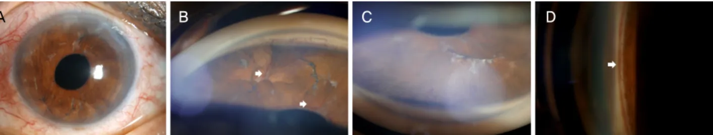
A B C D
Figure 1. Initial anterior segment photographs of the left eye. (A) Slit-lamp examination showing diffuse iris neovascularization. (B)
Note two polypropylene suture knots in the inferior part and (C) one in the superior part accompanying iris neovascularization (arrows). (D) Gonioscopic examination showing neovascularization (arrow) in all quadrants and the representative picture is shown in the temporal quadrant.분 안압하강치료와 함께 허혈성 변화를 유발한 원인 질환 에 대한 치료가 필수이다. 그럼에도 불구하고 인공수정체 홍채고정술 후 발생한 신생혈관녹내장환자에서 안압하강 치료만으로 홍채 및 전방각의 신생혈관이 호전된 증례가 있어 이에 대해 보고하고자 한다.
증례보고
74세 여자 환자가 3일 전부터 시작된 좌안 통증 및 좌측 편두통을 주소로 내원하였다. 초진 시 최대교정시력은 우 안 0.8, 좌안 0.9였고, 골드만압평안압계로 측정한 안압은 우안 14 mmHg, 좌안 38 mmHg였다. 당뇨 외에 전신 질환 은 없었으며, 약 1년 6개월 전에 타 병원에서 우안 백내장 수술, 좌안 부분 유리체절제술 및 인공수정체 홍채고정술 을 받은 병력이 있었다. 세극등현미경검사에서 우안은 낭내 인공수정체가 관찰되었고, 그 외 이상 소견은 없었다. 반면 좌안 검진 시 좌안 결막 충혈 및 경도의 각막부종이 관찰되 었고, 동공은 우안에 비해 약간 산동된 상태로, 동공 반응 은 다소 감소된 상태였다. 좌안에는 신생혈관이 홍채 전반 에 넓게 분포하고 있었고, polypropylene (Prolene®, Ethicon, Cornelia, GA, USA) 실매듭이 홍채의 윗부분에 1개, 아랫 부분에 2개 관찰되어, 좌안은 인공수정체 홍채고정술을 받 았음을 확인하였다(Fig. 1A). 굵고 선명한 홍채신생혈관은 동공연을 따라 분포하고 있었고, polypropylene 실매듭 주 변에도 분포해 있었다(Fig. 1B, C). 좌안 전방은 깊었고, 염 증세포(inflammatory cell)는 관찰되지 않았다. 전방각경검 사에서 우안은 개방각 소견이 관찰되었고, 좌안은 개방각 상태이긴 하였으나 모든 사분면에서 신생혈관이 관찰되었 다(Fig. 1D). 안저검사에서 우안은 정상 소견을 보였던 반 면, 좌안은 하측 시신경유두테의 얇아진 소견(Fig. 2A) 및 이에 상응하는 하부 망막신경섬유층 결손과 국소적 부위의 얇은 망막전막이 관찰되었다(Fig. 2B). 빛간섭단층촬영검사
(Spectralis®, Heidelberg Engineering, Heidelberg, Germany) 에서도 좌안 하측 부위의 얇아진 시신경유두주위 망막신경 섬유층 소견이 관찰되었으며(Fig. 2C), 이에 상응하는 하이 측 황반부 영역에서 망막신경절세포층 결손이 관찰되었다 (Fig. 2D). 자동시야계(Humphrey Field Analyzer II 750, Carl Zeiss Meditec, Dublin, CA, USA)에서 좌안 시신경손 상부위에 상응하는 영역인 상측 부위의 시야 결손이 관찰 되었고, 추가적으로 시행한 형광안저혈관조영술(Spectralis HRA®, Heidelberg Engineering)에서 좌안 망막의 미세동맥 류로 의심되는 병변 외에 망막의 허혈 상태를 시사하는 추 가 소견(망막출혈, 삼출물, 망막내미세혈관이상 등)은 관찰 되지 않았다(Fig. 2E). 이에 좌안 신생혈관녹내장으로 진단 하였으나, 환자가 추가 시술/수술을 거부하여, 일단 안압하 강제인 2% dorzolamide/0.5% timolol (Cosopt®, Merck &
Co, Inc., Whitehouse Station, NJ, USA)와 brimonidine tar- trate (Alphagan-P®, Allergan Pharmaceuticals, Irvine, CA, USA)를 하루 2회 점안, latanoprost (Xalatan®, Pfizer Inc., New York, NY, USA)를 저녁 1회 점안으로 처방 후 점안 제만 사용하면서 경과 관찰하기로 하였다.
치료 시작 1주일 후 내원 시 좌안 교정시력은 0.8이었고, 안압은 14 mmHg로 초진 시에 비해 63.2% 감소되었다. 세 극등현미경검사 시 좌안 각막은 깨끗하였고, 홍채 신생혈 관 및 전방각 신생혈관 분포 범위는 1주일 전에 비해 확연 히 감소하였음을 확인하였다. 환자가 여전히 추가 시술/수 술에 대한 거부감이 강하여 국소 안압하강제 점안만 그대 로 유지하며 경과 관찰하였다. 치료 시작 3개월 후 내원 시 좌안 교정시력은 0.8, 안압은 16 mmHg로 유지되었다. 좌 안 세극등현미경검사에서 각막은 정상 소견을 보였다. 홍 채의 상측 부위 국소 영역에서 홍채 신생혈관이 관찰되었 으나, 전방각 신생혈관은 관찰되지 않았다. 경과 관찰을 위 해 시행한 좌안 형광안저혈관조영술의 소견은 이전과 비슷 함을 확인하였다. 이에 좌안 안압하강제 그대로 유지하며
A B C
D E
Figure 2. Initial examination of the patient. (A) Disc photograph showing an inferior neuroretinal rim thinning (arrow) and (B)
red-free retinal nerve fiber layer (RNFL) photograph showing an inferior RNFL defect (arrowheads) in the left eye. Optical coher- ence tomography image presenting (C) inferior RNFL thinning and (D) corresponding retinal ganglion cell layer defect. (E) Fluorescein angiography image showing no definite ischemic retinal lesion, except several microaneurysms. NS = nasal-superior;TS = temporal-superior; T = temporal; TI = temporal-inferior; NI = nasal-inferior; G = global; N = nasal.
A B C D
Figure 3. Anterior segment photographs of the left eye at six months after topical intraocular pressure lowering treatment. (A)
Slit-lamp examination showing complete regression of the iris neovascularization. (B, C) The remaining polypropylene suture knots fixated in the iris with the compete regression of iris neovascularization. (D) Gonioscopic examination also showing complete re- gression of angle neovascularization.경과 관찰하였다. 치료 시작 6개월 후에도 좌안 안압은 10 mmHg로 유지되었고, 좌안 홍채 신생혈관(Fig. 3A-C) 및 전방각 신생혈관(Fig. 3D)은 관찰되지 않았다. 좌안 시 신경 및 시야 손상 범위는 초진 시와 비슷한 상태였고, 현 재 국소 안압하강제 그대로 유지하며 경과 관찰 중이다.
고 찰
망막질환 또는 전신질환 외에 신생혈관녹내장을 초래할 수 있는 안과적 수술로는 평면부 유리체절제술 또는 수정 체제거술, 공막돌륭술, 실리콘 오일 주입술, 유리체강내주 사, 인공수정체 공막고정술 등이 알려져 있다. 본 증례는
polypropylene 실을 이용하여 인공수정체의 홍채고정술 시 행 후 발생한 신생혈관녹내장환자로 홍채 표면(특히 동공 연 및 실매듭 주위) 및 전방각의 신생혈관이 특징적으로 관 찰되었으며, 이로 인한 방수 유출 저하로 안압상승 및 시신 경 손상을 유발하였을 것으로 생각된다. 본 증례를 통하여 홍채에 단단히 매어 있는 실로도 허혈 유발 및 전안부 신생 혈관 형성이 가능하며, 국소 안압하강제의 사용만으로 신 생혈관 소멸 및 안압하강 치료가 가능함을 보여주었다.
홍채신생혈관발생부터 신생혈관녹내장까지는 크게 신생 혈관 발생 전(prerubeosis), 신생혈관 발생(rubeosis), 개방각 녹내장, 폐쇄각녹내장의 4단계로 나뉜다.1,2 본 증례의 환자 는 초진 시 신생혈관 발생 후 개방각 상태에서 폐쇄각 상태 로 진행되는 단계에 있었을 것으로 생각된다. 홍채 및 전방 각에 신생혈관이 발생하고 섬유혈관조직(fibrovascular tis- sue)이 섬유주 위로 자라나면서 결국 방수 유출 저하 및 폐 쇄각 변화를 유발하여 안압이 상승하고, 이에 동반된 시신 경과 시야 손상이 야기되었을 것으로 생각된다. 이상의 소 견을 종합해 볼 때 본 증례의 경우 신생혈관녹내장 진단이 적합하다고 본다. 본 환자는 양안 모두 안저검사에서 좌안 의 국소적 망막전막 및 경미한 미세동맥류 외에 심한 당뇨 망막병증을 시사하는 소견은 관찰되지 않았고, 경동맥초음 파검사, 뇌 자기공명영상촬영 및 혈액검사 결과 모두 정상 으로 상공막정맥압 상승이나 안허혈증후군을 포함한 다른 전신 질환에 의한 신생혈관녹내장 의심 가능성이 낮았다.
뿐만 아니라 polypropylene 실매듭 주위로 신생혈관이 두드 러지게 분포해 있고 홍채 실질 파괴 및 위축 소견을 보이는 것으로 보아 인공수정체홍채고정술 후 발생한 홍채의 앞섬 모체동맥과 긴뒤섬모체동맥을 통한 혈류 공급 저하에 의한 허혈성 변화가 주된 요인이었을 것으로 생각된다. 본 증례 의 경우 후낭(posterior capsule)이 없는 상태임을 감안할 때 좌안에서 부분 유리체절제술 후 혈액망막장벽(blood-retinal barrier)이 파괴되면서 발생한 망막의 허혈성 변화로 인해 축적된 혈관내피성장인자가 동시에 전안부에 영향을 미쳤 을 가능성도 생각해 볼 수 있을 것이다. 하지만 형광안저혈 관조영술에서 망막의 허혈성 변화를 의심할 만한 소견이 뚜렷하지 않아 신생혈관 발생과 망막병변과의 직접적인 인 과관계를 논하기에는 부족한 측면이 있다. 마지막으로 본 증례에서 홍채섬모체염으로 인한 홍채혈관 확장으로 신생 혈관이 발생하였을 가능성에 대해서도 생각해 볼 수 있다.
하지만 환자의 과거력상 홍채섬모체염을 유발할 만한 전신 질환이 없었으며, 전방 염증세포 및 각막후면침착물, 유리 체 혼탁 등의 홍채섬모체염을 시사하는 소견이 뚜렷하지 않아 가능성이 낮다고 판단하였다.
일반적으로 신생혈관녹내장의 치료 시 안구내 허혈을 초
래한 기저 원인 질환 치료가 우선인 경우가 많다. 당뇨망 막병증, 망막중심정맥폐쇄, 안허혈증후군 등이 신생혈관 녹내장의 원인일 경우 신생혈관의 발생과 증식을 억제하 기 위해 범망막광응고술, 유리체강내 항-혈관내피성장인자 (anti-VEGF) 주입술 등을 우선적으로 시행한다.1,7-9 이와 동 시에 안압하강 치료를 병행하게 되는데 방수생성억제제 (beta adrenergic antagonists, alpha-2 agonists, carbonic an- hydrase inhibitors) 계열의 안압하강제 약물을 일차로 사용 하며, 프로스타글란딘 유사체(prostaglandin analogue)는 안 내 염증에 의한 허혈을 촉발시킬 우려가 있고, 폐쇄각의 경 우 포도막공막 방수 유출로의 기능이 원활하지 않아 약물 의 효능이 저하될 수 있으므로 상황에 따라 추가하도록 권 유되고 있다. 실제 임상에서는 신생혈관녹내장환자에서 안 압이 상승되었다 하더라도 신생혈관의 발생과 증식을 억제 하는 치료만으로도 신생혈관이 소실되면서 안압이 떨어지 는 경우도 종종 경험할 수 있으며 이에 대해 이미 많은 보 고가 이루어져 있다.8,10-12 Wakabayashi et al8이 신생혈관녹 내장 40안을 대상으로 발표한 결과에서도 폐쇄각녹내장 전 단계의 신생혈관 녹내장에서는 유리체강내 베바시주맙(be- vacizumab)주입술 만으로도 신생혈관이 소실되고, 안압이 조절될 수 있음을 보여주었다. 본 증례에서도 전방 내 또는 유리체강내 항-혈관내피성장인자(anti-VEGF)주입술을 시 행하거나 홍채에 팽팽하고 단단하게 고정되어 있는 poly- propylene 실을 제거하는 방법으로 허혈의 호전을 유도할 수 있었을 것으로 생각된다. 하지만 특이한 점은 홍채 앞면 및 전방각의 광범위한 영역에 발생한 신생혈관이 국소 안 압하강점안제만으로 호전되었다는 점이다. 가능한 원인으 로는 다음과 같은 이유를 들어 생각해 볼 수 있을 것이다. 첫 번째로 안압이 하강하면서 안관류압(ocular perfusion pressure)이 증가되어 신생혈관의 소멸을 촉진시켰을 가능 성이 있다. 안관류압은 혈압과 안압의 압력 차이를 뜻하며 안구에 공급되는 혈류량을 간접적으로 보여주는 지표이 다.13 환자의 혈압이 비슷한 정도로 유지되었다고 가정할 때 큰 폭의 안압하강이 이루어지면서 상대적으로 안관류압 이 증가하여 허혈상태를 개선하는 데에 도움을 주었다고 추론해 볼 수 있다. 두 번째로 약에 의한 직접적인 안혈류 량 증가 효과를 추측해 볼 수 있다. 사용한 세 가지 약물 중 dorzolamide/timolol 제제는 방수생성억제제로서의 작용 외에도 안혈류량 및 이완기 안관류압을 증가시킨다고 알려 져 있으며,14 Siesky et al15이 원발개방각녹내장환자를 대상 으로 한 연구에 의하면 dorzolamide는 녹내장환자에서 망 막의 산소포화도를 증가시킨다고 보고한 바 있다. 본 증례 에서 약물이 실제적으로 안구 내의 어느 부위의 혈류량을 증가시켰는지 직접적으로 확인하기는 어려우나 위의 이유
로 안내 허혈이 개선되고 신생혈관 감소에 기여한 결과 안 압이 조절되었을 가능성이 있다.
결론적으로 본 증례는 신생혈관녹내장환자에서 망막의 허혈성 질환이 직접적인 원인이 아니며, 완전 폐쇄각이 아 닌 경우 적절한 안압하강치료만으로도 안관류압 및 안혈류 량을 증가시켜 역설적으로 신생혈관 소멸이 가능할 수도 있음을 보여주었다고 생각한다. 따라서 이러한 경우의 환 자를 검진 및 치료 시 즉각적인 시술 및 침습적인 치료가 어려운 경우 위의 가능성을 염두에 두고 안압하강치료를 먼저 시행해 보는 것도 하나의 방법이 될 수 있겠다.
REFERENCES
1) Sivak-Callcott JA, O'Day DM, Gass JD, Tsai JC. Evidence-based recommendations for the diagnosis and treatment of neovascular glaucoma. Ophthalmology 2001;108:1767-76.
2) Shazly TA, Latina MA. Neovascular glaucoma: etiology, diagnosis and prognosis. Semin Ophthalmol 2009;24:113-21.
3) Hayreh SS. Neovascular glaucoma. Prog Retin Eye Res 2007;26:
470-85.
4) Jeong YC, Hwang YH. Etiology and features of eyes with rubeosis iridis among Korean patients: A Population-Based Single Center Study. PLoS One 2016;11:e0160662.
5) Cohen S, Kremer I, Yassur Y, Ben-Sira I. Peripheral retinal neo- vascularization and rubeosis iridis after a bilateral circular buck- ling operation. Ann Ophthalmol 1988;20:153-6.
6) Barile GR, Chang S, Horowitz JD, et al. Neovascular complica-
tions associated with rubeosis iridis and peripheral retinal detach- ment after retinal detachment surgery. Am J Ophthalmol 1998;126:
379-89.
7) Aref AA. Current management of glaucoma and vascular occlusive disease. Curr Opin Ophthalmol 2016;27:140-5.
8) Wakabayashi T, Oshima Y, Sakaguchi H, et al. Intravitreal bev- acizumab to treat iris neovascularization and neovascular glauco- ma secondary to ischemic retinal diseases in 41 consecutive cases.
Ophthalmology 2008;115:1571-80, 80.e1-3.
9) Wand M, Dueker DK, Aiello LM, Grant WM. Effects of panretinal photocoagulation on rubeosis iridis, angle neovascularization, and neovascular glaucoma. Am J Ophthalmol 1978;86:332-9.
10) Iliev ME, Domig D, Wolf-Schnurrbursch U, et al. Intravitreal bev- acizumab (Avastin) in the treatment of neovascular glaucoma. Am J Ophthalmol 2006;142:1054-6.
11) Batman C, Ozdamar Y. The effect of bevacizumab for anterior seg- ment neovascularization after silicone oil removal in eyes with pre- vious vitreoretinal surgery. Eye (Lond) 2010;24:1243-6.
12) Mason JO 3rd, Albert MA Jr, Mays A, Vail R. Regression of neo- vascular iris vessels by intravitreal injection of bevacizumab.
Retina 2006;26:839-41.
13) Costa VP, Harris A, Anderson D, et al. Ocular perfusion pressure in glaucoma. Acta Ophthalmol 2014;92:e252-66.
14) Rolle T, Tofani F, Brogliatti B, Grignolo FM. The effects of dorzo- lamide 2% and dorzolamide/timolol fixed combination on retinal and optic nerve head blood flow in primary open-angle glaucoma patients. Eye (Lond) 2008;22:1172-9.
15) Siesky B, Harris A, Cantor LB, et al. A comparative study of the ef- fects of brinzolamide and dorzolamide on retinal oxygen saturation and ocular microcirculation in patients with primary open-angle glaucoma. Br J Ophthalmol 2008;92:500-4.
= 국문초록 =
망막비허혈 신생혈관녹내장환자에서 안압하강치료만으로 신생혈관 소실
목적: 인공수정체홍채고정술 후 발생한 신생혈관녹내장환자에서 원인 질환에 대한 치료 없이 안압하강치료만으로 홍채 및 전방각의 신생혈관이 호전된 1예를 보고하고자 한다.
증례요약: 양안 백내장수술을 받은 74세 여자 환자가 3일 전부터 시작된 좌안 통증 및 편두통을 주소로 내원하였다. 초진 시 좌안 최대교정시력은 0.9, 안압은 38 mmHg였다. 세극등현미경검사 시 좌안 홍채 전반에 분포된 신생혈관과 홍채 위아래 부분에 polypropylene 실매듭이 관찰되었다. 전방각경검사 시 좌안 모든 사분면의 신생혈관 및 아래 사분면의 국소적 주변홍채앞유착이 관 찰되었다. 안저검사에서 시신경 하측 시신경유두테의 얇아진 소견 및 망막신경섬유층 손상이 관찰되었고, 형광안저혈관조영술에서는 망막미세동맥류 이외 특이 소견은 없었다. 좌안 신생혈관녹내장 진단하에 안압하강제만을 점안하며 경과 관찰하였고, 치료 시작 6개 월 후 안압은 10 mmHg였고, 홍채 및 전방각 신생혈관은 소실되었다.
결론: 망막의 허혈성 병변이 원인이 아닌 신생혈관녹내장환자에서 완전 폐쇄각 상태가 아니며 즉각적인 원인 제거 치료가 어려운 경우, 안압하강치료를 우선적으로 시행하며 경과 관찰할 수 있겠다.
<대한안과학회지 2020;61(3):307-312>
이상욱 / Sang Wook Lee
을지대학교 의과대학 노원을지대학교병원 안과학교실 Department of Ophthalmology,
Nowon Eulji Medical Center, Eulji University School of Medicine