접 수 일:2009. 7. 28.
채 택 일:2009. 11. 25.
교신저자:이귀세라 E-mail:[email protected]
전신성홍반성낭창과 관련된 폐고혈압 환자에서의 성공적인 임신
가톨릭대학교 의과대학 산부인과학교실
장동규 ․ 정윤지 ․ 이 영 ․ 이귀세라A successful pregnancy in patient with pulmonary hypertension associated with systemic lupus erythematosus
Dong Gyu Jang, M.D., Yoon Ji Chong, M.D., Young Lee, M.D., Guisera Lee, M.D.
Department of Obstetrics and Gynecology, School of Medicine, The Catholic University of Korea, Suwon, Korea
Pulmonary hypertension is a rare and potentially life-threatening complication of Systemic lupus erythematosus (SLE), and 5 cases has been previously documented in pregnancy. Four cases died after delivery and only one case was alive. We describe the case of a 28-year-old pregnant woman with pulmonary hypertension related to SLE with no previous history of immunologic disease including SLE. Diagnosis was made at 22 weeks of gestation. Medication including prednisolone and hydroxychloroquinone was commenced immediately and continued throughout the pregnancy. On fetal sonogram, the fetal growth was 3~10 percentile and diastolic notch of uterine arteries was noted. However, a healthy baby girl weighing 2,400 g was born in planned vaginal delivery at gestation week 38. There were no postpartum complications.
Key Words: Pregnancy, Systemic lupus erythematosus, Pulmonary hypertension, Intrauterine growth restriction
Pulmonary hypertension is a rare and potentially life-threatening complication of SLE, and 5 cases has been previously documented in pregnancy.1-3 Only one case was alive after delivery,1 other 4 cases died after delivery.1-3 Pulmonary hypertension is reported in 5%
to 14% of patients with SLE. Overall mortality is 25~50% two years after diagnosis of pulmonary hypertension.4 The most common symptoms of pulmo-
nary hypertension in patients with SLE are fatigue, dyspnea and syncope. Raynaud’s phenomena is present in 75% of patients with pulmonary hypertension re- lated to SLE, compared with 25% in patients with SLE without pulmonary hypertension.5 Echocardiography reveals right ventricular hypertrophy and dilatation.
Pulmonary hypertension in patients with SLE is asso- ciated with intimal hyperplasia, smooth muscle hyper- trophy and medial thickening, similar to the changes seen in primary pulmonary hypertension.4 The devel- opment of pulmonary hypertension is not necessarily consistent with the stage of the SLE and severity of
A B
Fig. 1. Chest X-ray and Electrocardiogram on admission. Chest X-ray revealed cardiomegaly (cardiothoracic ratio >0.66) and mild pulmonary congestion. Electrocardiogram showed low voltage with normal sinus rhythm.
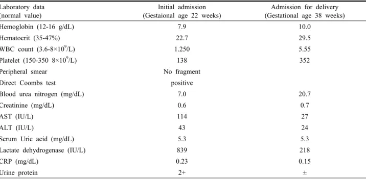
Table 1. Laboratory findings Laboratory data
(normal value)
Initial admission (Gestaional age 22 weeks)
Admission for delivery (Gestational age 38 weeks)
Hemoglobin (12-16 g/dL) 7.9 10.0
Hematocrit (35-47%) 22.7 29.5
WBC count (3.6-8×10
9/L) 1.250 5.55
Platelet (150-350 8×10
9/L) 138 352
Peripheral smear No fragment
Direct Coombs test positive
Blood urea nitrogen (mg/dL) 7.0 20.7
Creatinine (mg/dL) 0.6 0.7
AST (IU/L) 114 27
ALT (IU/L) 43 24
Serum Uric acid (mg/dL) 5.3 5.3
Lactate dehydrogenase (IU/L) 839 218
CRP (mg/dL) 0.23 0.15
Urine protein 2+ ±
the lupus activity. The diagnosis is frequently delayed because the symptoms tend to occur late by which time the pulmonary artery pressure may be severely ele- vated and cardiac output reduced.1
The case presented here is that of a pregnant wom- an with SLE and pulmonary hypertension which has not been hitherto documented in Korea.
Case Report
A 25-year-old multiparous woman, was referred to us at 22 weeks of gestation. The chief complaints were dyspnea, fatigue, anorexia, fevers and chills, and pain sensation of both legs for 2 weeks. A cyanotic face and clubbed fingers were noted at admission.
A B C
D E F
Fig. 2. Doppler imaging to evaluate the bilateral uterine artery, MCA and umbilical artery for assessment of fetal health. (A) The early diastolic notching is noted at right uterine artery. (B) The early diastolic notching is noted at left uterine artery. (C) The umbilical artery shows normal velocities at 24 weeks of gestation. RI=0.69. (D) MCA shows high resistance and low diastolic velocities at 24 weeks of gestation. RI=0.82. (E) The umbilical artery shows an increase in diastolic velocities and a decline in RI (=0.54) at 33 weeks of gestation compared as that at 24 weeks of gestation. (F) MCA shows high diastolic velocities at 33 weeks of gestation. R=0.66.
The patient’s history revealed no previous disease diagnosis including immunologic diseases. Two years previously, the patient had uneventfully given birth a 2.4 kg baby at term.
Chest X-ray revealed cardiomegaly (cardiothoracic ratio >0.66) and mild pulmonary congestion (Fig. 1A).
Electrocardiogram showed low voltage with normal si- nus rhythm (Fig. 1B). An echocardiogram showed an elevated right ventricular systolic pressure 87.4 mmHg (normal 20~25 mmHg) with mild tricuspid regur- gitation, an elevated right atrial pressure of 15 mmHg (normal 0~8 mmHg), right ventricular and atrial dila- tation, and a small amount of pericardial effusion.
Left–sided cardiac function was normal, and intra- cardiac shunt was not seen. Computed tomography (CT) of the lung was performed to exclude pulmonary thrombosis or embolus as a cause of pulmonary hypertension. CT did not reveal occlusion or thrombus of pulmonary arteries, and showed bilateral pleural
effusion and pericardial effusion. Complete blood count showed leucopenia and anemia (white blood cell count (WBC); 1,250/dL (66.3% neutrophil, 17.2% lym- phocyte), hemoglobin (Hb); 7.9 g/dL, hematocrit (Hct);
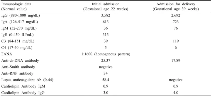
24.4, platelet count 138,000/μL). Urine analysis showed 2+ urine protein. The liver enzyme aspartate aminotransferase/alanine trasnsaminase (AST/ALT) were slightly increased (114/43 U/L) and blood urea nitrogen and creatinine (BUN/Cr) was normal range (5.0/7.0 mg/day) (Table 1). Immunologic laboratory results were FANA (+) (1:1600, homogenous type), Anti-ds DNA (+) (25.37), Lupus anticoagulant (+), Anti-RNP (3+), coombs direct test (+), hypocomple- tenemia (C3/C4: 39/5) (Table 2).
On fetal sonogram, fetal estimated weight was about the 3rd percentile size (0.42 kg) and early dia- stolic notch sign of bilateral uterine arteries was not- ed (Fig. 2A, B). Resistant index (RI) of the umbilical arteries was 0.69 (Fig. 2C) and RI of middle cerebral
Table 2. Immunologic Data Immunologic data
(Normal value)
Initial admission (Gestaional age 22 weeks)
Admission for delivery (Gestational age 39 weeks)
IgG (880-1800 mg/dL) 3,582 2,692
IgA (126-517 mg/dL) 613 723
IgM (52-270 mg/dL) 36 76
IgE (0-450 IU/mL) 313
C3 (84-151 mg/dL) 39 119
C4 (17-40 mg/dL) 5 6
FANA 1:1600 (homogenous pattern)
Anti-ds-DNA antibody 25.37 17.89
Anti-Smith antibody negative
Anti-RNP antibody 3+
Lupus anticoagulant Ab (0-44) 58.4 negative
Cardiolipin Antibody IgM 0.9 0.9
Cardiolipin Antibody IgG 3.0 4.0
artery (MCA) was 0.82 (Fig. 2D), which were both showed within normal range. No anomalies were found on the fetal echocardiogram. Based on these findings, the patient was diagnosed with severe pulmonary hy- pertension secondary to SLE in pregnancy, with intra- uterine growth restriction (IUGR)
Oral prednisolone (15 mg/day) and hydroxy- chloroquine (400 mg/day) was immediately commenced.
An echocardiogram obtained 1-week later showed reduced right ventricular systolic pressure (59 mmHg).
The patient was discharged on day 27 on medication of prednisolone and hydroxychloroquine. She visited ev- ery week at department of obstetrics and rheumato- logy.
During the remainder of pregnancy, other than mild dyspnea, the patient’s condition and course were unremarkable. A follow-up sonography revealed an estimated fetal weights of 1.16 kg at 29 weeks of gestation (10th percentile), 1.39 kg at 31 weeks of gestation (10th percentile) and 1.63 kg at 33 weeks of gestation (3rd percentile). No evidence of fetal compromise was evident. RI of umbilical artery at 33 weeks of gestation (0.54) was within the normal range
compatible with gestation (Fig. 2E), which shows an increase in diastolic velocities and a decline in RI (=0.69) compared as that at 24 weeks of gestation.
However, early diastolic notch of uterine artery persisted and the diastolic velocity of MCA increased (RI=0.66) (Fig. 2F).
The initial plan was to allow the pregnancy to proceed to at most 34 weeks and then induce labor for vaginal delivery. However, upon the insistence of the patient and her husband, the pregnancy was extended to 38 weeks while ensuring that the health of both patient and fetus was maintained. At the time of admission for labor induction, patient’s blood pressure was 120/80 mmHg and some mild dyspnea was noted.
The dilatation of cervix was about 2 cm. The degree of facial cyanosis and finger clubbing had not changed.
Chest X-ray did not demonstrate cardiomegaly or other abnormalities. Laboratory results were improved from the initial laboratory data. Hb 10.0 g/dL, WBC 5,500/dL, platelet count 352,000/μL, AST/ALT 27/24 IU/L, C3 119 mg/dL, anti-ds-DNA antibody 17.89 (Table 2).
Low dose oxytocin infusion was used for augmenta-
tion of labor and epidural blockade was done for analgesia. Labor was uneventful and a healthy female neonate weighing 2.40 kg was delivered vaginally.
Apgar scores at 1 and 5 min were 7 and 5, respec- tively, and the pH of umbilical artery and vein was 7.290.
The patient was managed on intensive care unit and after 72 hours she was transferred back to the maternity unit. The patient’s vital signs were normal and there was not profuse vaginal bleeding. The patient made an uneventful postpartum recovery and was discharged 6-days after delivery on an oral regimen of prednisolone and hydroxychloroquine. The pathology of placenta showed acute chorioamnionitis with microcalcification. Echocardiographic studies revealed dilatation of the right ventricle regurgitation of mild tricuspid was noted. PA systolic pressure of Right ventricle decreased from 87.5 mmHg in pregnancy to 50 mmHg at postpartum. Follow-up examination over the next 3-months conducted at rheumatology-cardiologic clinic and the obstetric clinic revealed no obvious deterioration.
Discussion
Respiratory involvement in SLE is not as well known as the cutaneous, rheumatological and renal manifestations. It can be classified in 5 groups based on the anatomy: pleural involvement, infiltrating pneumonia, airways involvement, vascular involve- ment, muscular and diaphragmatic involvement.
Pulmonary hypertension represents vascular involve- ment.6 Pulmonary hypertension is present when sys- tolic pulmonary arterial pressure exceeds 30 mmHg or the mean pulmonary artery pressure exceed 20 mmHg.7 The initial systolic pulmonary arterial pres- sure of this case’s patient was 87.4 mmHg. The causes of secondary pulmonary hypertension in pregnancy were Takayasu’s arteritis, pulmonary vasculitis of
connective tissue such as SLE or scleroderma, sickle cell disease, chronic pulmonary thromboembolism, hepatitis, dwarfism with congenital hypothyroidism and peripheral pulmonary stenoses.8 We performed CT of the lung to exclude pulmonary thrombosis or embo- lus as a cause of pulmonary hypertension.
The pathogenesis of pulmonary hypertension in SLE has not elucidated clearly yet. However, the fre- quently observed association with Raynaud’s phenom- enon is consistent with a chronic and diffuse vaso- spastic condition which leads to muscular necrosis and secondary inflammation.9 The mortality of pulmonary hypertension in SLE was exceeds 50%, and often in- volves sudden death. Five cases of pulmonary hyper- tension associated with SLE have been previously documented in pregnancy.1-3 All but one delivery ended in death within 96 h postpartum.1 The time of highest risk for death from pulmonary hypertension is immediately after delivery or within the first 72 h postpartum. The reason of highest risk for maternal death in the first 48~72 hours after delivery may be related to the rise in the cardiac output and total blood volume during the pregnancy and the immediate postpartum period. The heart has the added load of an increased cardiac output with a fixed vascular resist- ance in the first 48~72 hours after delivery. In a pa- tient with pulmonary hypertension, the increasing cardiac output and the already increased pulmonary vascular resistance above normal may result in re- duced left ventricular filling, hypoxic state, and fi- nally acute right heart failure as the cause of sudden maternal death.1,3
SLE and/or pulmonary hypertension are frequently associated with IUGR. IUGR has been reported in up to 33% of cases of pulmonary hypertension10 and 40%
of cases of SLE.11 All five neonates, who previously documented in the cases of pulmonary hypertension associated with SLE in pregnancy, were IUGR.1-3 Antiphospholipid antibodies, hypocomplementemia
correlates with IUGR in SLE.1,12 Bilateral uterine ar- tery notches at 22~24 weeks gestation in women with positive lupus anticoagulant and/or antiphospholipid antibody is a diagnostically accurate indicator of ad- verse pregnancy outcomes that including IUGR, pre- eclampsia, placental abruption and intrauterine fetal death.13,14 Thrombogenic action of lupus anticoagulant and/or antiphospholipid antibody leads to decreased placental perfusion and subsequent infarction. The antiphospholipid antibody-mediated inhibition of trophoblastic invasion and antiphospholipid anti- body-mediated vasculopathy in placental bed arteries result in abnormal uterine artery waveforms.14 In the present case, bilateral uterine artery notches were observed at gestation week 22 and were maintained until term. And hypocomplementenia and positive lu- pus anticoagulant antibody were shown in the im- munologic laboratory data. At term, MCA Doppler ve- locities showed increased pattern that means brain sparing effect in IUGR.15 The neonate’s birth weight corresponded to about the 3rd percentile,16 fortunately other adverse pregnancy outcome such as preeclampsia or intrauterine death did not occur.
Vaginal delivery may be appropriate if adequate
pain control is provided and fetal distress does not develop.1 Oxytocin should be used with care because of its systemic vasodilatation. However, low dose oxy- tocin infusion used for augmentation of labor dose not seem to be have detrimental effects.17 Adequate an- algesia is necessary for preventing from maternal hy- poxia and acidosis developed due to labor pain. The maternal intensive care should be necessary for 72 h after birth because this period is critical time for ma- ternal death. Pulmonary vasodilators such as nitric oxide and prostaglandin have some evidence that treatment with those medication improve symptoms and exercise capacity in the non-pregnant patient.18,19 However, there are a few cases in the use of prosta- glandin or nitric oxide in pregnancy.20,21
In summary, very few reports have been published on lupus-related pulmonary hypertension in pregnancy.
Most of the reported cases ended in the early post- partum death. Fortunately, in the present case, delivery was uneventful and both the patient and the infant con- tinue to thrive. It is hoped that early diagnosis and ad- mission and individualized care will lead more often to successful outcome.