대흉외지 2007;40:867-870 □ 증례보고 □
− 867 −
*울산대학교 의과대학 서울아산병원 흉부외과학교실
Department of Thoracic and Cardiovascular Surgery, Asan Medical Center, University of Ulsan College of Medicine
**울산대학교 의과대학 울산대학교병원 흉부외과학교실
Department of Thoracic and Cardiovascular Surgery, Ulsan University Hospital, University of Ulsan College of Medicine 논문접수일:2007년 8월 16일, 심사통과일:2007년 9월 13일
책임저자:이재원 (138-736) 서울시 송파구 풍납2동 388-1, 서울아산병원 흉부외과 (Tel) 02-3010-3580, (Fax) 02-3010-6966, E-mail: [email protected] 본 논문의 저작권 및 전자매체의 지적소유권은 대한흉부외과학회에 있다.
항인지질 증후군과 전신성 홍반성 루푸스 환자에게 발생된 만성 폐혈전색전성 폐동맥 고혈압에 대해 시행한 혈전내막제거술
강 필 제*ㆍ이 재 원*ㆍ김 정 원**
Pulmonary Thromboendarterectomy for Chronic Thromboembolic Pulmonary Hypertension in a Patient with Antiphospholipid Syndrome
and Systemic Lupus Erythematosus
Pil Je Kang, M.D.*, Jae-Won Lee, M.D.*, Jeong Won Kim, M.D.**
Antiphospholipid syndrome (APS) is defined as venous and/or arterial thromboses, recurrent fetal losses, thrombocy- topenia in combination with repeatedly positive tests for the lupus anticoagulant (LAC), and anticardiolipin antibodies (aCL). The pulmonary manifestations in APS are relatively rare. We report a rare case of antiphopholipid syndrome with systemic lupus erythematosus in a patient who presented with pulmonary hypertension secondary to a chronic pulmonary thromboembolism. A bilateral thromboendarterectomy was performed satisfactorily and the incision was extended to the left intrapleural pulmonary artery.
(Korean J Thorac Cardiovasc Surg 2007;40:867-870) Key words: 1. Pulmonary embolism
2. Thromboembolism 3. Thromboendarterectomy 4. Antiphospholipid syndrome 5. Systemic lupus eruthematosus
증 례
15세 남자 환자가 호흡곤란과 쉽게 멍드는 현상을 주소 로 2005년 3월 외부 병원 방문 후 본원 소아과로 전원되 었다. 환자는 심장초음파검사에서 폐동맥고혈압이 확인되 어 시행한 흉부 전산화 단층 촬영에서 양쪽 상폐동맥과 왼쪽 하폐동맥에 폐혈전색전증 소견이 관찰되었다. 혈액 검사에서 혈소판 73,000/μL로 감소되어 있었고, FANA 양 성, VDRL 1:1 양성, lupus anticoagulant (LAC) 양성, Anti- cardiolipin (aCL) antibodies IgG과 IgM은 각각 강양성(+++)
과 음성, Anti-dsDNA antibody 4.1, Protein C 88% (정상 70∼
140%), Protein S free 105% (정상 70∼130%), Antithrombin III level 68 mg/dL, C3 42.3 mg/dL, C4 4.7 mg/dL 등이었다.
환자의 증상과 증후 및 혈액검사를 통해 항인지질증후 군과 전신성 홍반성 루푸스로 진단하였고, 호흡곤란은 폐 혈전색전증에 의한 폐동맥고혈압으로 비롯된 것으로 생 각하여 prednisolone 충격요법(pulse therapy)을 시행하였다.
이후 외래에서 경과 관찰하던 중 운동시 호흡곤란 및 객 혈이 발생하여 싸이톡산 1g을 정맥주사하였으나, 운동시 호흡곤란이 악화되고 객혈, 가슴 통증 등이 심해져서 심
대흉외지 2007;40:867-870
− 868 −
Fig. 3. Left pulmonary arteriotomy is extended to the left pleural.
Fig. 2. Preoperative chest CT shows pulmonary thromboembolism of supe- rior, inferior segmental arteries of left lower lobe (arrows) and superior seg- mental artery of right lower lobe.
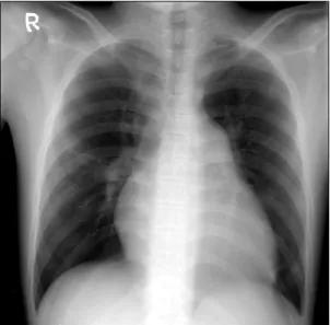
Fig. 1. Preoperative chest X-ray shows enlarge pulmonary conus and decreased pulmonary vascular marking in both lung fields.
부전, 폐혈전색전증의 진행을 의심하며 입원하였다. 단순 흉부방사선촬영에서 우측 중하엽에 간유리 양상을 보였 고 흉막액을 동반한 심비대가 있었다(Fig. 1). 흉부 전산화 단층촬영은 좌하엽의 상구역동맥에 있던 혈전색전증은 좀 더 근위부로 진행되었고 좌하엽에 하구역동맥과 우하 엽의 상구역동맥의 폐혈전색전증은 변화가 없었다(Fig. 2) 심장초음파검사에서 우심실이 커져 있었고 계산된 압력 은 88 mmHg로 매우 높았으며, 난원공 등을 통한 좌-우 단 락은 없었고 삼첨판 폐쇄부전(Grade 3/4)과 안정시 폐고혈 압(TR Vmax 4.7 m/s)이 관찰되었다. 하지 초음파검사에서 심정맥혈전증(deep vein thrombosis)은 관찰되지 않았으나 반복적인 폐동맥색전증을 예방하기 위하여 하대정맥 필 터를 삽입한 후 폐혈전색전제거술을 시행하기로 하였다.
수술은 정중 흉골절제한 후 상행대동맥과 상, 하대정맥 에 삽관하여 심폐바이패스를 시행하였다. 주폐동맥은 커 져 있었으며 대동맥 겸자 후 좌우폐동맥을 열고 관찰한 결과 우측 폐동맥은 우하엽 상구역 동맥에 만성적인 혈전 조직으로 막혀 있었다. 좌측 폐동맥은 거의 막혀 있었으 며, 좌상엽 및 좌하엽 모든 구역 동맥이 혈전으로 가득 차 있었다. Jamieson dissector를 이용하여 혈전을 제거하던 중 흘러나오는 혈액량이 많아 중등도 저체온 및 총혈류정지 상태(total circulatory arrest; TCA)로 수술을 진행하였다. 좌 측 폐동맥은 시야를 확보하기 위해 좌측 폐의 대열을 분 리하여 좌하엽 구역 동맥을 절게한 후 혈전색전 제거술을 시행하였다(Fig. 3). 총 심폐기 가동 시간은 168분이었으 며, TCA 시간은 40분이었다. 폐동맥혈압은 전신 혈압
강필제 외 Chronic Thromboembolic Pulmonary Hypertension with Antiphospholipid Syndrome and SLE
− 869 − Fig. 4. Immediate postoperative chest CT shows reduction of eccen- tric pulmonary thrombus below the level of both interlobar pulmonary artery.
90/60 mmHg일 때 수술 전 86/54 mmHg에서 수술 직후 55/32 mmHg로 감소되었다.
수술 후 2일째까지 NO 가스를 적용하였고, 수술 후 6일 째 호흡기 이탈을 한 후 8일째 일반 병실로 전동되었다.
수술 후 9일째 시행한 심장초음파검사에서 우심실압은 55 mmHg로 감소하였고, 삼첨판 폐쇄부전(Grade 1/4)과 폐고 혈압(TR Vmax 3.7 m/s)은 감소하였으며, 흉부 전산화단층 촬영에서 이전과 비교하여 호전된 폐동맥 혈류를 확인할 수 있었다(Fig. 4). 수술 후 18일째 퇴원하여 특별한 문제 없이 23개월째 외래 경과 관찰 중이다.
고 찰
항인지질 증후군은 정맥, 동맥의 혈전증, 혈소판 감소 증, 반복적인 유산 등과 함께 lupus anticoaculant (LAC)와 anticardiolipin antibody (aCL)가 반복적으로 양성 반응을 보이는 질환을 말한다[1-3]. 항인지질 증후군은 어느 관련 질병 없이 생길 수 있어서 전신성 홍반성 루푸스(SLE)나 다른 류마티스성 질환, 자가면역 질환을 가진 환자에서도 일어날 수 있다[2]. 혈전이 왜 발생하는지에 대해서는 아 직 정확히 밝혀지지 않았으나, 한 가설은 세포의 자멸사 과정에서 세포의 손상이 있어서, 이것이 세포막 인지질과 다양한 응고 단백질의 결합을 유도하여 인지질-단백질 결 합체가 형성되고 새로운 항원 결정기가 왔을 때 연속적으 로 자가항체에 목표가 된다고 한다[4]. 이 질환은 심근 경
색, 폐동맥 혈전색전증, 뇌졸중, 장간막 혈전증 등이 발생 하기 때문에 예후가 나쁘다[1,3]. 항 인지질 증후군의 호흡 기 증상 발현은 상대적으로 드물다. 폐동맥의 색전증과 경색, 폐고혈압, 급성호흡부전 증후군, 폐포 내 출혈, 크고 작은 폐혈관의 원발성 혈전 등이 있으며 이러한 경우
“antiphopholipid lung syndrome”이라고 칭한다[1].
항인지질 증후군을 가진 환자에서 급성 혈전색전증의 치료는 다른 질병군과 동일하여 헤파린 정맥주사에 이어 와파린으로 전환되어 평생 복용하여야 한다. 항인지질 증 후군에서 적절한 항응고 용량은 혈전증이 있었으면 고강 도(PT INR>3.0)의 치료를 해야 재발이 없다는 보고도 있 으나[4,5] 보통 PT INR을 2.5에서 3.0 정도 유지한다.
폐동맥색전증에 의한 폐동맥고혈압은 서서히 진행하여 우심실 기능부전으로 발전하여 사망에 이르는 질환이다.
비 특이적인 호흡곤란을 호소하며 절반 정도에서 흉통, 그리고 객혈을 동반하기도 한다. 신체검사에서 경정맥 확 장이나 간비대, 사지부종, 청색증 등 다양한 증후가 관찰 된다. 진단은 심장초음파검사를 통해 폐동맥고혈압, 우심 실기능부전, 삼첨판 역류 정도, 그리고 난원공 개존의 유 무 등을 확인할 수 있다. 과거에는 폐동맥조영술을 시행 하여 확진하였으나, 최근 컴퓨터단층촬영이 발달하면서 우심실비대나 폐동맥 확장의 정도, 혈전의 양상이나 위치, 폐분엽동맥과 폐실질의 변화 등 여러 정보를 얻을 수 있 게 되었다. 그러나 색전증이 진행한 경우 내과적인 치료 만 한다면 예후가 나쁘기 때문에 수술적인 제거술을 시행 한다[6].
급성 폐동맥색전증에 대한 치료는 1924년 Kirschner가 폐동맥색전 제거술을 시행하여 첫 생존 예를 보고하였고, Cooley 등이 1961년 심폐바이패스하에 폐동맥색전 제거술 을 처음 보고하였다[7]. 만성 폐동맥색전증은 1950년 Carroll에 의해 처음 시도되었으나 사망하였고, 1970년대 에 Carol 등이 심폐바이패스 없이 측면개흉을 통한 편측 폐 혈전내막제거술을 시행하여 37.5%의 사망률을 보고하 였다[5]. 이후 수술 사망률은 점차 감소되어 최근에는 Thistlethwaite 등[8]이 2005∼6년 동안 4.3%까지 보고하고 있다. 폐 혈전내막 제거술은 폐 혈전색전증으로 발생하는 폐동맥 고혈압에 의한 우측심장부전을 예방하여 환자의 생명을 구하는 시술이다.
항인지질증후군에서 발생한 급성 폐혈전색전증은 몇몇 보고가 있었지만, 만성 폐혈전색전증에 대한 수술은 매우 드물다[4]. 저자들은 항인지질 증후군 및 전신성 홍반성 루푸스 환자에서 발생한 만성적인 폐혈전색전증에 대하
대흉외지 2007;40:867-870
− 870 − 여 폐동맥 내막절제술을 성공적으로 시행하였고, 특히 원 위부에 있는 폐혈전색전증을 왼쪽 폐의 대열을 분리한 뒤 좌하엽 구역 동맥을 절제하여 혈전색전 제거술을 성공적 으로 시행하였기에 문헌고찰과 함께 국내에서 처음으로 보고하는 바이다.
참 고 문 헌
1. Stojanovich L. Pulmonary manifestations in antiphos- pholipid syndrome. Autoimmun Rev 2006;5:344-8.
2. Wilson WA, Gharavi AE, Koike T, et al. International consensus statement on preliminary classification criteria for definite antiphospholipid syndrome: report of an inter- national workshop. Arthritis Rheum 1999;42:1309-11.
3. Medina G, Vera-Lastra O, Barile L, Salas M, Jara LJ.
Clinical spectrum of males with primary antiphospholipid syndrome and systemic lupus erythematosus: a compara-
tive study of 73 patients. Lupus 2004;13:11-6.
4. Nakajima T, Ando H, Ueno Y, et al. Successful throm- boendarterectomy for chronic pulmonary embolism in a patient with systemic lupus erythematosus and antiphos- pholipid syndrome. Jpn Circ J 1997;61:958-64.
5. Rosove MH, Brewer PM. Antiphospholipid thrombosis:
clinical course after the first thrombotic event in 70 patients. Ann Intern Med 1992;117:303-8.
6. Song SH, Park PW, Jun TG, et al, Pulmonary throm- boendarterectomy for pulmonary hypertension caused by chronic pulmonary thromboembolism. Korean J Thorac Cardiocasc Surg 2006;39:626-32.
7. Cooley DA, Beall AC Jr, Alexander JK. Acute massive pulmonary embolism. Successful surgical treatment using temporary cardiopulmonary bypass. JAMA 1961;177:283-6.
8. Thistlethwaite PA, Madani M, Jamieson SW. Outcomes of pulmonary endarterectomy surgery. Semin Thorac Car- diovasc Surg 2006;18:257-64.
=국문 초록=
항인지질 증후군(antiphospholipid syndrome)은 정맥, 동맥의 혈전증, 혈소판 감소증, 반복적인 유산 등과 함께 lupus anticoagulant (LAC)와 anticardiolipin antibody (aCL)가 반복적으로 양성 반응을 보 이는 질환을 말하며 호흡기 증후 발현은 상대적으로 드물다. 저자들은 항인지질 증후군 및 전신성 홍 반성 루푸스 환자에서 발생한 만성적인 폐혈전색전증에 대하여 폐동맥 내막절제술, 특히 원위부에 있는 폐혈전색전증을 왼쪽 폐의 대열을 분리한 뒤 좌하엽 구역 동맥을 절제하여 혈전색전 제거술을 성공적으로 시행하였기에 문헌고찰과 함께 국내 최초로 보고하는 바이다.
중심 단어:1. 폐색전증 2. 혈전색전증 3. 혈전내막제거술 4. 항인지질 증후군 5. 전신성 홍반성 루푸스