25
ABBREVIATIONS: GDP-β-S, guanosine 5'-[β-thio]diphosphate trili- thium; ICC, interstitial cells of Cajal.
Corresponding Author: Jae Yeoul Jun, Department of Physiology, College of Medicine, Chosun University, 375, Seoseok-dong, Dong-gu, Gwangju 501-759, Korea. (Tel) 82-62-230-6412, (Fax) 82-62-232- 4943, (E-mail) jyjun@ chosun.ac.kr
Involvement of Thromboxane A
2in the Modulation of Pacemaker Activity of Interstitial Cells of Cajal of Mouse Intestine
Jin Ho Kim1, Soo Jin Choi2, Cheol Ho Yeum3, Pyung Jin Yoon3, Seok Choi3, and Jae Yeoul Jun3 Departments of 1Neurology and 3Physiology, College of Medicine, Chosun University, Gwangju 501-759, 2Department of Radiology, Gacheon University Gil Medical Center, Guwol-dong, Incheon 405-760, Korea
Although many studies show that thromboxane A2 (TXA2) has the action of gastrointestinal (GI) motility using GI muscle cells and tissue, there are no reports on the effects of TXA2 on interstitial cells of Cajal (ICC) that function as pacemaker cells in GI tract. So, we studied the modulation of pacemaker activities by TXA2 in ICC with whole cell patch-clamp technique. Externally applied TXA2
(5μM) produced membrane depolarization in current-clamp mode and increased tonic inward pacemaker currents in voltage-clamp mode. The tonic inward currents by TXA2 were inhibited by intracellular application of GDP-β-S. The pretreatment of ICC with Ca2+ free solution and thapsigargin, a Ca2+-ATPase inhibitor in endoplasmic reticulum, abolished the generation of pacemaker currents and suppressed the TXA2-induced tonic inward currents. However, chelerythrine or calphostin C, protein kinase C inhibitors, did not block the TXA2-induced effects on pacemaker currents. These results suggest that TXA2 can regulate intestinal motility through the modulation of ICC pacemaker activities.
This modulation of pacemaker activities by TXA2 may occur by the activation of G protein and PKC independent pathway via extra and intracellular Ca2+ modulation.
Key Words: Thromboxane A2 (TXA2), Interstitial cells of Cajal (ICC), Pacemaker currents, Intestinal motility
INTRODUCTION
The prostanoids are oxygenated derivatives of C20 fatty acids, principally arachidonic acid. Five pathways of arachidonic acid metabolism are currently recognized, and prostanoids are the products of the first of these to be discovered, the cyclooxygenase pathway. There are two major classes of prostanoids, the prostaglandins (PGs) and the thromboxanes (Needleman et al, 1986).
PGs have been shown to be widely distributed in the gastrointestinal (GI) tract. Both the mucosa and the muscle layer of the gut are capable of generating the major products of biotransformation of arachidonic acid via the cyclooxygenase pathway such as PGE2, PGF2α, PGI2, and thromboxanes (Bennet et al, 1968; Ferreira et al, 1976;
LeDuc & Needdleman, 1979; Robert, 1981; Sanders, 1981;
Sanders, 1984). Among the rest, thromboxane A2 (TXA2) is well known to alter the GI activity. For example, TXA2 was reported to be a potent constrictor of human stomach, ileum and colon (Bennett et al, 1981) and of the rat gastric fundus (Bennett & Sanger, 1982). However, it failed to contract guinea-pig ileum (Coleman et al, 1981). While TXA2 induced the contraction or relaxation in mouse ileum, suggesting some species difference (Okada et al, 2000).
Interstitial cells of Cajal (ICC) in most locations form gap junctions with muscle cells and also with each other (Daniel
& Posey-Daniel, 1984; Zhou & Komuro, 1992; Berezin et al, 1994). Indirect evidence suggests them, to be responsible for the generation of phasic contractions in GI tract (Thuneberg, 1982; Sanders, 1992). ICC generate rhythmic oscillations in membrane potential known as pacemaker potentials and this generation is due to spontaneous inward currents called pacemaker currents (Koh et al, 1998;
Thomsen et al, 1998). ICC also form close contacts with nerve terminals (Berezin et al, 1988, Torihashi et al, 1993) and express the receptors for, and respond to, a variety of neurotransmitters (Publicover et al, 1992; Shuttleworth et al, 1993; Young et al, 1993; Sternini et al, 1995; Portbury et al, 1996). Consequently, ICC have also been proposed to act as intermediaries in transmission between neurons and muscle cells (Thuneberg, 1982; Sanders, 1992). Recent evidence suggests that ICC may serve both functions.
There are many reports to indicate that TXA2 has function on intestinal motility by acting on smooth muscles, however no studies have so far been performed to determine the effects of TXA2 on electrical events in mouse ICC.
Therefore, the purpose of our study was to investigate the effects of TXA2 on pacemaker activity in cultured ICC.
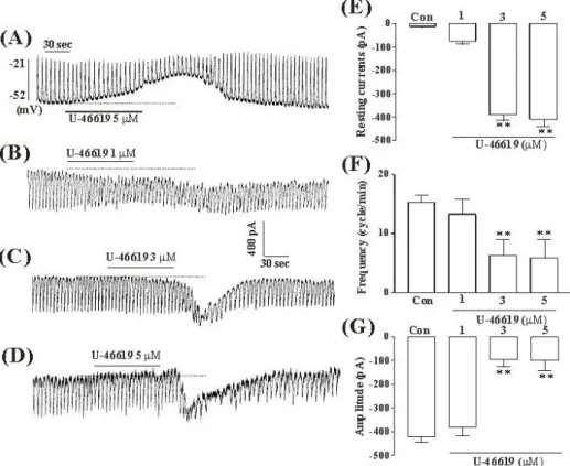
Fig. 1. The effects of TXA2 on pacemaker potentials and pacemaker currents recorded in cultured ICC from mouse small intestine (A) Pacemaker potentials of ICC which were exposed to TXA2 (5μM) in the current-clamping mode (I=0). Vertical solid line scales amplitude of pacemaker potential and horizontal solid line scales for duration of recording (s) pacemaker potentials. (B), (C), and (D) Pacemaker currents of ICC recorded at a holding potential of -70 mV, when exposed to various concentrations of TXA2 (1, 3, and 5μM). The dotted lines indicate zero current levels. Vertical solid line scales amplitude of pacemaker current and horizontal solid line scales duration of recording (s) pacemaker currents. The responses to TXA2 are summarized in (E), (F) and (G). The bars represent mean values±SE.
**Significantly different from the untreated control (Con) (p < 0.01).
METHODS Preparation of cells and tissues
Balb/C mice (3∼7 days old) of either sex were anesthetized with ether and sacrificed by cervical dislocation. The small intestines from 1 cm below the pyloric ring to the cecum were removed and opened along the mesenteric border. The luminal contents were washed away with Krebs-Ringer bicarbonate solution. The tissues were pinned to the base of a Sylgard dish, and the mucosa was removed by sharp dissection. Small stripes of intestinal muscle were equili- brated in Ca2+-free Hank's solution for 30 min and the cells were dispersed with an enzyme solution containing 1.3 mg/ml collagenase (Worthington Biochemical Co, Lakewood, NJ, USA), 2 mg/ml bovine serum albumin (Sigma Chemical Co., St. Louis, MO, USA), 2 mg/ml trypsin inhibitor (Sigma) and 0.27 mg/ml ATP. Cells were plated onto sterile glass coverslips coated with murine collagen (2.5μg/ml, Falcon/BD) in 35 mm culture dishes. The cells were then cultured at 37oC in a 95 % O2-5 % CO2 incubator in SMGM (smooth muscle growth medium, Clonetics Corp., San Diego, CA, USA) supplemented with 2% antibiotics/
antimycotics (Gibco, Grand Island, NY, USA) and murine
stem cell factor (SCF, 5 ng/ml, Sigma). Interstitial cells of Cajal (ICC) were identified immunologically with a monoclonal antibody for Kit protein (ACK2) labelled with Alexa Fluor 488 (molecular Probe, Eugene, OR, USA).
Patch clamp experiments
The whole-cell configuration of the patch-clamp technique was used to record membrane currents (voltage clamp) and membrane potentials (current clamp) from cultured ICC.
Currents or potentials were amplified by use of an Axopatch 1-D (Axon Instruments, Foster, CA, USA). Command pulse was applied using an IBM-compatible personal computer and pClamp software (version 6.1; Axon Instruments). The data were filtered at 5 kHz and displayed on an oscilloscope, a computer monitor, and a pen recorder (Gould 2200, Gould, Valley view, OH, USA).
Results were analyzed using pClamp and Sigma plot (version 9.0) software. All experiments were performed at 30oC.
Solutions and drugs
The cells were bathed in a solution containing (mM): 5 mM KCl, 135 mM NaCl, 2 mM CaCl2, 10 mM glucose, 1.2 mM MgCl2, and 10 mM HEPES, pH adjusted to 7.2 with
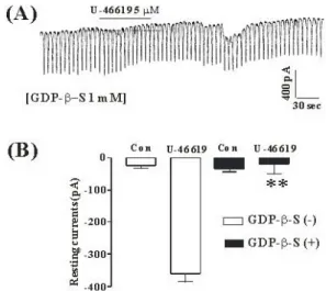
Fig. 2. The effects of GDP-β-S in response to TXA2 induced pacemaker currents from ICC of mouse small intestine (A) Pacemaker currents from ICC exposed to TXA2 (5μM) in the presence of GDP-β-S (1 mM) in the pipette. The tonic inward currents with suppressed amplitudes and frequency induced by TXA2 were blocked by internally applied GDP-β-S (1 mM). The dotted lines indicate the zero current levels. The effects of TXA2
in the presence of GDP-β-S are summarized in (B). Vertical solid line scales amplitude of pacemaker current and horizontal solid line scales for duration of recording (s) pacemaker currents. Bars represent mean values ± SE. The effects of GDP-β-S on TXA2-induced pacemaker currents were significantly different from the TXA2-induced pacemaker currents (p < 0.01). The bars represent mean values±SE. **Significantly different from the untreated control (Con) (p < 0.01).
Tris. The pipette solution contained 140 mM KCl, 5 mM MgCl2, 2.7 mM K2ATP, 0.1 mM Na2GTP, 2.5 mM creatine phosphate disodium, 5 mM HEPES, 0.1 mM EGTA, pH adjusted to 7.2 with Tris.
Drugs used were: U-46619, guanosine 5'-[-thio]diphosphate trilithium salt (GDP-β-S), calphostin C, chelethrine, and thapsigargin. All drugs were purchased from Sigma Chemical Co (St. Louis, MO, USA).
Statistical analysis
Data are expressed as mean±standard error. Differences in the data were evaluated by Student's t test. A p-values less than 0.05 were taken as a statistically significant difference. The n-values reported in the text refer to the number of cells used in the patch-clamp experiments.
RESULTS
Effect of TXA2 on pacemaker potentials and currents in cultured ICC
ICC were identified with immunofluorescence using Kit antibody, and had a distinctive morphology easily recognizable in cultures. We performed the electrophysiological recording from cultured ICC under current (I=0) and voltage clamp mode. Under current clamp mode, ICC spontaneously showed pacemaker potentials. The resting membrane potential was
-48±3 mV and amplitude was 25±6 mV. In the presence of U46619 (5μM), a thromboxane receptor agonist, membrane potentials were depolarized to -19±2.4 mV, and the amplitude of pacemaker potentials was decreased to 4.7±1.4 mV (n=5, Fig. 1A, bar graph not shown). These results suggested that ICC have the spontaneous pacemaker activity, and that U46619 has an effect on this electrical activity of ICC.
Under a voltage clamp at a holding potential of -70 mV, the ICC generated spontaneous inward currents. Treatment of cultured ICC with various concentrations of U46619 (1, 3, and 5μM) produced tonic inward currents and decreased the frequency and the amplitude of pacemaker currents in a dose-dependent manner (Fig. 1B, C, and D). As shown in Fig 1E, F, and G, the values of frequency, amplitude and resting currents about pacemaker currents in control condition were significantly different from those obtained in the presence of U46619 (3 and 5μM).
Involvement of G proteins in the TXA2- induced tonic inward currents in cultured ICC
The effects of GDP-β-S, a nonhydrolysable guanosine 5'-diphosphate analogue which permanently inactivates GTP binding proteins, were examined to determine whether the G-protein is involved in the effects of TXA2
on ICC. When GDP-β-S (1 mM) was in the pipette, U46619 (5μM) did not show the tonic inward currents (Fig. 2A).
In the presence of GDP-β-S in the pipette, the resting currents in control were -19±8 pA. The resting currents by treatment with U46619 in the presence of GDP-β-S were -22.6±5 pA (n = 4, Fig. 2B), which were significantly different from those obtained by treatment with U46619 in the absence of GDP-β-S.
External Ca2+-free solution and Ca2+-ATPase inhibitor of endoplasmic reticulum suppress TXA2 effects in cultured ICC
To investigate the role of external Ca2+ or internal Ca2+, TXA2 was tested under external Ca2+-free conditions and in the presence of thapsigargin, a Ca2+-ATPase inhibitor of endoplasmic reticulum. The application of external Ca2+-free solution completely inhibited the pacemaker currents in voltage clamp mode at a holding potential of -70 mV, and U46619 (5μM)-induced effects on pacemaker currents in this condition were blocked (n=5, Fig. 3A). The value of resting currents with U46619 (5μM) in Ca2+-free solution was significantly different, compared with control value obtained in normal solution (Fig. 3B). In addition, the treatment of thapsigargin (5μM) inhibited the pacemaker currents in ICC and blocked the U46619-induced tonic inward currents (Fig. 3C). In the presence of thapsigargin, the value of resting currents by treatment with U46619 was significantly different from those obtained by treatment with U46619 in the absence of thapsigargin (n=6, Fig. 3D).
Effects of protein kinase C inhibitor on TXA2-induced responses in cultured ICC
We tested the effects of chelerythrine or calphostin C, an inhibitor of protein kinase C (PKC), to investigate whether TXA2-induced responses to pacemaker currents are mediated by the activation of PKC. Chelerythrine (1μM) or calphostin
Fig. 3. The effects of an external Ca2+-free solution or thapsigargin on the TXA2-induced response on pacemaker currents in cultured ICC from mouse small intestine (A) External Ca2+-free solution abolished the generation of pacemaker currents. Under this condition, the TXA2 (5μM)-induced tonic inward currents were blocked. (C) Thapsigargin (5μM) abolished the generation of pacemaker currents.
Thapsigargin also blocked the TXA2 (5μM)-induced tonic inward currents. The dotted lines indicate the zero current levels. Responses to the TXA2 in the external Ca2+-free solution and in the presence of thapsigargin are summarized in (B) and (D). Vertical solid line scales amplitude of pacemaker current and horizontal solid line scales duration of recording (s) pacemaker current. The bars represent mean values ± SE. **Significantly different from the untreated Control (Con) (p < 0.01).
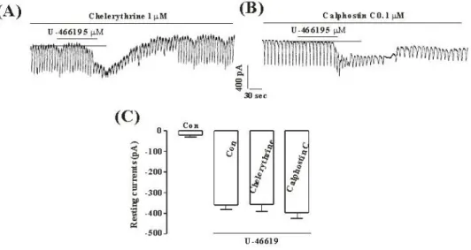
Fig. 4. The effects of chelerythrine or calphostin C on the TXA2-induced response on pacemaker currents in cultured ICC from mouse small intestine (A), (B) Pacemaker currents of ICC exposed to TXA2 (10μM) in the presence of chelerythrine (1μM) or calphostin C (0.1μM).
In this condition, the TXA2 caused tonic inward currents. The dotted lines indicate the zero current levels. Responses to the TXA2 in the presence of chelerythrine or calphostin C are summarized in (C). Vertical solid line scales amplitude of pacemaker current and horizontal solid line scales duration of recording (s) pacemaker current. The bars represent mean values ± SE.
C (0.1μM) did not have an effect on tonic inward currents by U46619 (5μM) (Fig. 4A and C), and the value was not also significantly different, compared with the tonic inward currents by U46619 obtained in the absence of chelerythrine or calphostin C (n=5, Fig. 4B and D).
DISCUSSION
Although the effects TXA2 on the intestinal motility in
tissue and smooth muscle cells have amply been demon- strated, this is the first study to determine the effects of TXA2 on electrical activity in small intestine.
Present results demonstrate that TXA2 regulates intestinal motility by modulating the pacemaker currents of ICC, and that this modulation is mediated via acting on G protein and extra- and intracellular Ca2+ mobilization in a PKC-independent manner.
While contractile actions of TXA2 on airway and vascular smooth muscles are well known, those on smooth muscle
of gastrointestinal tract have not yet been fully characterized.
Also, TXA2 have some species difference in GI tract.
Recently, it was suggested that TXA2 has strong contractile actions on both the fundus and ileum, which were mediated mainly via the thromboxane receptors in mice (Okada, 2000). Moreover, the above study showed that TXA2 has the highest potency among the prostaglandin E2, D2, I2, and F2α, suggesting physiological or pathophysiological roles of TXA2 played in the direct or indirect actions on gastroin- testinal smooth muscles, which has largely been attributed to its action on vasculature and blood platelets. Namely, the above study indicates the possibility that TXA2 may have stimulatory or inhibitory functions on electrical activity of ICC. In the present study, we observed that TXA2 evoked the depolarization of pacemaker potentials and tonic inward currents of pacemaker currents in ICC, implying that the regulation of electrical activity in ICC may be involved in the contractile effects of TXA2 in intestinal tract.
TXA2 induces the effects by increasing the cytosolic Ca2+
concentration which is independent of extracellular Ca2+
and does not involve changes in inositol (1,4,5) trisphosphate (IP3) levels, suggesting that this TXA2 action involves mobilization of Ca2+ from intracellular stores via an IP3-independent process in platelet (Nakano et al, 1988;
Arita et al, 1989). And many reports suggested that TXA2
leads to phospholipase C (PLC) activation, indicating that TXA2 receptors are coupled in part to a pertussis toxin- insensitive G protein (Pollock et al, 1984; Watson et al, 1985; Brass et al, 1987; Sage & Rink, 1987; Siess et al, 1986; Shenker et al, 1991). Overall, these studies suggest that TXA2 receptors couple directly to G protein, resulting in PLC activation, IP3-dependent Ca2+ mobilization, and activation of PKC activation through diglyceride formation, further coupling TXA2 receptors to a second G protein. It is not clear whether coupling to the G protein is involved in IP3-independent Ca2+ mobilization and/or a Ca2+ channel that mediates the entry of extracellular Ca2+. In this study, the tonic inward currents of pacemaker currents were blocked by TXA2, when GDPβS was present in the pipette.
This means that the effects of TXA2 on electrical activity in ICC may be related with G proteins. Because many suggestions have been made that TXA2 may have biological activity through mobilization of intracellular Ca2+, we used thapsigargin, a potent endoplasmic reticulum Ca2+-ATPase inhibitor, and found that those inhibitors suppressed the TXA2-induced tonic inward currents. These results strongly suggest that the release of Ca2+ from internal storage by TXA2 is essential to produce tonic inward currents, in correspondence with previous suggestions. In addition, it is well known that the generation of pacemaker currents is dependent upon intracellular Ca2+ oscillation and the periodic release of Ca2+ from endoplasmic reticulum is essential for generation of pacemaker currents. Also, in the present study, chelerythrine or calphostin C, specific and potent PKC inhibitors, did not block TXA2-induced effects, suggesting that PKC is not involved on the actions of TXA2
in ICC.
In conclusion, this study describes the effects of TXA2 on ICC in the mouse small intestine. TXA2 depolarized the membrane with increased tonic inward currents, which was via external Ca2+ influx and internal Ca2+ mobilization in a PKC-independent manner. Thus, the action of TXA2 on ICC may explain the excitatory action of TXA2 in GI motility.
ACKNOWLEDGEMENT
This work was supported by a research fund from Chosun University 2003.
REFERENCES
Arita H, Nakano T, Hanasaki K. Thromboxane A2: its generation and role in platelet activation. Prog Lipid Res 28: 273-301, 1989 Bennett A, Elev KG, Scholes GB. Effects of prostaglandins E1 and
E2 on human, guinea pig and rat isolated small intestine. Br J Pharmacol 34: 630-638, 1968
Bennett A, Hensy CN, Sanger GJ, Stamford IF. Metabolites of arachidonic acid formed by human gastrointestinal tissues and their actions on the muscle layers. Br J Pharmacol 74: 435-444, 1981
Bennett A, Sanger GJ. Pinane thromboxane A2 analogues are non-selective prostanoid antagonists in rat and human stomach muscle. Br J Pharmacol 77: 591-596, 1982
Berezin I, Daniel EE, Huizinga JD. Ultrastructure of interstitial cells of Cajal in the canine distal esophagus. Can J Physiol Pharmacol 72: 1049-1059, 1994
Berezin I, Huizinga JD, Daniel EE. Interstitial cells of Cajal in the canine colon: a special communication network at the inner border of the circular muscle. J Comp Neurol 273: 42-51, 1988 Brass LF, Shaller CC, Belmonte EJ. Inositol 1,4,5-triphosphate
induced granule secretion in platelets, Evidende that the activation of phospholipase C mediated by platelet thromboxane receptors involves a guanine nucleotide binding protein-dependent mechanism distinct from that of thrombin. J Clin Invest 79: 1269
-1275, 1987
Coleman RA, Humphrey PP, Kennedy I, Levy GP, Lumley P.
Comparison of the actions of U-46619, a prostaglandin H2-analogue, with those of prostaglandin H2 and thromboxane A2 on some isolated smooth muscle preparations. Br J Pharmacol 73: 773-
778, 1981
Daniel EE, Posey-Daniel V. Neuromuscular structures in opossum esophagus: role of interstitial cells of Cajal. Am J Physiol 246:
G305-G315, 1984
Ferreira SH, Herman AG, Vane JR. Prostaglandin production by rabbit isolated jejunum and its relationship to the inherent tone of the preparation. Br J Pharmacol 56: 469-477, 1976 Koh SD, Sanders KM, Ward SM. Spontaneous electrical rhythmicity
in cultured interstitial cells of Cajal from the mouse small intestine. J Physiol 513: 203-213, 1998
LeDuc LE, Needleman P. Regional localization of prostacyclin and thromboxane synthesis in dog stomach and intestinal tract. J Pharmacol Exp Ther 211: 181-188, 1979
Nakano T, Hanasaki K, Arita H. Different effects of two thromboxane A2/prostaglandin H2 receptor ligand, U46619 and S-14, on rabbit platelets. FEBS Lett 234: 309-312, 1988 Needleman P, Turk J, Jakschik BA, Morrison AR, Lefkowith JB.
Arachidonic acid metabolism. Annu Rev Biochem 55: 69-109, 1986
Okada Y, Hara A, Ma H, Xiao CY, Takahata O, Kohgo Y, Narumiya S, Ushikubi F. Characterization of prostanoid receptors mediating contraction of the gastric fundus and ileum: studies using mice deficient in prostanoid receptors. Br J Pharmacol 131: 745-755, Pollock WK, Armstrong RA, Brydon LJ, Jones RL, Macintyre DE. 2000 Thromboxane-induced phosphatide formation in human platelets.
Relationship to receptor occupancy and to changes in cytosolic free calcium. Biochem J 219: 833-842, 1984
Portbury AL, Furness JB, Young HM, Southwell BR, Vigna SR.
Localization of NK1 receptor immunoreactivity to neurons and interstitial cells of the guinea-pig gastrointestinal tract. J Comp Neurol 367: 342-351, 1996
Publicover NG, Horowitz NN, Sanders KM. Calcium oscillations in
freshly dispersed and cultured interstitial cells of from canine colon. Am J Physiol 262: C589-C597, 1992
Robert A. Prostaglandins and the gastrointestinal tract. In:
Hohnson LR ed, Physiology of the Gastrointestinal Tract. 1st ed.
Raven, New York, p 1407-1434, 1981
Sage SO, Rink TJ. The kinetics of changes in intracellular calcium concentration in fura-2-loaded human platelets. J Biol Chem 262: 16364-16369, 1987
Sanders KM. Evidence that endogenous prostacyclin modulates the electrical and mechanical activities of canine ileal circular muscle. J Gastroenterol 19: 401, 1981
Sanders KM. Evidence that prostaglandins are local regulatory agents in canine ileal circular muscle. Am J Physiol 246: G361-
G371, 1984
Sanders KM. Ionic mechanisms of electrical rhythmicity in gastrointestinal smooth muscles. Annu Rev Physiol 54: 439-
453, 1992
Shenker A, Goldsmith P, Unson CG, Spiegel AM. The G protein coupled to the thromboxane A2 receptor in human platelets is a member of the novel Gq family. J Biol Chem 266: 9309-9313, 1991
Shuttleworth CWR, Xue C, Ward SM, de Vente J, Sanders KM.
Immunohistochemical localization of 3',5'-cyclic guanosine monophosphate in the canine proximal colon: responses to nitric oxide and electrical stimulation of enteric inhibitory neurons.
Neuroscience 56: 513-522, 1993
Siess W, Stifel M, Binder H, Weber PC. Activation of V1-receptors by vasopressin stimulates inositol phospholipids hydrolysis and arachidonate metabolism in human platelets. Biochem J 233: 83
-91, 1986
Sternini C, Su D, Gamp PD, Bunnett NW. Cellular sites of expression of the neurokinin-1 receptor in the rat gastrointestinal tract. J Comp Neurol 258: 531-540, 1995
Thomsen L, Robinson TL, Lee JCF, Farraway L, Hughes MJH, Andrews DW, Huizinga JD. Interstitial cells of Cajal generate a rhythmic pacemaker current. Nature Med 4: 448-451, 1998 Thuneberg L. Interstitial cells of Cajal: intestinal pacemakers? Adv
Anat Embryol Cell Biol 71: 11-30, 1982
Torihashi S, Kobayashi S, Gerthoffer WT, Sanders KM. Interstitial cells in deep muscular plexus of canine small intestine may be specialized smooth muscle cells. Am J Physiol 265: G638-G645, Watson SP, Reep B, McConnell RT, Lapetina EG. Collagen stimulates 1993 [3H]inositol trisphosphate formaton in indomethacin-treated human platelets. Biochem J 226: 831-837, 1985
Young HM, McConalogue K, Fruness JB, de Vente J. Nitric oxide targets in the guinea-pig intestine identified by induction of cyclic GMP immunoreactivity. Neuroscience 55: 583-596, 1993 Zhou DS, Komuro T. Interstitial cells associated with the deep
muscular plexus of the guinea-pig small intestine, with special reference to the interstitial cells of Cajal. Cell Tissue Res 268:
205-216, 1992