J Korean Soc Radiol 2015;73(4):252-258 http://dx.doi.org/10.3348/jksr.2015.73.4.252
INTRODUCTION
A choledochal cyst is a rare congenital anomaly of the biliary system, manifested as the cystic dilatation of bile ducts, usually occurring in the common bile duct (1-4). The Todani classifica- tion system is widely used to classify choledochal cysts; however, not all cases fit into this classification system. Here, we present a case of a choledochal cyst that did not fit into this Todani clas- sification system: saccular cystic dilatation of the confluent por- tion of both intrahepatic ducts (IHDs).
CASE REPORT
A 45-year-old male patient presented with a sudden onset of severe pain in the upper right abdomen. On physical examina- tion, there was tenderness in the upper right abdominal area, and Murphy’s sign was positive. There was no increase in inflam-
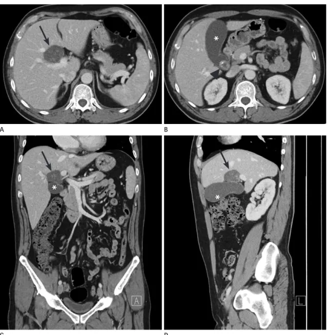
matory markers in the blood tests, except a mild increase of ala- nine aminotransferase (43 IU/L) and gamma glutamyl transpep- tidase (103 IU/L). In the contrast-enhanced abdominal CT per- formed to rule out biliary colic and diseases such as acute cho- lecystitis, we detected a 4.5 cm thin-walled unilocular cystic le- sion containing multiple calcified stones. The lesion abutted the gallbladder in the porta hepatis without apparent origin (Fig. 1).
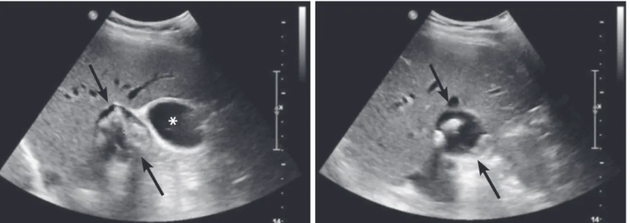
There was a calcified gallstone without the mural thickening of the gallbladder. Both intrahepatic and common bile ducts were undilated. At this point, the differential diagnosis included the duplication anomalies of the gallbladder with cholelithiasis, an exophytic complicated hepatic cyst originating from segment 4 of the liver, and a choledochal diverticulum with choledocholi- thiasis. In the abdominal ultrasonography, we detected a 4.5 cm unilocular thin-walled cystic lesion containing multiple echo- genic stones, which abutted the gallbladder. The lesion did not show the anatomical layering of its wall, which was detected in
A Case Report of an Unusual Type of Choledochal Cyst
with Choledocholithiasis: Saccular Dilatation of the Confluent Portion of Both Intrahepatic Ducts
비전형적 유형의 총담관낭과 총담관결석증의 증례: 양 간내담관 합류지점의 낭성확장
Jin Young Kim, MD, Hee Jin Kim, MD*, Hyun Young Han, MD
Department of Radiology, Eulji University Hospital, Daejeon, Korea
A choledochal cyst is a rare congenital anomaly of the biliary system manifested as the cystic dilatation of bile ducts, usually occurring in the common bile duct. Here, we describe an unusual type of choledochal cyst in a 45-year-old male that did not fit into the most widely accepted Todani classification of these cysts. The lesion mim- icked duplication anomalies of the gallbladder and was finally diagnosed as a chole- dochal cyst involving the confluent portion of both intrahepatic ducts.
Index terms Choledochal Cyst Todani Classification Congenital Biliary Disorder Unusual Type
Received April 6, 2015 Revised May 21, 2015 Accepted June 10, 2015
*Corresponding author: Hee Jin Kim, MD Department of Radiology, Eulji University Hospital, 95 Dunsanseo-ro, Seo-gu, Daejeon 35233, Korea.
Tel. 82-42-611-3567 Fax. 82-42-611-3590 E-mail: [email protected]
This is an Open Access article distributed under the terms of the Creative Commons Attribution Non-Commercial License (http://creativecommons.org/licenses/by-nc/3.0) which permits unrestricted non-commercial use, distri- bution, and reproduction in any medium, provided the original work is properly cited.
confluent portion of both IHDs at the level of the porta hepatis with multiple intracystic stones. Both IHDs directly arose from
A choledochal cyst is a rare congenital anomaly of the biliary system, involving any part of the bile duct. It is manifested as the
Fig. 1. Axial (A, B), coronal (C), and sagittal (D) views of a contrast-enhanced abdominal CT showing a round cystic lesion (arrow) with multiple internal calcified stones, at the porta hepatis (just superior to the gallbladder). Gallbladder (asterisk) showing a calcified stone (arrowhead) with- out mural thickening as an inflammation.
A
C
B
D
*
* *
cystic dilatation of bile ducts, usually occurring in the common bile duct (1-4). It was first reported by Douglas in 1852 (2). The detection of congenital anomalies, including choledochal cysts, may avoid diagnostic errors, help to plan surgery, and prevent unnecessary ductal injury (5).
The symptoms associated with a choledochal cyst are non- specific, which can lead to a delay in the diagnosis. It usually pre- sents with abdominal pain, jaundice, and a palpable right upper quadrant mass. The symptoms are more prevalent in children than in adults (1). Babbitt et al. (2), described that the symptoms of a choledochal cyst may be similar to those of Mirizzi’s syndro- me at presentation. The incidence of choledochal cysts is 100 times more common in Asian countries, particularly Japan (one in 1000), than in Western countries (one in 100000–150000). It is more common in women, with a female:male ratio of 4:1 (1, 3, 6). Approximately 2/3 of cases are diagnosed before the age of ten (1, 7), and it is relatively rare for it to occur in an adult, as in the present case.
The Todani classification system is the most widely accepted classification of choledochal cysts based on their anatomical lo- cation and cholangiographic morphology. This classification sys- tem was expanded from the work of Along-Lej et al., and de- scribes five main types of choledochal cysts, with several sub- types: type I is the most common type (80–90%), involving the dilatation of the entire common hepatic or common bile duct or segments of each. It is subclassified into type IA (cystic dila- tation of the common bile duct), type IB (focal segmental dila- tation of the distal common bile duct), and type IC (fusiform dilatation of the common hepatic and common bile ducts). Type
II is rare (2%), and is a true diverticulum anywhere in the extra- hepatic duct. Type III (also called choledochocele) is also rare (1.4–5%), and is confined to the intraduodenal portion of the common bile duct. Type IV is more common (19%) and may be further subclassified into type IVA, which involves both the in- tra- and extrahepatic bile ducts, and the least common type IVB, where only extrahepatic cysts are observed. Type V, or Caroli’s disease, is characterized by the dilatation of single or multiple intrahepatic bile ducts (1, 3, 6).
Although most of the choledochal cysts can be classified us- ing the Todani classification system, a few cases have been previ- ously reported that did not fit into the classification system (Table 1). Loke et al. (6), described a case of an atypical choledochal cyst characterized by the cystic dilatation of the cystic duct. They de- scribed this as a type VI choledochal cyst. Choledochal cysts in- volving only the IHD were reported by Prekop et al. (right IHD) (7), Salles et al. (right IHD) (8), and Sadiq et al. (IHD at the level of the porta hepatis) (3). Michaelides et al. (1), described a case of a choledochal cyst (with dilatation of the common bile duct, common hepatic duct, and the proximal portion of the cystic duct as a bicornal configuration); they proposed the classification of this atypical type as a new subtype of Todani I cyst, namely type ID. Similar to these cases, our case of a choledochal cyst also did not fit into the typical Todani classification system because the cyst involved the confluent portion of both IHDs.
Many theories have been proposed regarding the etiology of choledochal cysts, however, none of these theories explain all the different types of choledochal cyst. A few theories have attempt- ed to explain the cause of the common types of choledochal cysts.
Fig. 2. Abdominal ultrasonography showing a thin-walled cystic lesion (arrows) abutting the gallbladder (asterisk) near the porta hepatis. It con- tains multiple echogenic foci with posterior shadowing, suggesting calcified stones. The lesion does not show the anatomical layering of its wall that is detected in the gallbladder.
*
The first theory, which is the most widely accepted, is that there is an abnormal common channel related to the anomalous union of the pancreaticobiliary duct (AUPBD). This anomalous union has two features relevant to the formation of choledochal cysts.
One is that the union of the pancreatic duct and common bile duct is located far from the duodenum, creating a long common channel of a length greater than 1.5 cm. The other is the angle of this junction. According to this theory, the abnormal pancre- aticobiliary duct union allows the chronic reflux of pancreatic enzymes into the biliary system. The chemical and enzymatic de- struction of the ductal wall results in the weakening and dilata-
tion of the involved bile duct, which subsequently forms a cyst.
However, this theory can be mainly applied in Todani classifi- cation type I, and does not explain the occurrence of choledochal cysts in the presence of a normal pancreaticobiliary ductal union, as seen in our case. A few previous articles have reported cases of choledochal cysts without accompanied AUPBD. Another study proposed an alternative theory that choledochal cysts rep- resent a spectrum of embryonic malformation of the pancreati- cobiliary system, one of which may be an anomalous junction (1, 2, 4, 5).
The histopathology of choledochal cysts shows a fibrotic wall Fig. 3. Magnetic resonance cholangiopancreatography images.
A. Maximal intensity projection image showing the gallbladder (asterisk) and a round cystic lesion (arrows) in the confluent portion of both intra- hepatic ducts (IHDs). There is no anomalous pancreaticobiliary ductal union. A schematic representation is also shown (right).
B, C. Left IHD (arrow in B) and right IHD (arrow in C) directly arose from the cyst, which is representative of the choledochal cyst. Multiple filling defects suggesting cholelithiasis and choledocholithiasis are shown in the gallbladder and choledochal cyst.
A
B C
varying from a few millimeters up to 10 millimeters in thickness.
Dense collagenous connective tissue makes up the cystic wall, without a complete epithelial lining. Elastic fibers and smooth muscle bundles can also be occasionally found as constituents of the wall. Choledochoceles (Todani classification type III) are dif- ferent in that they are lined by either duodenal or bile duct mu- cosa as an exception. An inflammatory reaction caused by the free flow of pancreatic juice into the common bile duct is often present. The recurrent episodes of inflammation result in thick- ening of the wall of the involved bile duct (1, 2).
A choledochal cyst can be confused with other cystic lesions.
The differential diagnosis includes hepatic cyst, enteric duplica- tion cyst, pancreatic pseudocyst, hepatic artery aneurysm, du- plication anomalies of the gallbladder, recurrent pyogenic chol-
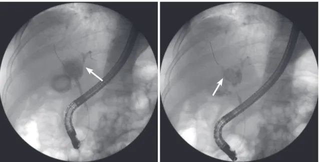
angitis, and spontaneous perforation of the common bile duct (9). Sahoo and Kumar (10), described a case report of a double gallbladder masquerading as a choledochal cyst, contrary to the diagnosis in our case. We had a differential diagnosis of several possible diseases, such as duplication anomalies of the gallblad- der with cholelithiasis, an exophytic complicated hepatic cyst originating from segment 4 of the liver, and a choledochal di- verticulum with choledocholithiasis. An ultrasonography of the lesion did not depict the mural layering that is detected in the gallbladder, therefore, we were able to rule out the possibility of duplication anomalies of the gallbladder with cholelithiasis. Re- current pyogenic cholangitis is caused by intrahepatic ductal strictures and intraductal calculi, and is characterized by the re- current attacks of abdominal pain, fever, and jaundice with mild- Fig. 4. Endoscopic retrograde cholangiopancreatography shows sequential contrast filling of the common bile duct, cystic lesion, and both intra- hepatic ducts (IHDs) (arrows), which are consistent with a choledochal cyst occupying a confluent portion of both IHDs.
Table 1. Previous Reports of Atypical Type of Choledochal Cyst
Year Authors Age Sex Symptoms Location of Atypical Type
of Choledochal Cyst
1999 Loke et al. 29 years F Jaundice Distal part of cystic duct
2007 Prekop et al. 14 months Asymmetrically enlarged abdomen
without symptoms
Within the entire right ductus hepaticus
2009 Sadiq et al. Neonate F - Intrahepatic duct at the level of porta
hepatis 2011 Michaelides et al. 3–67 years
(6 patients)
4 F and 2 M Jaundice, abdominal pain and palpable mass
Cystic dilatation of common hepatic, common bile ducts, and proximal cystic duct (bicornal configuration)
2013 Salles et al. Neonate M - Right intrahepatic duct near the
confluence of both intrahepatic ducts
des et al. (1), described the importance of the surgeon’s knowl- edge regarding the details of the cyst, such as the exact location and length of the common channel.
Common complications of choledochal cysts, including cho- lecystitis, biliary stricture, recurrent cholangitis, recurrent acute pancreatitis, cholelithiasis, choledocholithiasis, and even malig- nancy have been reported. Choledocholithiasis is one of the most common complications as seen in our case. Savader et al. (4), reported rare complications such as ectopic pancreas (jejunum, one case), intrahepatic abscesses (one case), and cystolithiasis (one case). Due to the fact that the frequency of biliary malign- ancy is more common in adults than in children, adult patients with choledochal cysts should be cautiously evaluated. Malig- nancy may occur in the remaining stump after resection, al- though the excision of the cyst greatly decreases the risk of ma- lignancy (1, 5).
In conclusion, we described an unusual type of choledochal cyst involving the confluent portion of both IHDs with choled- ocholithiasis. Our report may help to increase the awareness of the unusual types of choledochal cysts that do not fit into the To- dani classification system.
REFERENCES
1. Michaelides M, Dimarelos V, Kostantinou D, Bintoudi A, Tzi-
cholangiographic appearance. AJR Am J Roentgenol 1991;
156:327-331
5. Yu J, Turner MA, Fulcher AS, Halvorsen RA. Congenital anomalies and normal variants of the pancreaticobiliary tract and the pancreas in adults: part 1, Biliary tract. AJR Am J Roentgenol 2006;187:1536-1543
6. Loke TK, Lam SH, Chan CS. Choledochal cyst: an unusual type of cystic dilatation of the cystic duct. AJR Am J Roent- genol 1999;173:619-620
7. Prekop I, Sevcik L, Jakubicka J, Bakos E, Bakos M, Durcansky D. Atypical cyst of the right ductus hepaticus. Bratisl Lek Listy 2007;108:474-476
8. Salles A, Kastenberg ZJ, Wall JK, Visser BC, Bruzoni M. Com- plete resection of a rare intrahepatic variant of a chole- dochal cyst. J Pediatr Surg 2013;48:652-654
9. Durgun AV, Gorgun E, Kapan M, Ozcelik MF, Eryilmaz R.
Choledochal cysts in adults and the importance of differen- tial diagnosis. J Hepatobiliary Pancreat Surg 2002;9:738- 741
10. Sahoo MR, Kumar TA. Double gallbladder masquerading as a choledochal cyst: a case report. Int J Case Rep Image 2013;
4:283-286
비전형적 유형의 총담관낭과 총담관결석증의 증례: 양 간내담관 합류지점의 낭성확장
김진영 · 김희진* · 한현영
총담관낭은 담관의 낭성확장으로 발현되는 담도계의 드문 선천성 기형으로, 보통 총담관에 발생한다. 본 증례보고에서는 45세 남성 환자에서 발생한 Todani 분류에 부합하지 않는 비전형적 유형의 총담관낭의 케이스를 보고하고자 한다. 이 병 변은 중복담낭 등을 모방하였으나, 양 간내담관의 합류지점에 발생한 총담관낭으로 진단되었다.
을지대학교병원 영상의학과