CASE REPORT
췌장 종괴를 형성하고 스테로이드에 호전을 보인 2형 자가면역 췌장염
전연주, 장재혁, 이인석, 이장은, 정문경, 정찬권
1, 최명규, 정인식
가톨릭대학교 의과대학 내과학교실, 병리학교실1
Steroid Responsive Pancreatic Mass-Forming Type 2 Autoimmune Pancreatitis
Yeon Joo Chun, Jae Hyuck Chang, In Seok Lee, Jang Eun Lee, Mun Kyung Chung, Chan Kwon Jung1, Myung-Gyu Choi and In-Sik Chung
Departments of Internal Medicine and Pathology1, The Catholic University of Korea College of Medicine, Seoul, Korea
Autoimmune pancreatitis (AIP) has two distinct subsets. Type 1 AIP or lymphoplasmacytic sclerosing pancreatitis is systemic disease with the elevation in serum levels of the IgG4. Type 2 AIP, also called duct-centric pancreatitis, features granulocyte epithelial lesions with duct obstruction in the pancreas without systemic involvement. Here, we report a case of type 2 AIP diagnosed by pathology, which is the first report in Korea. The case is a 56-year-old woman who presented with anorexia and vomiting. Computed tomography revealed mass-like lesion in the pancreatic head and the compression of the distal common bile duct and the head portion of the main pancreatic duct. Serum levels of the IgG4 were normal. Histologic examination revealed a dense neutrophil infiltration in the pancreatic parenchyme associated with extensive fibrosis, thereby confirming the diagnosis of type 2 AIP. The abnormalities in the clinical, laboratory, and radiological findings improved after oral steroid treatment. (Korean J Gastroenterol 2011;58:53-57)
Key Words: Pancreatitis; Autoimmune diseases
Received February 12, 2011. Revised April 28, 2011. Accepted May 3, 2011.
CC This is an open access article distributed under the terms of the Creative Commons Attribution Non-Commercial License (http://creativecommons.org/licenses/
by-nc/3.0) which permits unrestricted non-commercial use, distribution, and reproduction in any medium, provided the original work is properly cited.
교신저자: 장재혁, 420-717, 경기도 부천시 원미구 소사동 2, 가톨릭대학교 부천성모병원 내과
Correspondence to: Jae Hyuck Chang, Department of Internal Medicine, Bucheon St. Mary’s Hospital, The Catholic University of Korea, 2, Sosa-dong, Wonmi-gu, Bucheon 420-717, Korea. Tel: +82-32-340-7086, Fax: +82-32-340-2255, E-mail: wwjjaang@catholic.ac.kr
Financial support: None. Conflict of interest: None.
서 론
자가면역 췌장염은 1961년 Sarles 등1이 복통, 폐쇄성 황 달, 고감마글로불린 혈증 등과 관련된 원인미상의 만성 췌장 염을 소개하면서 처음 기술되었고, 1995년 Yoshida 등2이 폐 쇄성 황달, 미만성 췌장종대 및 췌관의 불규칙적인 협착, 고감 마글로불린 혈증, 췌장 섬유화를 보인 68세 여자 환자를 자가 면역 췌장염이라고 처음 명명한 이후 많은 보고가 있었다. 이 러한 자가면역 췌장염은 임상적, 조직학적 특징에 따라 1형과 2형으로 구분할 수 있다.3 1형 자가면역 췌장염은 일본에서 보고된 바와 같이2,4 전통적인 자가면역 췌장염의 특징에 부합 하는 것으로 림프형질세포 침윤 경화성 췌장염(lymphoplas-
macytic sclerosing pancreatitis)이라고도 하며 그 이름과 같이 췌관 주위 림프형질세포의 침윤과 소용돌이 모양 혹은 나선형의 섬유화, 폐쇄성 소정맥염 등을 특징으로 한다.3 또한 특징적으로 혈청 면역글로불린 G4의 상승을 동반한다.5 2형 자가면역 췌장염은 비알콜성 췌관 파괴성 췌장염(non-alco- holic duct-destructive pancreatitis),6 특발성 췌관 중심 췌 장염(idiopathic duct-centric pancreatitis),7 혹은 과립구 상 피 병변 양성 췌장염(granulocyte epithelial lesion-positive pancreatitis)8으로 기술된 바 있고, 중성구의 췌관벽 침윤과 그로 인한 췌관벽 상피의 파괴를 특징으로 한다. 또한 폐쇄성 정맥염은 두드러지지 않으며 면역글로불린 G4의 췌장 침윤도 거의 없다고 알려져 있다.9 따라서 비교적 젊은 환자에서 특발
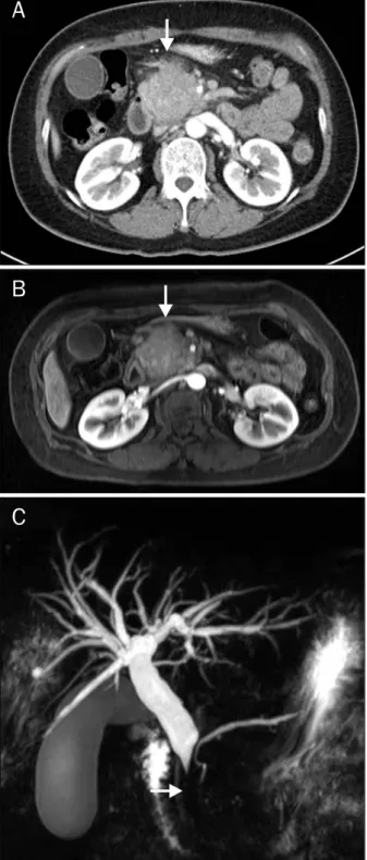
Fig. 1. Abdominal CT and MRI/MRCP findings. (A) Abdominal CT showed bulging contour, mass-like lesion in the head of the pancreas (arrow). (B, C) Pancreas and bile duct MRI/MRCP showed diffuse swelling of the pancreas head and compression of the distal common bile duct and proximal pancreatic duct, resulting in dilatation of the extrahepatic bile duct and pancreatic duct (arrows).
선학적 검사, 혈청 검사 및 조직검사를 통해 2형 자가면역 췌 장염을 진단하고 스테로이드 치료로 호전을 보였기에 문헌 고 찰과 함께 보고하는 바이다.
증 례
56세의 여자 환자가 일주일간의 구토 및 식욕부진을 주소 로 내원하였다. 내원 당시 혈압은 140/70 mmHg, 맥박 79회/
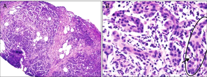
분, 체온 36.1oC였으며 급성 병색의 모습을 보였다. 환자의 과거력, 가족력 및 사회력에서 특이소견은 없었다. 신체 검사 에서 상복부에 통증 및 압통이 있었으나 반발통은 없었다. 말 초혈액 검사에서 백혈구 5,830/mm3, 혈색소 11.5 gm/dL, 혈 소판 319,000/uL이었고, 혈청 생화학 검사에서 총단백 7.5 g/dL, 알부민 3.8 g/dL, amylase 38 U/L로 정상이었으나 AST 119 IU/L, ALT 175 IU/L, alkaline phsphatase 238 IU/L, γ-GTP 884 U/L, 총빌리루빈 4.40 mg/dL, 직접빌리루 빈 3.20 mg/dL로 증가되어 있었으며, CA 19-9도 165.08 U/mL로 상승되어 있었다. 혈청 면역검사에서 항핵항체와 류 마티스 인자는 음성이었고, 면역글로불린 G4 1.19 g/dL, 면 역글로불린 G 1,404 mg/dL로 정상이었다. 복부 CT에서 췌 장 두부에 51×42 mm 크기의 종괴 음영이 관찰되었고, 이것 은 정상 췌장과 거의 비슷하게 조영 증강되어 종괴의 변연이 뚜렷하게 그려지지 않았다(Fig. 1A). 자기공명영상 및 자기공 명담도췌관조영술에서 역시 췌장 두부의 미만성 종대와 이로 인한 총담관 말단의 압박소견을 관찰할 수 있었다(Fig. 1B, C). 피부간경유담관조영술을 실시하여 하부 담관의 폐색과 간 외담관의 확장을 확인하고 담도배액관을 설치하였다. 췌장 종 괴에 대한 정확한 진단을 위하여 복강경하 조직검사를 실시하 였다. 조직소견은 중성구의 침윤이 두드러지나 림프구의 침윤 은 많지 않았으며 섬유화가 두드러진 소견이었고, 일부의 소 췌관(ductule)에 중성구의 침윤이 관찰되었다(Fig. 2A, B). 내 시경 역행성 담췌관조영술을 실시하여 담도 협착부위를 통과 하는 7 Fr, 7 cm 플라스틱 스텐트(Zimmon stent, Wilson- Cook Medical, Winston-Salem, NC, USA)를 삽입하고 경피 담도배액관은 제거하였다. 2형 자가면역 췌장염으로 진단하
Fig. 3. Abdominal CT findings. (A) Follow up CT scan after 15 days from the start of prednisolone. Globular swelling of the pancreas head de- creased comparing with Fig. 1A. (B) Follow up CT scan after 2 and half months from the start of predni- solone showed marked decrease of pancreas swelling.
Fig. 2. Pathological findings. (A) Biopsy showed focal fibrosis and parenchymal destruction of pancreas (H&E stain, ×100). (B) Higher magnification showed diffuse infiltration of neutrophils in the parenchyme of the pancreas (arrow) in the epithelial cells of the ductule (circle) (H&E stain,×400).
L, lumen.
고 prednisolone을 하루 40 mg씩 경구 투여를 시작하였으며 1주마다 5 mg씩 감량하였다. Prednisolone 치료 15일 뒤 촬 영한 복부 CT에서 췌장의 종괴는 33×27 mm로 그 크기가 감소하였고(Fig. 3A), 혈청 CA 19-9도 21.86 U/mL으로 감소 하였다. Prednisolone 치료 1달 반 후부터는 하루 5 mg으로 용량을 유지하였다. Prednisolone 경구 투여 2달 반 뒤 촬영 한 복부 CT에서 이전의 췌장 종괴는 더 감소하여 크기가 25×20 mm로 측정되었다(Fig. 3B). 이후 환자는 담도의 플라 스틱 스텐트를 제거하고 특별한 증상 없이 외래에서 경과 관 찰 중이다.
고 찰
1995년 일본에서 자가면역 췌장염을 처음 명명한 이후2 국 내에서도 2002년 국내에서 자가면역 췌장염의 증례가 처음 보고되었다.11 그 이후 자가면역 췌장염의 진단과 치료에 대해 논의가 활발히 되고 있으며 관련 보고 또한 점차 증가하고
있다.10,12 국내 보고들은 자가면역 췌장염의 아형 구분 없이
보고하였으며 대부분 1형 자가면역 췌장염에 관하여 보고하 였다. 2형 자가면역 췌장염의 가능성이 있는 증례도 소수 제 시하였지만 1형의 아형인지 2형인지 감별하지 않았다.10 한 국내 보고에서 단기 스테로이드 치료에 반응한 혈청음성 주췌 관 협착성 만성췌장염이 소개되었으나 조직소견이 2형 자가 면역 췌장염으로 진단하기에는 어려움이 있었다.13
기존의 자가면역 췌장염에 대한 진단기준은 1형 자가면역 췌장염에 대한 내용을 중심으로 하고 있으며, 2형 자가면역 췌장염에 대한 기준은 아직 명확히 확립되어 있지 않다. 2003 년부터 종례의 자가면역 췌장염의 조직소견과 다른 양상을 보 이는 자가면역 췌장염의 증례가 지속적으로 보고되었으며,7 Zamboni 등14과 Klöppel 등15이 이를 정리하여 과립구 상피 병변을 동반한 자가면역 췌장염의 전형적인 조직소견을 보고 하였다. 그리고 이러한 특징을 가진 자가면역 췌장염이 전통 적인 자가면역 췌장염의 임상적인 특징과는 차이가 있음이 알 려졌으며, 이를 2형 자가면역 췌장염으로 분류하게 되었다.
비알콜성 췌관 파괴성 췌장염,6 특발성 췌관 중심 췌장염,7 혹 은 과립구 상피 병변 양성 췌장염8으로 불리며, 중성구의 췌관 벽 침윤이 두드러져서 내강을 막거나 췌관벽 상피를 파괴하게 된다. 조직학적 특징 외에 중요한 차이는 2형 자가면역 췌장 염에서는 혈청 면역글로불린 G4의 증가가 뚜렷하지 않고, 담 관, 신장, 후복막, 침샘, 임파선 등 다른 장기를 침범하는 전신 질환인 1형 자가면역 췌장염과 달리 다른 장기의 침범 소견을 보이지 않는다는 점이다.17,18 다만, 2형 자가면역 췌장염의 20-30%에서는 염증성 장질환을 동반할 수 있다고 보고되었 다.7,14
자가면역 췌장염의 치료는 스테로이드의 경구 복용을 근간 으로 이루어진다. 구체적인 스테로이드의 투여 방법은 아직 확립되어 있지 않지만, 2009년 개정된 일본의 가이드라인에 서는 초기 용량으로 경구 prednisolone을 하루 0.6 mg/kg부 터 시작하여 2-4주 유지하다가 임상 증상이나 혈청학적 검사, 방사선학적 소견 등의 변화에 따라 1-2주에 5 mg씩 2-3개월 에 걸쳐서 서서히 감량하며, 재발 방지를 위하여 2.5-5 mg/day의 용량으로 유지요법을 시행하는 것을 추천하고 있 다.19 하지만 이는 주로 1형 자가면역 췌장염의 치료 결과를 바탕으로 정립된 것으로, 2형 자가면역 췌장염의 치료에 차이 가 있을지는 명확하지 않다. 이번 증례에서는 기존의 치료와 동일하게 prednisolone 40 mg을 초기 용량으로 투여하였으 며, 1주마다 5 mg씩 감량하였다. 스테로이드 투여 2주 후에 60% 정도의 췌장 종괴의 크기 감소를 보였고 2달 반 후에는 초기보다 75% 정도의 크기 감소를 보여 빠르고 뚜렷한 췌장 종대의 호전을 보였다. 이번 증례와 같이 2형 자가면역 췌장 염이 스테로이드에 대해 좋은 반응을 보이는 지는 향후 추가 적인 연구가 필요하며, 또한 재발에 대한 연구도 필요하다.
1형과 2형 사이에 임상적인 차이가 있기는 하지만, 조직학 적 소견 외에 임상적 소견이나 혈청학적 소견, 타장기 침범 등의 차이로 둘을 구별하기란 매우 어렵다. 과거에 췌장의 조 직검사는 합병증을 우려하여 극히 제한적으로 시행되었다. 현 재까지 1형에 비해 2형 자가면역 췌장염의 증례 보고가 매우 적은 점도 이러한 이유로 설명될 수 있다. 이번 증례에서는 복강경하 조직검사를 시행하였지만, 최근 활발히 시행되고 있
않은 경우가 있어, 보다 적극적인 조직 검사가 필요할 것이다.
이번 증례에서도 복부 CT에서 췌장의 종괴가 관찰되었고, 면 역글로불린 G4를 비롯한 혈청학적 검사에서 음성을 보이면서 CA19-9이 증가하는 등 췌장의 악성 종양과 감별이 쉽지 않은 상황에서 적극적인 조직검사를 통해서 불필요한 수술을 피할 수 있었다.
이번 증례의 제한점은 조직검사에서 뚜렷하게 췌관이 관찰 되지 않았다는 점이다. 그러나 일부에서 소췌관이 관찰되며 이 소췌관의 상피세포 내로 중성구가 침윤되어 소췌관염이 발 생한 소견이 보이면서, 췌장실질에 많은 중성구의 침윤이 뚜 렷하였기 때문에 1형과 구분되는 2형 자가면역 췌장염의 진 단을 할 수 있었다.
저자들은 췌장의 두부에 종괴를 형성한 56세의 여자에서 혈청 검사 및 조직검사를 통해 국내에서 처음으로 2형 자가면 역 췌장염을 진단하였고 스테로이드 치료로 호전을 보였기에 문헌 고찰과 함께 보고하는 바이다.
REFERENCES
1. Sarles H, Sarles JC, Muratore R, Guien C. Chronic inflammatory sclerosis of the pancreas--an autonomous pancreatic disease?
Am J Dig Dis 1961;6:688-698.
2. Yoshida K, Toki F, Takeuchi T, Watanabe S, Shiratori K, Hayashi N. Chronic pancreatitis caused by an autoimmune abnormality.
Proposal of the concept of autoimmune pancreatitis. Dig Dis Sci 1995;40:1561-1568.
3. Park DH, Kim MH, Chari ST. Recent advances in autoimmune pancreatitis. Gut 2009;58:1680-1689.
4. Okazaki K, Chiba T. Autoimmune related pancreatitis. Gut 2002;51:1-4.
5. Hamano H, Kawa S, Horiuchi A, et al. High serum IgG4 concen- trations in patients with sclerosing pancreatitis. N Engl J Med 2001;344:732-738.
6. Ectors N, Maillet B, Aerts R, et al. Non-alcoholic duct destructive chronic pancreatitis. Gut 1997;41:263-268.
7. Notohara K, Burgart LJ, Yadav D, Chari S, Smyrk TC. Idiopathic chronic pancreatitis with periductal lymphoplasmacytic infiltra- tion: clinicopathologic features of 35 cases. Am J Surg Pathol 2003;27:1119-1127.
8. Klöppel G. Chronic pancreatitis, pseudotumors and other tu- mor-like lesions. Mod Pathol 2007;20(Suppl 1):S113-S131.
9. Zhang L, Notohara K, Levy MJ, Chari ST, Smyrk TC. IgG4-positive plasma cell infiltration in the diagnosis of autoimmune pancreatitis. Mod Pathol 2007;20:23-28.
10. Kim KP, Kim M, Lee YJ, et al. Clinical characteristics of 17 cases of autoimmune chronic pancreatitis. Korean J Gastroenterol 2004;43:112-119.
11. Kim JY, Chang HS, Kim MH, et al. A case of autoimmune chronic pancreatitis improved with oral steroid therapy. Korean J Gastroenterol 2002;39:304-308.
12. Park SJ, Kim MH, Moon SH, et al. Clinical characteristics, re- currence features, and treatment outcomes of 55 patients with autoimmune pancreatitis. Korean J Gastroenterol 2008;52:
230-246.
13. Lee JY, Park DH, Park SH, et al. Two cases of seronegative, main pancreatic ductal narrowing, chronic pancreatitis that were re- sponsive to short-term steroid treatment. Korean J Med 2007;
72(Suppl 2):S103-S109.
14. Zamboni G, Lüttges J, Capelli P, et al. Histopathological features of diagnostic and clinical relevance in autoimmune pancreatitis:
a study on 53 resection specimens and 9 biopsy specimens.
Virchows Arch 2004;445:552-563.
15. Klöppel G, Lüttges J, Löhr M, Zamboni G, Longnecker D.
Autoimmune pancreatitis: pathological, clinical, and immuno- logical features. Pancreas 2003;27:14-19.
16. Yadav D, Notahara K, Smyrk TC, et al. Idiopathic tumefactive chronic pancreatitis: clinical profile, histology, and natural his- tory after resection. Clin Gastroenterol Hepatol 2003;1:129- 135.
17. Kamisawa T, Egawa N, Nakajima H. Autoimmune pancreatitis is a systemic autoimmune disease. Am J Gastroenterol 2003;98:
2811-2812.
18. Takahashi N, Kawashima A, Fletcher JG, Chari ST. Renal involve- ment in patients with autoimmune pancreatitis: CT and MR imaging findings. Radiology 2007;242:791-801.
19. Okazaki K, Kawa S, Kamisawa T, et al. Japanese clinical guide- lines for autoimmune pancreatitis. Pancreas 2009;38:849- 866.
20. Detlefsen S, Mohr Drewes A, Vyberg M, Klöppel G. Diagnosis of autoimmune pancreatitis by core needle biopsy: application of six microscopic criteria. Virchows Arch 2009;454:531-539.