submit.radiology.or.kr
대한영상의학회지 2011;65(1):77-8077
서론염증거짓종양은 드문 양성질환으로 다양한 정도의 세포 외 콜 라겐과 림프구 및 형질세포가 방추상 세포와의 혼합으로 구성 된다(1, 2). 영상의학적 소견과 침범 부위는 다양하게 나타날 수 있으며, 다병소로 발생하거나 종괴의 재발 및 전이가 나타나기도 한다(3). 저자들은 한 환자에서 간, 췌장, 담관, 신장, 신우, 복 강 및 중격동 내의 림프절, 기관지를 동시에 침범한 전신성의 염 증거짓종양으로 조직검사와 임상적 소견으로 진단된 1예를 초 음파와 전산화단층촬영술(CT) 소견을 중심으로 보고한다.
증례 보고
67세 남자 환자로 황달을 주소로 내원하였다. 내원 당시 혈액 검사 상 총 빌리루빈은 13.0 mg/dL (정상 0.1~0.2 g/dL), 직 접빌리루빈은 7.9 mg/dL (정상 0.1~0.4 mg/dL) 및, 종양 표 지인자인 CA 19-9가 1200 U/mL 이상으로(정상 0~37 U/
mL) 증가하였으며 C 반응성 단백질(CRP)이 1.84 mg/dL (정 상 0~0.5 mg/dL)로 약간 상승하였다. 상복부 초음파 소견에
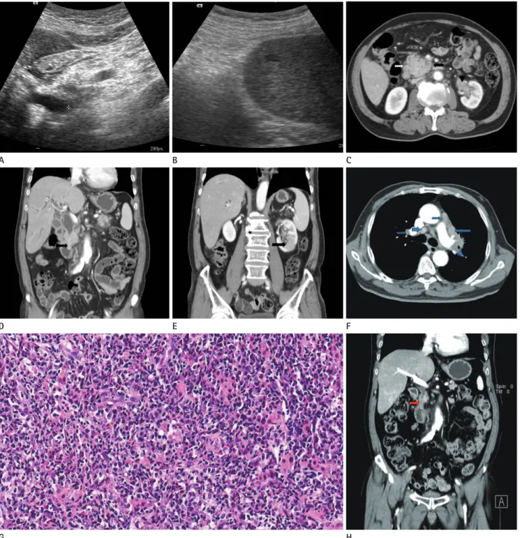
서 총담관은 12 mm로 확장되어 있었으며 총담관의 원위부가 두꺼워져 있었고(Fig. 1A), 간 내에도 두 군데의 작은 저에코성 결절이 관찰되었다(Fig. 1B). 전산화단층촬영술(Computed Tomograrphy) 소견으로는 췌장의 두부가 직경 3.5 cm 정도로 커져 있었고(Fig. 1C), 췌관 및 담관이 확장되어 있었으며 원위 부 담관의 급격한 협착과 비후가 보였다(Fig. 1D). 간 내에는 두 개의 저음영 결절이 있었으며, 신우가 두껍게 조영되면서 신 장 내에도 경계가 불분명한 종괴가 보였다(Fig. 1E). 이외에도 복강 및 종격동 내 여러 군데에 림프절의 비후와 좌상부 기관지 의 비후 및 종괴가 있었다(Fig. 1F). 환자는 간 내 병변에 대한 초음파유도하 조직검사를 실시하여 림프구와 형질세포 및 섬유 성 조직으로 구성된 염증거짓종양으로 진단하였으며(Fig 1G), 항생제를 제외하고 특별한 치료 없이 20일 후 임상 및 영상학적 소견이 현저한 호전을 보여(Fig. 1H) 최종적으로 다발성 장기 를 동시에 침범한 염증거짓종양으로 진단하였다.
고찰
염증거짓종양은 흔하게는 폐나 안와에 발생하지만, 어느 부
Case Report
pISSN 1738-2637
J Korean Soc Radiol 2011;65(1):77-80
Received April 11, 2011; Accepted May 18, 2011 Corresponding author: Hee Jin Kwon, MD Department of Radiology, College of Medicine, Dong-A University,
1 Dongdaesin-dong 3-ga, Seo-gu, Busan 602-103, Korea.
Tel. 82-51-240-5367 Fax. 82-51-253-4931 E-mail: [email protected]
Copyrights © 2011 The Korean Society of Radiology
Inflammatory pseudotumors are benign soft tissue tumors that in rare cases can also manifest in multiple organs. We report here on the radiologic findings of a case of inflammatory pseudotomor mimicking malignant lymphoma involving the liver, pancreas, common bile duct, kidney, renal pelvis and lymph nodes of the abdomen and mediastinum, as well as the bronchus in an adult.
Index terms
Granuloma, Plasma Cell Liver
Pancreas Biliary Tract Urogenital System
Tomography, X-Ray Computed
Multisystemic Organ Involvement by an Inflammatory Pseudotumor: A Case Report
다수의 장기를 침범한 염증거짓종양: 증례 보고
Woo Jeong Kim, MD, Hee Jin Kwon, MD, Jin Han Cho, MD, Jong Yeong Oh, MD, Kyung Jin Nam, MD, Dong Ho Ha, MD
Department of Radiology, College of Medicine, Dong-A University, Busan, Korea
다수의 장기를 침범한 염증거짓종양
submit.radiology.or.kr
대한영상의학회지 2011;65(1):77-80
78
Fig. 1. A 67-year-old man presenting with jaundice and poor oral intake.
A. Ultrasonography showing hyperechoic thickening in the distal CBD with proximal biliary tree dilatation.
B. Ultrasonography in the axial plane at the S6 segment region demonstrating a small oval hypoechoic mass.
C. Axial CT image shows the swelling of pancreatic head (arrows) and thickening of the left renal pelvis and upper ureter (arrowhead).
D. Coronal reformatted CT image showing thickening and stenosis of the distal common bile duct (arrow) and dilatation of proximal biliary tree.
E. Coronal reformatted CT image showing an ill-defined, low-attenuation mass lesions (arrow) in the left kidney.
F. Axial CT image showing multiple lymph node enlargement with homogeneous enhancement in both the perihilar and mediastinal area (ar- rows), with LUL bronchial wall thickening (arrowhead).
G. Histologic specimen obtained by core needle biopsy from the hepatic lesion. Photomicrograph of the histologic preparation (hematoxylin-eo- sin, original magnification x 400) showing a broad fibro-inflammatory lesion, composed of dense lymphoplasma cell infiltration and fibrosis.
H. Follow-up CT scan images obtained after 20 days later, showing the improvement of thickening of the distal CBD (arrow), left upper ureter, and bronchus, and decreased in size of the ill-defined mass in the left kidney and pancreas head (not shown).
A
D
G
C
F
H B
E
김우정 외
submit.radiology.or.kr
대한영상의학회지 2011;65(1):77-8079
참고문헌
1. Narla LD, Newman B, Spottswood SS, Narla S, Kolli R. In- flammatory pseudotumor. Radiographics 2003;23:719- 729
2. Walsh SV, Evangelista F, Khettry U. Inflammatory myofi- broblastic tumor of the pancreaticobiliary region: mor- phologic and immunocytochemical study of three cases.
Am J Surg Pathol 1998;22:412-418
3. Difiore JW, Goldblum JR. Inflammatory myofibroblastic tumor of the small intestine. J Am Coll Surg 2002;194:
502-506
4. Voss SD, Kruskal JB, Kane RA. Chronic inflammatory pseu- dotumor arising in the hepatobiliary-pancreatic system:
progressive multisystemic organ involvement in four pa- tients. AJR Am J Roentgenol 1999;173:1049-1054
5. Lopez-Tomassetti Fernandez EM, Luis HD, Malagon AM, Gonzalez IA, Pallares AC. Recurrence of inflammatory pseudotumor in the distal bile duct: lessons learned from a single case and reported cases. World J Gastroenterol 2006;12:3938-3943
6. Kamisawa T, Funata N, Hayashi Y, Tsuruta K, Okamoto A, Amemiya K, et al. Close relationship between autoimmune pancreatitis and multifocal fibrosclerosis. Gut 2003;52:
683-687
7. Park SB, Cho KS, Kim JK, Lee JH, Jeong AK, Kwon WJ, et al.
Inflammatory pseudotumor (myoblastic tumor) of the genitourinary tract. AJR Am J Roentgenol 2008;191:1255- 1262
8. Toosi MN, Heathcote J. Pancreatic pseudotumor with scle- rosing pancreato-cholangitis: is this a systemic disease?
Am J Gastroenterol 2004;99:377-382
9. Slavotinek JP, Bourne AJ, Sage MR, Freeman JK. Inflam- matory pseudotumour of the pancreas in a child. Pediatr Radiol 2000;30:801-803
10. Lévy S, Sauvanet A, Diebold MD, Marcus C, Da Costa N, Thiéfin G. Spontaneous regression of an inflammatory pseudotumor of the liver presenting as an obstructing malignant biliary tumor. Gastrointest Endosc 2001;53:371- 374
위에서나 발생할 수 있으며, 성별의 차이는 없다. 환자는 증상이 없는 경우도 있으나 일부에서는 설명되지 않는 발열이나 체중감 소, 종괴 효과가 나타나기도 하며 임상적으로나 영상학적으로 악성 종양과의 감별이 힘든 경우도 있다(1, 4). 원인과 발병기전 은 여전히 논란의 여지가 있으나, 조직학적으로 급성 혹은 만성 염증세포와 다양한 정도의 섬유성 조직으로 구성된다(1, 2). 세 계건강보건기구에서는 염증거짓종양을 불확실한 성격을 갖는 경계성 종양으로 분류하였고, 정확하게 알려진 바는 없으나 감 염, 자가면역, 전신의 염증성 질환의 급성악화 등으로 설명하고 있다. 최근에는 자가면역성 질환으로 염증거짓종양을 침범범위 에 따라 분류하기도 하는데(5), 전신성의 염증거짓종양의 형태 를 보고하였으며, 인체 내 대부분의 장기가 포함될 수 있다(6).
다발성으로 한 환자에서 간담도 및 췌장, 비뇨기관 등의 장기를 함께 침범한 경우는 보고되어 있으나(4, 7, 8) 저자들이 조사한 바로는 아직 국내에서 한 환자에게 간, 췌장, 담도계, 비뇨기계, 복강 및 종격동 내 림프절, 기관지를 동시에 침범하고 특별한 치료 없이 자연적으로 호전된 예는 보고된 바가 없다.
영상학적 소견은 매우 비특이적이다. 초음파에서는 다양한 에 코를 보이는데, 분명한 혹은 불분명한 경계의 저에코 혹은 고에 코성 종괴로 나타난다(7). 전산화단층촬영술에서는 균질 혹은 비균질의 저음영 혹은 고음영의 종괴로 강한 조영증강이 특징적 인 소견으로 보고하였으나, 실제로는 종괴 내 세포성분과 섬유 화의 정도에 따라 다양한 영상학적 소견을 보일 수 있으며, 조영 증강이 특이적인 소견은 아니다(1, 4, 7). 실제로 다양한 형태의 조영증강이 보고된 바 있는데, 조기 테두리 조영증강과 후기 구 심성 조영증강, 불균질 혹은 균질 조영증강 및 전혀 조영이 되지 않는 경우도 있다(9).
예후는 악성종양과 비교하면 좋은 임상경과를 보이지만, 때 때로 확진을 얻거나 증상완화를 위해 수술적인 절제를 하기도 한다. 그러나 일부에서는 보존적 약물 치료나 치료 없이 자연적 으로 호전된 경우도 있다(10). 본 증례는 한 환자에서 다양한 장기를 침범한 비특이적 종괴로 악성 림프종으로 오인하였으나 자연적으로 호전된 경우로, 최종적으로 염증거짓종양으로 진단 되었던 경우이다.
결론적으로 염증거짓종양은 비특이적인 종괴로 나타나고 다 양한 장기를 침범할 수 있으므로 다양한 장기 내에 다발성 종양 이 있는 환자에서 방사선학적으로 전형적인 악성 림프종이나 범 발성 악성 전이가 아닌 경우라면, 감별 진단에 반드시 포함해야 할 것으로 생각하며 이러한 영상 소견을 인지함으로써 조기진단 과 불필요한 근치적 절제를 막을 수 있을 것이다.
다수의 장기를 침범한 염증거짓종양
submit.radiology.or.kr
대한영상의학회지 2011;65(1):77-80
80
다수의 장기를 침범한 염증거짓종양: 증례 보고
김우정
·
권희진·
조진한·
오종영·
남경진·
하동호염증거짓종양은 특별한 원인 없이 발생하는 양성 연조직 종양으로 매우 드물게 다양한 장기를 침범할 수 있다. 저자들은 성 인에서 간, 췌장, 담관, 신장 및 신우, 복강 및 종격동 내 림프절, 기관지를 동시에 침범하여 악성 림프종으로 오인하였으나, 조직검사와 함께 임상적으로 염증거짓종양으로 진단된 1예의 영상 소견을 문헌 고찰과 함께 보고한다.
동아대학교 의과대학 영상의학과학교실