• 검색 결과가 없습니다.

Multicenter Validation Study of a Prognostic Index for Portal Vein Tumor Thrombosis in Hepatocellular Carcinoma

N/A
N/A
Protected

Academic year: 2021

Share "Multicenter Validation Study of a Prognostic Index for Portal Vein Tumor Thrombosis in Hepatocellular Carcinoma"

Copied!
10
0
0

로드 중.... (전체 텍스트 보기)

전체 글

(1)

Multicenter Validation Study of a Prognostic Index for

Portal Vein Tumor Thrombosis in Hepatocellular Carcinoma

Purpose

We previously reported on a staging system and prognostic index (PITH) for portal vein tumor thrombosis (PVTT) in hepatocellular carcinoma (HCC) patients treated with radiotherapy (RT) at a single institution. The aim of this study is to validate the PITH staging system using data from patients at other institutions and to compare it with other published staging systems.

Materials and Methods

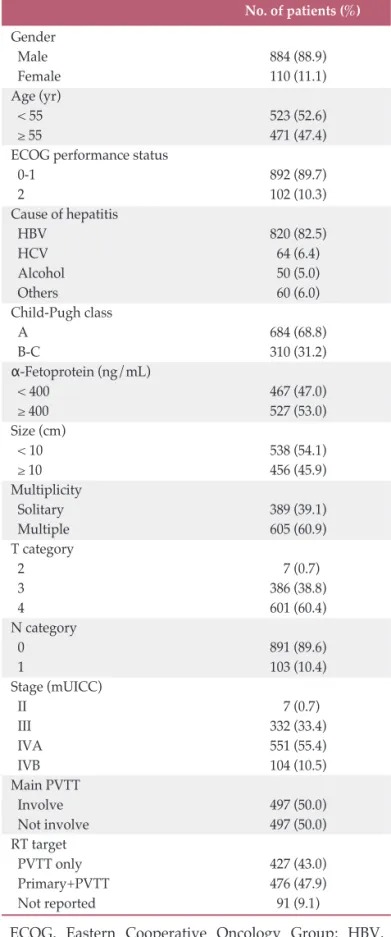
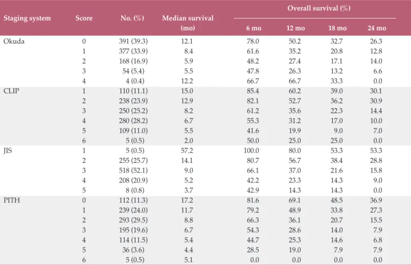
A total of 994 HCC patients with PVTT who were treated with RT between 1998 and 2011 by the Korean Radiation Oncology Group were analyzed retrospectively. All patients were staged using the Cancer of the Liver Italian Program (CLIP), Japanese Integrated Staging (JIS), Okuda, and PITH staging systems, and survival data were analyzed. The likelihood ratio, Akaike information criteria (AIC), time-dependent receiver operating characteristics, and prediction error curve analysis were used to determine discriminatory ability for comparison of staging systems.

Results

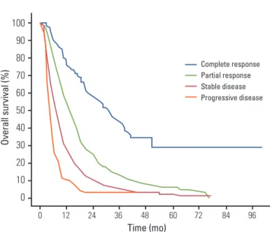
The median survival was 9.2 months. Compared with the other staging systems, the PITH score gave the highest values for likelihood ratio and lowest AIC values, demonstrating that PITH may be a better prognostic model. Although the values were not significant and differences were not exceptional, the PITH score showed slightly better performance with respect to time-dependent area under curve and integrated Brier score of prediction error curve.

Conclusion

The PITH staging system was validated in this multicenter retrospective study and showed better stratification ability in HCC patients with PVTT than other systems.

Key words

Hepatocellular carcinoma, Portal vein, Radiotherapy, Multicenter study, Validation + + + + + + + + + + + + + + + + + + + +

+ + + + + + + + + + + + + + + + + + + + + + + + + + + + + + + + + + + + + + + + + + + + + + + + + + + + + + + + + + + + + + + + + + + + + + + + + + + + + + + + + + + + + + + + + + + + + + + + + + + + + + + + + + + + + + + + + + + + + + + + + + + + + + + + + + + + + + + + + + + + + + + + + + + + + + + + + + + + + + + + + + + + + + + + + + + + + + + + + + + + + + + + + + + + + + + + + + + + + + + + + + + + + + + + + + + + + + + + + + + + + + + + + + + + + + + + + + + + + + + + + + + + + + + + + + + + + + + + + + + + + + + + + + + + + + + + + + + + + + + + + + + + + + + + + + + + + + + + + + + + + + + + + + + + + + + + + + + + + + + +

Correspondence: Hee Chul Park, MD, PhD Department of Radiation Oncology, Samsung Medical Center,

Sungkyunkwan University School of Medicine, 81 Irwon-ro, Gangnam-gu, Seoul 135-710, Korea Tel: 82-2-3410-2612

Fax: 82-2-3410-2619

E-mail: [email protected]

Received August 7, 2013 Accepted October 16, 2013

*Jeong Il Yu and Sang Min Yoon contributed equally to this work.

Jeong Il Yu, MD

1

Sang Min Yoon, MD, PhD

2

Hee Chul Park, MD, PhD

1

Jong Hoon Kim, MD, PhD

2

Tae Hyun Kim, MD, PhD

3

Joong-Won Park, MD, PhD

3

Jinsil Seong, MD, PhD

4

Ik Jae Lee, MD, PhD

4

Hong Seok Jang, MD, PhD

5

Chul Seung Kay, MD, PhD

6

Chul Yong Kim, MD, PhD

7

Eui Kyu Chie, MD, PhD

8

Jin Hee Kim, MD, PhD

9

Mi-Sook Kim, MD, PhD

10

Young Min Choi, MD, PhD

11

1

Department of Radiation Oncology, Samsung Medical Center,

Sungkyunkwan University School of Medicine, Seoul,

2

Department of Radiation Oncology, Asan Medical Center,

University of Ulsan College of Medicine, Seoul,

3

Research Institute and Hospital, National Cancer Center, Goyang,

4

Department of Radiation Oncology, Yonsei University College of Medicine, Seoul,

5

Department of Radiation Oncology,

The Catholic University of Korea College of Medicine, Seoul,

6

Department of Radiation Oncology, Incheon St. Mary Hospital,

The Catholic University of Korea College of Medicine, Incheon,

7

Department of Radiation Oncology, Korea University College of Medicine, Seoul,

8

Department of Radiation Oncology,

Seoul National University College of Medicine, Seoul,

9

Department of Radiation Oncology, Keimyung University Dongsan Medical Center, Keimyung University School of Medicine, Daegu,

10

Department of Radiation Oncology,

Korea Institute of Radiological and Medical Sciences,

Seoul,

11

Department of Radiation Oncology,

Dong-A University College of Medicine, Busan, Korea

(2)

Introduction

Portal vein tumor thrombosis (PVTT), a common compli- cation of hepatocellular carcinoma (HCC) with a reported incidence of 34% to 50% in patients with advanced HCC, is one of the most negative prognostic factors [1,2]. However, survival duration in patients with PVTT is very heteroge- neous, depending on other clinical characteristics and/or hepatic function [2,3].

Several staging systems have already been used in determination of prognosis and identification of optimal treatment modalities for HCC patients [1,4-7]. However, HCC patients with PVTT cannot be stratified using these systems [8-10]. More precise stratification would be helpful in identification of patients who might benefit from specific treatments.

To address this shortcoming, in a previous study we proposed a prognostic index for PVTT in patients with HCC (PITH) who were treated with radiotherapy (RT) [11].

However, it was a retrospective single-institution study, which was inherently influenced by selection bias. Therefore, the PITH staging system is currently not considered reliable enough for direct application in clinical situations. The purpose of the current study is to validate the PITH staging system and compare it with other known systems in order to determine which system enables the most accurate prediction of survival through a retrospective analysis of patients from the Korean Radiation Oncology Group (KROG).

Materials and Methods

1. PITH staging system

In our previous study we proposed the PITH staging system based on the clinical outcomes of 281 patients with combined HCC and main and/or first branch PVTT who were treated with RT at Samsung Medical Center [11]. This system is based on seven pretreatment factors: Eastern Cooperative Oncology Group (ECOG) performance status, Child-Pugh classification, tumor size, tumor multiplicity, site of PVTT involvement, degree of occlusion of portal flow, and the presence of lymph node metastasis. Of those factors, degree of occlusion of portal flow was excluded in the current study due to diagnostic obscurity and differences among institutional imaging work-up protocols. We used the remaining six pretreatment factors in validation of the PITH staging system. This study was approved and exempted

from permission by the Institutional Review Board of Samsung Medical Center, Sungkyunkwan University School of Medicine (IRB no. 2011-10-057).

2. Eligibility criteria and data collection

To validate the PITH staging system we enrolled patients with HCC combined with main and/or first branch PVTT who were treated with RT from January 1998 to October 2011 at 10 Korean institutions of the KROG.

Because other local treatment modalities can be considered when PVTT is confined to segmental branches (i.e., portal vein branches other than the main or first branch), we excluded those patients in order to maintain homogeneity of the study.

The diagnosis of HCC was based on histology of tumor tissue or reliable clinical criteria, largely in accordance with guidelines proposed by the Korean Liver Cancer Study Group (KLCSG), as follows: liver nodules in a high-risk group (association with liver cirrhosis or viral hepatitis) with one typical dynamic imaging finding and a trend of increas- ing elevated serum α-fetoprotein (AFP) level above 400 ng/mL, or two typical dynamic imaging findings without AFP increase. After 2009, the cutoff value of AFP was lowered to 200 ng/mL.

PVTT was identified using helical computed tomography (CT) scans with contrast enhancement, magnetic resonance imaging scans, or angiography. On contrast-enhanced CT scans, PVTT was identified by the presence of a low-attenu- ation intraluminal filling defect adjacent to the primary tumor with contrast enhancement. Thrombi located in the bilateral first branches and the main trunk were categorized as main PVTT. Tumor size was defined as the length of the longest diameter of the primary tumor with PVTT.

CT simulation was used in planning RT. The RT protocol, which included fractionated 3D conformal radiation therapy (3D-CRT; 1.8 Gy or 2.0 Gy/fraction), hypofractionated RT (2.0 to 5.0 Gy/fraction), or stereotactic body RT (SBRT, more than 5.0 Gy/fraction with fewer than five fractions) was not a criterion for eligibility for evaluation by the PITH system provided that the other eligibility criteria of this study were met. Recurrence and type of treatment before and after RT were also not considered in determining eligibility.

Data from 1,050 patients were gathered from the 10 institutions of the KROG. Among these, 56 patients could not be analyzed due to missing data that were requisite for this study (24 patients due to inaccessibility of RT history and 32 patients due to lack of information on performance status).

Consequently, 994 patients were enrolled in this study.

(3)

3. Comparison of staging systems

All patients were categorized according to the Cancer of the Liver Italian Program (CLIP), Japanese Integrated Staging (JIS), Okuda, and PITH staging systems based on their clinical and laboratory characteristics before RT.

Because all patients enrolled in this study were stage C according to the Barcelona Clinic Liver Cancer (BCLC) staging system, we did not compare the BCLC with the PITH system.

4. Assessment of RT response and statistical analysis

PVTT response after RT was determined using serial CT scans 4 to 12 weeks after completion of treatment, and the modified Response Evaluation Criteria in Solid Tumors (mRECIST) were used for measurement of response [12].

Patient overall survival (OS) was the single end point used to assess the performance of the different staging systems.

OS was measured from the date of the start of RT to the date of death or the last follow-up visit. OS data were plotted using the Kaplan-Meier method and compared using the log-rank test. Homogeneity of the staging systems (i.e., a small difference in survival among patients in the same classification within each system) was determined by the likelihood ratio and Akaike information criterion (AIC) was also used to evaluate the discriminatory ability of the given models by the Cox proportional hazards model, which was tested using the Schoenfeld residuals method [13]. Finally, time-dependent receiver operating characteristics (ROC) curve estimation and prediction error curve analysis were used to evaluate the discriminatory and stratification abilities in the different staging systems [14,15]. A staging system is considered to have a better performance if there is a higher likelihood ratio and time-dependent area under curve (AUC), and a lower AIC. Low integrated Brier score (IBS), which is the area under the curve of the prediction error curve analysis, is regarded as a prerequisite for a good staging system. All analyses were performed using PASW ver. 18.0 (SPSS Inc., Chicago, IL) except for time-dependent ROC curve estimation and prediction error curve analysis, which were performed using R statistical package 1.0.4.

Results

1. Patient characteristics

Table 1 summarizes the characteristics of the 994 patients

ECOG, Eastern Cooperative Oncology Group; HBV, hepatitis B virus; HCV, hepatitis C virus; mUICC, modified International Union Against Cancer; PVTT, portal vein tumor thrombosis; RT, radiation therapy.

Table 1. Characteristics of 994 patients from the Korean Radiation Oncology Study Group

No. of patients (%) Gender

Male 884 (88.9)

Female 110 (11.1)

Age (yr)

< 55 523 (52.6)

≥ 55 471 (47.4)

ECOG performance status

0-1 892 (89.7)

2 102 (10.3)

Cause of hepatitis

HBV 820 (82.5)

HCV 64 (6.4)

Alcohol 50 (5.0)

Others 60 (6.0)

Child-Pugh class

A 684 (68.8)

B-C 310 (31.2)

α-Fetoprotein (ng/mL)

< 400 467 (47.0)

≥ 400 527 (53.0)

Size (cm)

< 10 538 (54.1)

≥ 10 456 (45.9)

Multiplicity

Solitary 389 (39.1)

Multiple 605 (60.9)

T category

2 7 (0.7)

3 386 (38.8)

4 601 (60.4)

N category

0 891 (89.6)

1 103 (10.4)

Stage (mUICC)

II 7 (0.7)

III 332 (33.4)

IVA 551 (55.4)

IVB 104 (10.5)

Main PVTT

Involve 497 (50.0)

Not involve 497 (50.0)

RT target

PVTT only 427 (43.0)

Primary+PVTT 476 (47.9)

Not reported 91 (9.1)

(4)

from the 10 institutional databases who were used for external validation. The median age of the patients was 54 years (range, 23 to 84 years). The male:female ratio was 8:1.

There were 102 patients (10.3%) with an ECOG performance status of 2 or higher. Main PVTT was observed in 50.0% of the patients, and the primary tumor and PVTT were irradiated simultaneously in 476 patients (47.9%).

2. Treatment before RT

Of the 994 patients, one or more treatments were adminis- tered in 822 patients (82.7%). The distribution of specific treatment modalities administered prior to RT was as follows (patients who received two or more treatment modalities were counted separately for each one): surgical resection (n=45, 4.5%), radiofrequency ablation (RFA; n=54, 5.4%), percutaneous ethanol injection (n=25, 2.5%), transcatheter arterial embolization or chemoembolization (TAE or TACE;

n=713, 71.7%), transcatheter arterial chemoinfusion (TACI;

n=67, 6.7%), chemotherapy (n=27, 2.7%). Six patients (0.6%) had undergone previous RT.

3. Radiotherapy

The primary gross tumor and PVTT were simultaneously irradiated in 476 patients (47.9%), and only PVTT was targeted in 427 patients (43.0%) because of their hepatic and tumor condition (e.g., large tumor [more than two-thirds of the liver] with severe liver cirrhosis, Child-Pugh class C, large tumor with main PVTT in both lobes of the liver, numerous intrahepatic metastases). Target volume definition was not clarified in the remaining 91 patients (9.2%).

A median daily dose of 2.5 Gy (range 1.8 to 17.0 Gy) was administered, yielding a total dose of 45.0 Gy (range 7.2 to 66.0 Gy), which translates to a biologic effective dose of 56.25 Gy

10

(8.5 to 137.7 Gy

10

as the α/β=10).

Table 2. Distribution and survival of the 994 validation patients as determined by Okuda, CLIP, JIS, and PITH scores Overall survival (%)

Staging system Score No. (%) Median survival

(mo) 6 mo 12 mo 18 mo 24 mo Okuda 0 391 (39.3) 12.1 78.0 50.2 32.7 26.3

1 377 (33.9) 8.4 61.6 35.2 20.8 12.8

2 168 (16.9) 5.9 48.2 27.4 17.1 14.0

3 54 (5.4) 5.5 47.8 26.3 13.2 6.6

4 4 (0.4) 12.2 66.7 66.7 33.3 0.0

CLIP 1 110 (11.1) 15.0 85.4 60.2 39.0 30.1

2 238 (23.9) 12.9 82.1 52.7 36.2 30.9

3 250 (25.2) 8.2 61.2 35.6 22.3 14.4

4 280 (28.2) 6.7 55.3 31.2 17.0 10.0

5 109 (11.0) 5.5 41.6 19.9 9.0 7.0

6 5 (0.5) 2.0 50.0 25.0 25.0 0.0

JIS 1 5 (0.5) 57.2 100.0 80.0 53.3 53.3

2 255 (25.7) 14.1 80.7 56.7 38.4 28.8

3 518 (52.1) 9.0 66.1 37.0 21.6 15.8

4 208 (20.9) 5.2 42.2 23.3 14.3 9.0

5 8 (0.8) 3.7 42.9 14.3 14.3 0.0

PITH 0 112 (11.3) 17.2 81.6 69.1 48.5 36.9

1 239 (24.0) 11.7 79.2 48.9 33.8 27.3

2 293 (29.5) 8.8 66.3 36.1 20.7 15.5

3 195 (19.6) 6.7 54.3 28.6 14.0 7.9

4 114 (11.5) 5.4 44.7 25.3 14.6 6.8

5 36 (3.6) 4.4 28.5 19.0 7.9 7.9

6 5 (0.5) 5.1 0.0 0.0 0.0 0.0

CLIP, Cancer of the Liver Italian Program; JIS, Japanese Integrated System; PITH, prognostic index of the portal vein tumor

thrombosis in hepatocellular carcinoma.

(5)

Conventional fractionated 3D-CRT (1.8-2.0 Gy/fraction) was used in 392 patients (39.4%). Hypofractionated RT (2.22-5.0 Gy/fraction) was the most common dosing scheme (n=581, 58.5%). SBRT (more than 5.0 Gy/fraction with fewer than five fractions) was administered to 21 patients (2.1%).

Chemotherapy was administered concomitantly with RT in 110 patients (intra-arterial in 75, sorafenib in 21, systemic in 4).

4. Treatment after RT

After RT, among patients who received two or more treatment modalities, 49 patients (4.9%) received other local treatments (such as surgery, RFA, percutaneous ethanol injection therapy) and TAE/TACE/TACI were performed on 534 patients (53.7%). Chemotherapy (arterial or systemic) was administered in 111 patients (11.2%) and 62 (6.2%) received sorafenib.

5. RT responses and OS

The median survival was 9.2 months and the actuarial 6-, 12-, 18-, and 24-month OS rates were 65.7%, 40.0%, 25.2%, and 18.1%, respectively.

With the exception of 46 patients (4.6%) who were not evaluated due to lack of follow up, complete response of the PVTT was observed in 50 patients (5.0%), partial response in 405 patients (40.7%), stable disease in 408 patients (41.0%), and progressive disease in 85 patients (8.6%), yielding an objective response rate of 45.7%. The median survival duration of the patients who showed complete response and partial response was 32 months and 13.1 months, respec- tively (Fig. 1).

6. Comparison of the Okuda, CLIP, JIS, and PITH staging systems

Patient distribution and survival rate according to each staging system are shown in Table 2 and Fig. 2. The CLIP and PITH staging systems showed a more even patient distribu- tion than the other staging systems.

Fig. 2A shows survival curves according to PITH scores.

The subgroup of patients with a score of zero experienced a significantly longer survival (median, 17.2 months) than the Table 3. Comparison of staging systems with respect to likelihood ratio, Akaike information criteria (AIC), time-dependent area under curve (AUC), and integrated Brier score (IBS)

Staging system Likelihood ratio AIC AUC IBS

Okuda 8.073 10,058.740 0.557 0.084

CLIP 16.193 10,052.621 0.590 0.083

JIS 10.394 10,056.419 0.560 0.084

PITH 28.812 10,042.002 0.593 0.082

CLIP, Cancer of the Liver Italian Program; JIS, Japanese Integrated System; PITH, prognostic index of the portal vein tumor thrombosis in hepatocellular carcinoma.

Ov er al l s ur vi va l ( % )

100 90 80 70 60 50 40 30 20 10 0

0 12 24 36 48 60

Time (mo)

72 84 96

Complete response Partial response Stable disease Progressive disease

Fig. 1. Overall survival according to radiotheraphy

response. Median survival for complete response, partial

response, stable disease, and progressive disease was 32

months, 13.1 months, 7.1 months, and 4.2 months, respec-

tively.

(6)

subgroup with a score of 4 to 6, who experienced a median survival of less than six months.

Comparison between the best prognostic subgroups of each system showed that a JIS score of 1 was associated with superior survival (median 57.2 months and 80.0% OS at 12 months); however, only five patients from the current group were classified as this subgroup.

With the exception of this result, the PITH staging system stratified subgroups better than any other staging system.

Nearly 70% of patients with a PITH score of zero had survival durations longer than 12 months and more than one-third of these patients had survival duration longer than two years. In addition, it clearly classified the worst prognos- tic subgroup; patients with a PITH score of 4 to 6 had median survival durations shorter than six months. Furthermore, significant differences in the survival curves were observed among PITH stage I, II, III, and IV, whereas no difference was observed between Okuda stage II and III (Fig. 3).

A B

C D

p-value p-value

p-value

p-value

Ov er al l s ur vi va l ( % )

100 90 80 70 60 50 40 30 20 10 0

0 12 24 36 48 60

Time (mo)

72 84 96

PITH 0 PITH 1 PITH 2 PITH 3 PITH 4 PITH 5 PITH 6

0.007

< 0.001 0.01 0.378 0.239 0.669

Ov er al l s ur vi va l ( % )

100 90 80 70 60 50 40 30 20 10 0

0 12 24 36 48 60

Time (mo)

72 84 96

Ov er al l s ur vi va l ( % )

100 90 80 70 60 50 40 30 20 10 0

0 12 24 36 48 60

Time (mo)

72 84 96

Ov er al l s ur vi va l ( % )

100 90 80 70 60 50 40 30 20 10 0

0 12 24 36 48 60

Time (mo)

72 84 96

CLIP 1 CLIP 2 CLIP 3 CLIP 4 CLIP 5 CLIP 6

0.390

< 0.001 0.053 0.02 0.794

Okuda 0 Okuda 1 Okuda 2 Okuda 3 Okuda 4

< 0.001 0.108 0.305 0.811

JIS 1 JIS 2 JIS 3 JIS 4 JIS 5

0.228

< 0.001

< 0.001 0.285

Fig. 2. Overall survival according to the score from each staging system. (A) Prognostic index of the portal vein tumor throm-

bosis in hepatocellular carcinoma (PITH). (B) Okuda. (C) Cancer of the Liver Italian Program (CLIP). (D) Japanese Integrated

System (JIS).

(7)

As shown in Table 3, the likelihood ratio (28.812) for the PITH staging system was higher than those of the other staging systems, indicating that PITH has better homogene- ity, and the AIC (10,042.002) was the lowest for the PITH staging system, indicating that PITH is more informative than other systems with regard to prediction of survival.

Time-dependent ROC curve and IBS of prediction error curve analysis were also analyzed, and the AUC and IBS values were compared. Although not all values reached generally accepted discriminatory levels, PITH showed the highest AUC (0.593) and lowest IBS (0.082).

Discussion

As reported in several studies, the presence of PVTT in HCC patients remains one of the most negative prognostic factors [1-3]. In the absence of any treatment modality, patient survival in this setting is less than three months [3].

As a result, most staging systems include this factor as a criterion and assign these patients to a poor prognostic group [1,4-6,16].

However, the survival duration in these patients has been

Fig. 3. Overall survival according to the stage from each staging system. (A) Prognostic index of the portal vein tumor throm- bosis in hepatocellular carcinoma (PITH). (B) Okuda. (C) Cancer of the Liver Italian Program (CLIP).

A

B C

p-value p-value

p-value

Ov er al l s ur vi va l ( % )

100 90 80 70 60 50 40 30 20 10 0

0 12 24 36 48 60

Time (mo)

72 84 96

PITHⅠ

(score 0)

PITHⅡ

(score 1, 2)

PITHⅢ

(score 3, 4)

PITHⅣ

(score 5, 6)

< 0.001

< 0.001 0.023

Ov er al l s ur vi va l ( % )

100 90 80 70 60 50 40 30 20 10 0

0 12 24 36 48 60

Time (mo)

72 84 96

Ov er al l s ur vi va l ( % )

100 90 80 70 60 50 40 30 20 10 0

0 12 24 36 48 60

Time (mo)

72 84 96

CLIPⅠ

(score 1-3)

CLIPⅡ

(score 4-6)

< 0.001 OkudaⅠ

(score 0)

OkudaⅡ

(score 1, 2)

OkudaⅢ

(score 3, 4)

< 0.001

< 0.001

(8)

shown to vary widely, from less than five months to more than five years depending on patient and tumor characteris- tics [9,10,17,18]. Previous staging systems do not take into account these variations [1,4-7]. In the BCLC staging system, all of these patients are designated as stage C, and sorafenib is the only recommended treatment [6,19-21].

Because PVTT is a heterogeneous entity with significant prognostic variation, there are likely to be some patients who will benefit from local therapy rather than sorafenib.

Theoretically, locoregional therapies such as TACE, radioem- bolization, and/or RT could suppress PVTT progression and delay intravascular tumor growth and the deterioration of liver function by maintaining adequate portal flow [22,23].

In fact, RT has been reported to yield fair outcomes with an objective response rate of approximately 40% to 50% and a median survival duration of 9 to 10 months [24,25]. These results suggest that RT is effective in patients with HCC and PVTT, and, based on these findings, the Korean Liver Cancer Study Group recommends sorafenib and RT as standard therapies for HCC with PVTT.

To address the lack of an effective staging system for HCC combined with PVTT, we conducted this validation study in order to obtain a more definitive conclusion regarding the clinical utility of the PITH staging system, which was originally developed using pretreatment tumors and PVTT characteristics obtained retrospectively from a single institu- tion [11]. From 10 Korean institutions within the KROG, 994 patients with HCC and main and/or first branch PVTT who were treated with RT from January 1998 to October 2011 were analyzed.

During the validation process, we found that patients were more evenly distributed by the CLIP and PITH staging systems than by other systems. The patient distribution within each PITH score, an important feature of a good prognostic system, was much better than that in the other staging systems.

Fairly good stratification was achieved based on PITH score compared with other systems. Specifically, PITH showed a clearer division of the subgroups with the best and worst prognosis. Based on statistical analysis of likelihood ratio, AIC, time-dependent ROC curve estimation, and prediction error curve, the PITH system showed better homogeneity and discriminatory ability compared with other systems.

Compared with the other staging systems, the PITH staging system classified patients more clearly based on survival duration. Two-thirds of patients with a PITH score of zero had survival durations longer than 12 months. In contrast, patients with a PITH score of 4 to 6 had survival durations shorter than six months, although PITH could not stratify them specifically. In the former group, there might be the potential to extend survival by combining treatment

modalities, whereas in the latter group, it might be appropri- ate to reconsider RT. Our findings indicate that the PITH staging system might be helpful when planning RT in patients with HCC combined with PVTT.

This study has some limitations. First, due to the retrospec- tive nature of the study, the potential for selection bias cannot be excluded. However, we attempted to minimize this bias by the large multicenter design. Second, the wide hetero- geneity in RT regimens and additional treatment after RT could be confounding factors. However, such heterogeneity is an essential component of treatment decisions in actual practice, and additional treatments after RT may be admin- istered when they offer the possibility of improving clinical outcome. The PITH system could be used more comfortably in such situations because specific restrictions were not applied in the current study to exclude patients receiving RT.

Third, we only included patients who received RT; therefore, it is difficult to generalize our finding to patients undergoing other treatment modalities.

Like other staging systems, PITH included performance status (ECOG), liver function (Child-Pugh class), and extent of primary tumor. However, PITH also considered the extent and position of the PVTT. The status of PVTT might be particularly important in patients treated with RT, and the slightly better performance of the PITH system in these patients could reflect consideration of PVTT. Conduct of a larger prospective validation study is required in order to broaden the applicability of this staging system.

Conclusion

This study successfully validated the PITH staging system in a study group obtained in a multicenter retrospective fashion. PITH showed better stratification ability than the Okuda, CLIP, and JIS systems. Compared with previous systems, PITH may be helpful to oncologists in more accurate determination of prognosis and selection of more appropriate treatment options through more effective stratification of patients with HCC and PVTT based on risk.

Conflicts of Interest

Conflict of interest relevant to this article was not reported.

(9)

1. A new prognostic system for hepatocellular carcinoma: a retrospective study of 435 patients: the Cancer of the Liver Italian Program (CLIP) investigators. Hepatology. 1998;28:

751-5.

2. Carr BI, Pancoska P, Branch RA. Tumor and liver determi- nants of prognosis in unresectable hepatocellular carcinoma:

a large case cohort study. Hepatol Int. 2010;4:396-405.

3. Llovet JM, Bustamante J, Castells A, Vilana R, Ayuso Mdel C, Sala M, et al. Natural history of untreated nonsurgical hepatocellular carcinoma: rationale for the design and evaluation of therapeutic trials. Hepatology. 1999;29:62-7.

4. Kudo M, Chung H, Osaki Y. Prognostic staging system for hepatocellular carcinoma (CLIP score): its value and limitations, and a proposal for a new staging system, the Japan Integrated Staging Score (JIS score). J Gastroenterol.

2003;38:207-15.

5. Leung TW, Tang AM, Zee B, Lau WY, Lai PB, Leung KL, et al.

Construction of the Chinese University Prognostic Index for hepatocellular carcinoma and comparison with the TNM staging system, the Okuda staging system, and the Cancer of the Liver Italian Program staging system: a study based on 926 patients. Cancer. 2002;94:1760-9.

6. Llovet JM, Bru C, Bruix J. Prognosis of hepatocellular carcinoma: the BCLC staging classification. Semin Liver Dis.

1999;19:329-38.

7. Okuda K, Ohtsuki T, Obata H, Tomimatsu M, Okazaki N, Hasegawa H, et al. Natural history of hepatocellular carcinoma and prognosis in relation to treatment: study of 850 patients. Cancer. 1985;56:918-28.

8. Katamura Y, Aikata H, Takaki S, Azakami T, Kawaoka T, Waki K, et al. Intra-arterial 5-fluorouracil/interferon combi- nation therapy for advanced hepatocellular carcinoma with or without three-dimensional conformal radiotherapy for portal vein tumor thrombosis. J Gastroenterol. 2009;44:492-502.

9. Peng ZW, Guo RP, Zhang YJ, Lin XJ, Chen MS, Lau WY.

Hepatic resection versus transcatheter arterial chemoem- bolization for the treatment of hepatocellular carcinoma with portal vein tumor thrombus. Cancer. 2012;118:4725-36.

10. Yoon SM, Lim YS, Won HJ, Kim JH, Kim KM, Lee HC, et al.

Radiotherapy plus transarterial chemoembolization for hepatocellular carcinoma invading the portal vein: long-term

patient outcomes. Int J Radiat Oncol Biol Phys. 2012;82:

2004-11.

11. Yu JI, Park HC, Lim DH, Park W, Yoo BC, Paik SW, et al. Prog- nostic index for portal vein tumor thrombosis in patients with hepatocellular carcinoma treated with radiation therapy. J Ko- rean Med Sci. 2011;26:1014-22.

12. Lencioni R, Llovet JM. Modified RECIST (mRECIST) assess- ment for hepatocellular carcinoma. Semin Liver Dis.

2010;30:52-60.

13. Akaike H. A new look at statistical model identification. IEEE Trans Automat Contr. 1974;19:716-23.

14. Gerds TA, Schumacher M. Efron-type measures of prediction error for survival analysis. Biometrics. 2007;63:1283-7.

15. Heagerty PJ, Zheng Y. Survival model predictive accuracy and ROC curves. Biometrics. 2005;61:92-105.

16. Henderson JM, Sherman M, Tavill A, Abecassis M, Chejfec G, Gramlich T. AHPBA/AJCC consensus conference on staging of hepatocellular carcinoma: consensus statement. HPB (Oxford). 2003;5:243-50.

17. Inarrairaegui M, Thurston KG, Bilbao JI, D'Avola D, Rodriguez M, Arbizu J, et al. Radioembolization with use of yttrium-90 resin microspheres in patients with hepatocellular carcinoma and portal vein thrombosis. J Vasc Interv Radiol.

2010;21:1205-12.

18. Kasai K, Ushio A, Kasai Y, Sawara K, Miyamoto Y, Oikawa K, et al. Combination therapy of intra-arterial 5-fluorouracil and systemic pegylated interferon alpha-2b for advanced hepato- cellular carcinoma. Int J Clin Oncol. 2011;16:221-9.

19. Bruix J, Sherman M; American Association for the Study of Liver Diseases. Management of hepatocellular carcinoma: an update. Hepatology. 2011;53:1020-2.

20. Cheng AL, Kang YK, Chen Z, Tsao CJ, Qin S, Kim JS, et al.

Efficacy and safety of sorafenib in patients in the Asia-Pacific region with advanced hepatocellular carcinoma: a phase III randomised, double-blind, placebo-controlled trial. Lancet Oncol. 2009;10:25-34.

21. Llovet JM, Ricci S, Mazzaferro V, Hilgard P, Gane E, Blanc JF, et al. Sorafenib in advanced hepatocellular carcinoma. N Engl J Med. 2008;359:378-90.

22. Burgmans MC, Irani FG, Chan WY, Teo TK, Kao YH, Goh AS, et al. Radioembolization after portal vein embolization in a

References

Acknowledgments

This research was supported by a Samsung Medical Center

grant [GFO1130081], the Korean Radiation Oncology Group

(KROG), and funded by the Korean Liver Cancer Study

Group.

(10)

patient with multifocal hepatocellular carcinoma. Cardiovasc Intervent Radiol. 2012;35:1519-23.

23. Zeng ZC, Fan J, Tang ZY, Zhou J, Qin LX, Wang JH, et al. A comparison of treatment combinations with and without radiotherapy for hepatocellular carcinoma with portal vein and/or inferior vena cava tumor thrombus. Int J Radiat Oncol Biol Phys. 2005;61:432-43.

24. Kim DY, Park W, Lim DH, Lee JH, Yoo BC, Paik SW, et al.

Three-dimensional conformal radiotherapy for portal vein thrombosis of hepatocellular carcinoma. Cancer. 2005;10 3:2419-26.

25. Meng MB, Cui YL, Lu Y, She B, Chen Y, Guan YS, et al.

Transcatheter arterial chemoembolization in combination with radiotherapy for unresectable hepatocellular carcinoma: a systematic review and meta-analysis. Radiother Oncol.

2009;92:184-94.

수치

Table 1 summarizes the characteristics of the 994 patients
Table 2. Distribution and survival of the 994 validation patients as determined by Okuda, CLIP, JIS, and PITH scores Overall survival (%)
Fig.  1.  Overall  survival  according  to  radiotheraphy response. Median survival for complete response, partial response, stable disease, and progressive disease was 32 months, 13.1 months, 7.1 months, and 4.2 months,  respec-tively
Fig. 2.  Overall survival according to the score from each staging system. (A) Prognostic index of the portal vein tumor throm- throm-bosis in hepatocellular carcinoma (PITH)
+2

참조

관련 문서

Conclusion: On Three-phase spiral CT , HCC showed different enhancing patterns according tumor type and the presence of portal vein thrombosis. Index Words: Liver

A Study of Staging System for Hepatocellular Carcinoma: Prognostic Factors in Korean Patients.. Kew MC and

Hepatocellular carcinoma (HCC) with obstructive jaundice that is caused by bile duct tumor thrombi (BDT) is a rare finding and the appropriate treatment has not yet been

Survival outcomes of hepatic resection compared with transarterial chemoembolization or sorafenib for hepatocellular carcinoma with portal vein tumor thrombosis.. Surgical

Key Words: Associating liver partition and portal vein ligation for staged hepatectomy; Hepatocellular carcinoma; Portal vein anomaly; Liver cirrhosis; Future liver remnant;

Associating liver partition and portal vein ligation for staged hepatectomy (ALPPS) procedure for hepatocellular carcinoma with.. chronic liver disease: a case report and review

The presence of portal vein thrombosis is considered to be one of the risk factors that make liver transplantation in liver cirrhosis patients difficult.. Portal

In the 7 patients who did not receive anticoagulation therapy, thrombosis developed at the portal vein stump or peripheral branch of the portal vein as evidenced on a