Prognostic Index for Portal Vein Tumor Thrombosis in Patients with Hepatocellular Carcinoma Treated with Radiation Therapy
We performed a retrospective review of 281 hepatocellular carcinoma (HCC) patients with portal vein tumor thrombosis (PVTT) treated with radiation therapy (RT) between 1998 and 2008 to develop a prognostic model for those patients. Of the 281 patients, PVTT and intrahepatic main masses completely disappeared in 10 patients (3.6%), and shown a partial response in 141 patients (50.2%). The median survival was 11.6 months. Patients who had more than PR have shown significantly longer survival than the others (22.0 months vs 5.0 months, P < 0.001). On the multivariate analysis, pre-treatment poor prognosticators for overall survival were ECOG performance status, Child-Pugh class, multiple tumors, main PVTT, complete portal vein occlusion, lymph node metastasis, and primary tumor size. Prognostic index of RT for PVTT of HCC (PITH) scores were defined as the number of pre-treatment poor prognostic factors. PITH scores correlated well with overall survival. In the analysis of 1 and 2 yr overall survival rate, patients who had PITH scores of 3 or greater showed a significantly lower rate of overall survival than the others (33.0%, 17.3% vs 70.1%, 40.8%, respectively, P < 0.001). The PITH scoring model, proposed in the current study in HCC patients with PVTT, reliably predict overall survival.
Key Words: Carcinoma, Hepatocellular; Portal Vein Tumor Thrombosis; Radiotherapy;
Prognostic Index Jeong Il Yu1, Hee Chul Park1,
Do Hoon Lim1, Won Park1,
Byung Chul Yoo2, Seung Woon Paik2, Kwang Cheol Koh2 and Joon Hyuk Lee2 Departments of 1Radiation Oncology and 2Medicine, Samsung Medical Center, Sungkyunkwan University School of Medicine, Seoul, Korea
Received: 2 March 2011 Accepted: 10 June 2011 Address for Correspondence:
Hee Chul Park, MD
Department of Radiation Oncology, Samsung Medical Center, Sungkyunkwan University School of Medicine, 81 Irwon-ro, Gangnam-gu, Seoul 135-710, Korea
Tel: +82.2-3410-2612, Fax: +82.2-3410-2619 E-mail: [email protected]
This research was supported by Basic Science Research Program through the National Research Foundation of Korea (NRF) funded by the Ministry of Education, Science and Technology (2010-0011107).
DOI: 10.3346/jkms.2011.26.8.1014 • J Korean Med Sci 2011; 26: 1014-1022
INTRODUCTION
Hepatocellular carcinoma (HCC) is the third leading cause of cancer-related deaths worldwide and especially in East Asia, and sub-Saharan Africa, where hepatitis B virus in endemic (1). Sur- gical resection is the first choice of treatment for HCC, but ap- proximately 80% of cases are unresectable because of poor he- patic functional reserve associated with cirrhosis and multifo- cality of its presentation (2). Especially, portal vein tumor throm- bosis (PVTT), which is a common complication in patients with advanced-stage HCC, with a reported incidence of 34% to 50%
of these patients (3, 4), is a major determinant of patients’ prog- nosis (5, 6). Because of a decrease in portal blood flow, consen- sus treatments for HCC, including surgical resection, and trans- catheter arterial chemo-embolization (TACE) lack efficacy. Some- times major treatment-related complications are inevitable in patients with PVTT.
Recently, several studies have shown that radiation therapy (RT), particularly, three-dimensional conformal radiation ther- apy (3D-CRT) is effective not only for tumor responses but also for survival in HCC patients with PVTT (7, 8). Median survival time on those patients was reported as long as 6 to 13 months.
But, large differences were shown in the post-RT prognosis be- tween individual HCC patients who were treated with RT. Some
patients have shown sustained local control without further treat- ment measures after RT. Sometimes, RT provides good results to successive additional local treatment, like TACE, radiofrequency ablation or surgical resection. But there are no established guide- line which divides patient group on which RT provides good treat- ment response and better prognosis.
The purpose of this study is to design prognostic model for HCC patients with PVTT who are treated with RT, and to divide those patients into prognostic groups.
MATERIALS AND METHODS Diagnosis and clinical data
We conducted a retrospective study of all HCC patients with PVTT who were treated with RT at Samsung Medical Center between January 1998 and December 2008. During this period, total 742 patients were received RT because of primary liver can- cer in our institution. Of these patients, 710 patients had been diagnosed as HCC. And 320 patients (45.1%) had vascular inva- sion from HCC. Without PVTT, inferior vena cava or hepatic vein was involved in 19 (2.7%). And 20 patients (2.8%) had PVTT in small branches of portal vein (segmental/section branches ex- cept main, left or right hemiliver portal vein). If the PVTT is con- fined to small branches only, the other local modality (surgery,
radiofrequency ablation, TACE, etc) should be considered. In the current study, we analyzed 281 patients (39.6%) who had tu- mor thrombosis in the main, right or left hemiliver portal vein.
The diagnosis of HCC was made based on the guidelines pro- posed by the Korea Liver Cancer Study Group (9). Using these criteria, a patient is diagnosed as HCC if who has one or more risk factors (hepatitis B or C virus infection, and/or cirrhosis) and one of the following: a serum α-fetoprotein (AFP) level of > 400 ng/mL and a positive result with at least one of the three typical imaging techniques (triple phase computerized tomography [CT], contrast enhanced dynamic magnetic resonance imaging [MRI] or hepatic angiography); or a serum AFP level of < 400 ng/
mL and positive findings with at least two of three imaging tech- niques. A positive finding for typical HCC with dynamic CT or MRI is indicative of arterial enhancement followed by venous washout in the delayed portal/venous phase.
PVTT was demonstrated using helical CT scans with contrast enhancement, MRI scans, or angiography. On contrast-enhanced CT scans, PVTT was identified by the presence of a low-attenu- ation intra-luminal filling defect adjacent to the primary tumor.
If thrombi were located in both hemiliver branches and the main trunk, we categorized the thrombi as main PVTT because of the retrograde invasion of portal vein thrombosis. Differentiating malignant PVTT and benign PVTT may be difficult. Intrathrom- bus vascularity, observed in the arterial phase of imaging stud- ies after the administration of contrast, has been reported to be a sign that is specific for PVTT on both CT and MRI (10). We as- sessed malignant PVTT based on these enhancing and/or grow- ing pattern, response to treatment, and relationship with prima- ry tumor.
The following clinical data were collected from the medical records: patient demographics; complete blood count; chemis- try profiles; liver function test; AFP; size of the tumor and PVTT;
number of the tumor; involved side of the liver by the tumor; in- volved site of the portal vein by the PVTT; length of the PVTT;
completeness of the portal vein occlusion; presence of lymph nodes or distant metastasis; Eastern Cooperative Oncology Group (ECOG) performance status; Child-Pugh score and class; date of diagnosis; type of treatment; treatment response; date of last follow-up; live status; and cause of death.
Previous treatment
Two hundred forty two patients of 281 in the current series had received one or more treatments with surgical resection (19, 6.8%), radiofrequency ablation (35, 12.5%), percutaneous etha- nol injection (3, 1.1%), TACE (232, 82.6%), RT (0.7%), chemo- therapy (0.4%) before they were diagnosed as HCC with PVTT.
Radiation therapy
Before simulation, shallow-breathing during the procedure was asked and all patients got education and training. Then, respi-
ratory motion of the liver was checked by fluoroscopy, and these data were used for determination of the margin. After that, all patients underwent contrast enhanced CT scans in a supine position for RT planning, with both arms raised above the head to facilitate use of lateral RT portals. CT scan data were trans- ferred to a 3D-CRT treatment planning system (from 1998 to 2003, PROWESS, Alliant Medical Technology, Chico, CA, USA;
from 2004 to 2008, PINNACLE, The Philips Medical System, Best, Netherland).
The PVTT, main mass, the normal liver, kidneys, duodenum and spinal cord were contoured and reconstructed to form a 3D representation. The RT beam angles were designed to minimize critical organ injury. A dose-volume histogram (DVH) was also generated.
The gross tumor volumes (GTV) were defined as the radio- graphically abnormal areas (main mass and PVTT, usually) not- ed on the CT images refer to the diagnostic CT and/or MRI. But, in several cases (large tumor [more than 2/3 of the liver] with severe liver cirrhosis, Child-Pugh class C, huge bilateral intrahe- patic tumor with main PVTT, numerous intrahepatic metastasis, etc), only PVTT was delineated as GTV. The clinical target vol- umes (CTV) were regarded as same as GTV. In designing the PTV, the margins were individualized by observing liver position as well as liver movement at the time of simulation. In determin- ing the cranial-caudal margins, the distance of diaphragmatic excursion by respiration, which was observed fluoroscopically, was added to the cranial-caudal margins (minimum 1.5 cm + motion). In the same way, anterio-posterior, and lateral margins (minimum 1.0 cm + motion) were decided.
A median daily dose of 3 gray (Gy, range 1.8 to 4.5 Gy) was ad- ministered using 6, 10 or 15 MV X-rays, at 5 fractions per week, to deliver a total dose of 30-54 Gy, which translates to a biologic effective dose (BED) of 39-70.2 Gy10 as the α/β = 10. BED was cal- culated using a linear quadratic model to be equivalent to 3 Gy fraction treatments in respect of acute effects on normal tissues and tumors. 3D-CRT planning was designed under tentative guidelines so that the normal liver volume irradiated with more than one-half of the prescription dose should not be 50% of the total liver volume.
Four-dimensional gated CT with RPM signal and gated verifi- cation in the treatment room were conducted after March 2008, and 44 patients were enrolled in this study.
Post-RT treatment
Additional treatment, mostly TACE, was planned and adminis- tered after the RT without considering of RT response after 2004. In 202 patients who received RT after 2004, TACE in 122 patients (60.4%), re-irradiation in 7 patients, radiofrequency ablation in 4 patients, chemotherapy (sorafenib) in 4 patients, and hepatectomy in 2 patients was conducted. By contrast, be- fore 2004, TACE was considered mainly in patients who showed
good response to RT. In 79 patients, 23 patients (29.1%) received TACE.
Assessment of response and toxicity
PVTT and/or tumor response (according to RT target) was as- sessed using World Health Organization criteria by serial CT scans, 4-8 weeks after completion of treatment and then every 2-3 months. Complete disappearance of PVTT and tumor was defined as a complete response (CR), a more than 50% reduc- tion of thrombus in the greatest cross-sectional area and/or per- fusion normalization of the area involved by tumor thrombosis was defined as a partial response (PR), a less than 50% reduc- tion of thrombus and tumor (including no change) was defined as stable disease (SD), and tumor growth was defined as pro- gressive disease (PD). Responders were patients with CR or PR, whereas non-responders were patients with SD or PD.
Acute morbidity was evaluated weekly during treatment and 1 month after the treatment. Late morbidity was defined as oc- curring after 3 months. RT induced liver disease (RILD) was de- fined as anicteric ascites and elevation of alkaline phosphatase (ALP) levels > 2 times the pre-treatment values in the absence of PD.
If patients complained of melena or upper abdominal pain persisting for longer than 2 weeks during the follow-up period, fiberoptic gastroduodenoscopy was conducted for clarifying the gastrointestinal (GI) toxicity. Toxicity was scored using the Com- mon Terminology Criteria for Adverse Events (CTCAE; version 3.0) at every visit.
Statistical analysis
Overall survival (OS) and PVTT progression free survival (PVTT PFS) were estimated using Kaplan-Meier product-limit method.
OS was measured from the RT start date to the date of death or the last follow-up visit, and PVTT PFS from the RT start date to the date of PVTT progression or the last follow-up visit.
Univariate logistic regression analysis was used to evaluate the association between OS and various parameters. For multi- variate analysis to evaluate the relation between the OS and var- ious parameters, the stepwise procedure was performed using a logistic regression model containing all variables that attained or had a trend toward statistical significance on univariate anal- ysis. P < 0.05 was considered statistically significant. All calcula- tions were performed with PASW 17.0 for Windows (SPSS, Chi- cago, IL, USA).
Ethics statement
This study was approved of permission by the institutional re- view board of Samsung Medical Center, Sungkyunkwan Univer- sity (IRB No. 2011-03-033). Informed consent was waived by the board.
RESULTS
Clinical characteristics
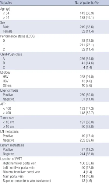
Table 1 describes the tumor characteristics of 281 patients. The median age of patients was 54 yr (range, 26-80 yr). Among the patients, 249 (88.6%) were male, and the male-to-female ratio was 7.8:1. The median size of tumor plus PVTT was 7 cm (range, 2-18 cm). Serum hepatitis B virus antigen markers were positive in 258 patients (91.8%) and hepatitis C virus antigen in 13 pa- tients (4.6%). Specific sites of PVTT were as follows: left hemili- ver portal vein (n = 50, 17.8%), right hemiliver portal vein (n = 100, 35.6%), bilateral hemiliver branch portal vein (n = 4, 1.4%), main portal vein (n = 114, 40.6%), extended to superior mesen- teric vein (n = 13, 4.6%).
Most patients had Child-Pugh classification A (84.0%) or B (14.0%), and only 2.0% had Child-Pugh class C. Thirty two pa-
Table 1. Characteristics of patients and tumor
Variables No. of patients (%)
Age (yr) ≤ 54
> 54 143 (50.9)
138 (49.1) Sex
Male Female
249 (88.6) 32 (11.4) Performance status (ECOG)
0 1 2
38 (13.5) 211 (75.1) 32 (11.4) Child-Pugh class
A B C
236 (84.0) 41 (14.6) 4 (1.4) Etiology
HBV HCV Others
258 (91.8) 13 (4.6) 10 (3.6) Liver cirrhosis
Positive
Negative 250 (89.0)
31 (11.0) AFP
< 400
≥ 400 133 (47.3)
148 (52.7) Tumor size
< 10 cm
≥ 10 cm 191 (68.0)
90 (32.0) LN metastasis
Positive
Negative 49 (17.4)
232 (82.6) Distant metastasis
Positive
Negative 37 (13.2)
244 (86.8) Location of PVTT
Right hemiliver portal vein Left hemiliver portal vein Bilateral hemiliver portal vein Main portal vein
Superior mesenteric vein involvement
100 (35.6) 50 (17.8) 4 (1.4) 114 (40.6)
13 (4.6)
ECOG, Eastern Cooperative Oncology Group; AFP, alpha-feto protein; LN, lymph node;
PVTT, portal vein tumor thrombosis.
tients (11.4%) presented poor performance status (ECOG 2).
Eighty-four patients (29.9%) who showed one or more of these characteristics (large tumor [more than 2/3 of the liver] with se- vere liver cirrhosis, Child-Pugh class C, huge bilateral intrahe- patic tumor with main PVTT, numerous intrahepatic metasta- sis, etc.) received RT only on PVTT.
Treatment responses
Four to eight weeks after the completion of the RT, an objective response was observed in 151 patients (53.8%). Of these patients, CR was observed in 10 patients (3.6%), PR in 141 patients (50.2
%), and SD in 72 patients (25.6%), and PD in 37 patients (13.2%).
There were 21 unevaluable patients (7.5%), who were lost to fol- low-up. AFP decrement was observed in 163 patients (58.0%) at 1 month follow up after RT.
Complication
Table 2 shows the complication profile. During the RT, the most common adverse effects were anorexia and nausea (152 patients,
54.1%), but only 1 patient showed grade III nausea, the others were grade I or II. Elevation of liver enzymes was also common, but generally it was less than a five-fold of the upper normal lim- it. And these elevations were self-limited in most cases. After the RT, 260 patients (92.5%) were evaluable for toxicities, except 21 patients who were lost to follow up. In 15 patients (5.3%), grade II or III clinical hepatic dysfunction was observed after the RT.
Among them, PD was observed in 9 of 15 patients (3.2%), and there was no classic RILD. But, in 2 patients (0.7%), transami- nases (aspatate aminotransferase [AST], alanine aminotransfer- ase [ALT]) were elevated more than five times the upper limit of the normal or of pre-treatment level. For gastrointestinal compli- cations, 8 patients (2.8%) were scored as grade II and 1 patient (0.4%) as grade III resulting from gastric or duodenal ulcers in- side the RT field.
Survival outcomes
The follow-up period ranged from 0 to 103.2 (median 8.0, mean 11.1) months and 111 of 281 (39.5%) patients were still alive at the time of last follow-up. The median survival was 11.6 (range 1.0 to 103.2) months and the actuarial 6-month, 12-month, 18- month, and 24-month overall survival rates were 65.2%, 48.1%, 35.1%, and 26.9%, respectively (Fig. 1A). In the cases of PVTT, the median PVTT PFS was 11.2 (range 0.2 to 103.2) months (Fig. 1B).
In responders, the median survival duration was 22.0 months and the 6-month, 12-month, 18-month, and 24-month overall survival rates were 85.3%, 70.0%, 53.9% and 41.4%, respectively.
In non-responders, the median survival duration was 5.0 months and the 6-month, 12-month, 18-month, and 24-month overall survival rates were 34.3%, 13.8%, 2.9%, and 0.0%, respectively (Fig. 2A). The responders had a significantly higher overall sur- vival rate than the non-responders (P < 0.001).
Prognostic factors for overall survival
The results of univariate and multivariate analyses to evaluate
Overall survival rate (%) PVTT progression free survival rate (%)
Months Months
Median 11.6 months Median 11.2 months
0 12 24 36 48 60 72 84 96 108 120 0 12 24 36 48 60 72 84 96 108 120
100 90 80 70 60 50 40 30 20 10 0
100 90 80 70 60 50 40 30 20 10 0
A B
Fig. 1. Overall survival and portal vein tumor thrombosis progression free survival of all patients: Kaplan-Meier curves for overall survival (A) and portal vein tumor thrombosis (PVTT) progression free survival (B) rate of all patients with hepatocellular carcinoma (HCC) and PVTT treated with radiation therapy (RT).
Table 2. Acute and chronic complication after radiation therapy
Complication No. of evaluable
patients
CTCAE Grade
0 I II III IV V
During RT Anorexia Nausea Vomiting Diarrhea AST ALT ALP
281 174 166 197 270 48 113 151
84 85 68 9 173 131 121
23 29 16 2 60 22 6
- 1
- - 35 12 2
- - - - 5 3 1
- - - - - - - After RT
Clinical liver dysfunction Gastro-duodenal ulcer
260 245
251 -
8 7 1 8
- -
- -
-
CTCAE, common terminology criteria for adverse events; RT, radiation therapy; AST, aspartate aminotransferase; ALT, alanine aminotransferase; ALP, alkaline phosphatase.
the relation between OS and various parameters are summa- rized in Table 3. Of the pre-treatment parameters, significant prognostic factors for OS in univariate analysis were performance
status (ECOG), Child-Pugh class, tumor size, multiple tumor, main PVTT involvement, bilateral involvement of PVTT, size of the PVTT, completeness of obstruction, lymph node metastasis, distant metastasis, pre-treatment AST/ALT/ALP/PIVKA-II level, and so on, while age, sex, hemoglobin, and pre-treatment AFP level (P = 0.053) were not. On the multivariate analysis, perfor- mance status (ECOG, P = 0.001), Child-Pugh class (P = 0.002), tumor size (P = 0.029), multiple tumors (more than 2, P = 0.016), main PVTT involvement (P = 0.022), completeness of obstruc- tion (P = 0.001), and lymph node metastasis (P = 0.001) showed statistically significant effects on OS.
Most significant prognostic factor after treatment was radia- tion response as discussed above. AFP decrement after RT was also important post-treatment prognostic factor. The patients who achieved AFP decrement (163 patients, 58.0%) showed su- perior median survival duration than the others (14.7 months vs 5.5 months, P = 0.009). And the other significant prognostic fac- tors in post-treatment parameters were decrement of the PIVKA- II level (P < 0.001) and total dose of the radiation delivered (me-
Overall survival rate (%)
Months
P < 0.001
Median 5.0 months Median 22.0 months
0 12 24 36 48 60 72 84 96 108 120
100 90 80 70 60 50 40 30 20 10 0
Fig. 2. Overall survival according to the RT response: Kaplan-Meier curves for overall survival rate according to the RT response.
Response (+) Response (-)
Table 3. Prognostic factors for overall survival rate
Variables No.
Median survival (month)
P value Univariate Multivariate Age (yr)
≤ 54
> 54 152
159 11.4
12.0 0.981 0.310
Sex Male Female
249 32
12.0 8.7
0.522 0.642
Performance status (ECOG) 0-1
≥ 2
249 32
13.0 5.0
< 0.001 0.001
Child-Pugh class A
B-C
236 45
13.0 5.3
< 0.001 0.002
HBV Positive Negative
258 23
11.4 11.9
0.462 0.517
Liver cirrhosis Positive Negative
250 31
11.6 10.6
0.836 0.559
Additional treatment before RT Positive
Negative
242 39
13.3 5.3
< 0.001 0.843
AST ≤ 80 > 80
195 86
16.3 5.1
< 0.001 0.380
ALT ≤ 80 > 80
241 40
12.0 5.8
0.037 0.422
ALP ≤ 150
> 150 158
123 14.7
7.1 < 0.001 0.232 AFP
< 400
≥ 400 133
148 12.6
10.9 0.053 0.07
ECOG, Eastern Cooperative Oncology Group; AST, aspartate aminotransferase; ALT, alanine aminotransferase; ALP, alkaline phosphatase; AFP, alpha-feto protein; LN, lymph node; PVTT, portal vein tumor thrombosis; RT, radiation therapy; 4D, four-dimensional; TACE, transcatheter arterial chemo-embolization.
Variables No.
Median survival (month)
P value Univariate Multivariate LN metastasis
Positive
Negative 49
232 5.3
13.3 < 0.001 0.001 Distant metastasis
Positive Negative
37 244
5.6 12.6
0.002 0.575
Tumor size < 10 cm ≥ 10 cm
191 90
13.8 6.0
< 0.001 0.029
Number of tumor ≤ 2 > 2
130 151
13.9 5.3
< 0.001 0.016
Involvement of main portal vein Positive
Negative
127 154
7.7 17.4
< 0.001 0.022
Bilaterality of PVTT Positive Negative
204 77
13.6 9.1
< 0.001 0.077
Length of PVTT < 5 cm ≥ 5 cm
128 153
16.3 8.0
0.001 0.089
Porta vein occlusion Complete Incomplete
125 156
17.0 7.7
< 0.001 0.001 RT dose (α/β = 10)
< 40 Gy10
40-50 Gy10
> 50 Gy10
29 117 135
4.2 10.4 13.8
0.032 0.202
Additional treatment after RT Positive
Negative 149
132 4.7
19.1 < 0.001 0.573 RT response
Positive
Negative 151
130 22.0
5.0 < 0.001 < 0.001
dian survival duration; less than 40 Gy10 4.2 months, 40 Gy10 to 50 Gy10 10.4 months, more than 50 Gy10 13.8 months, P = 0.031) in univariate analysis. Additional treatments before and/or af- ter RT showed a statistically significant effect on OS. But, those differences might be related with selection bias of the patients who live longer could receive additional treatments. It is biased on that additional treatments showed no differences to OS in multivariate analysis and closely related with RT response (P <
0.001).
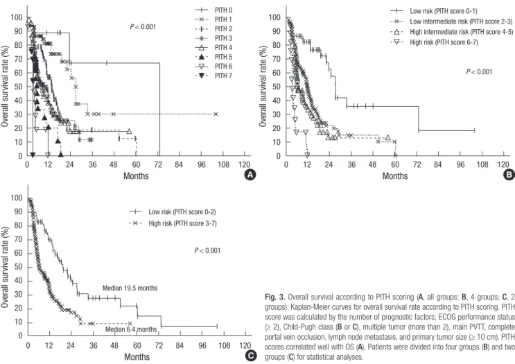
A Predictive model for overall survival
Based on clinical relevance and availability of the information, we used seven pre-treatment factors that showed statistical sig- nificance in both univariate and multivariate analysis to con- struct a Predictive Index for PVTT of the HCC (PITH) and calcu- late a PITH score. The included factors were grade II or more of performance status (ECOG), Child-Pugh classification B or C, 10 cm or more of tumor size, multiplicity of the tumor, main PVTT involvement, complete occlusion of portal flow and lymph node metastasis. PITH score is defined as the number of the above factors. We estimated OS from the PITH score, and statistically significant differences were detected in each group (P < 0.001, Fig. 3A). We divided the patients into low, low intermediate, high
intermediate and high risk groups according to the PITH score.
The low risk group with score 0 or 1 consisted of 54 patients (19.2
%) and had 82.5% OS at one year, 27.2 months median survival.
The low intermediate risk group with score 2 or 3 consisted of 133 patients (47.3%) and had 45.9% OS at one year, 11.4 months median survival. The high intermediate risk group with score 4 or 5 consisted of 76 patients (27.0%) and high risk group who had 6 or more score are made up of 18 patients (6.4%), and 33.0%, 0.0% OS at one year, and 6.4 months, 3.3 months median sur- vival, respectively (P < 0.001, Fig. 3B). And also, when divided them into two groups by the score 0 to 2 and 3 or more (low risk group 110 patients, high risk group 171 patients), OS showed dif- ference (70.1% vs 33.0% at one year, P < 0.001, Fig. 3C).
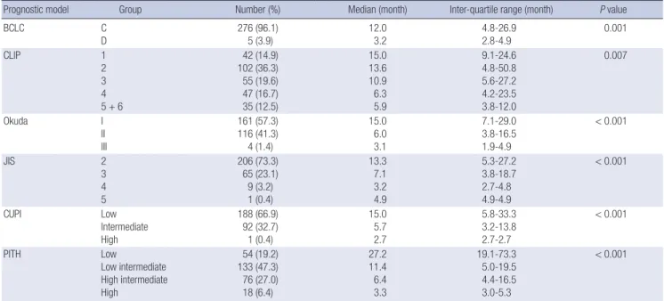
Comparison of the other stage system
The prognostic ability of the PITH score was compared with Bar- celona Clinic Liver Cancer (BCLC), Cancer of the Liver Italian Program (CLIP), Okuda, Japan Integrated Staging (JIS), and Chi- nese University Prognostic Index (CUPI) staging systems. For each score, this performance was evaluated by comparing by log rank test the survival curves of the single categories, calculated using the Kaplan-Meier method. In Table 4, median values and interquartile ranges of the OS for the six prognostic systems are
Fig. 3. Overall survival according to PITH scoring (A, all groups; B, 4 groups; C, 2 groups): Kaplan-Meier curves for overall survival rate according to PITH scoring. PITH score was calculated by the number of prognostic factors; ECOG performance status (≥ 2), Child-Pugh class (B or C), multiple tumor (more than 2), main PVTT, complete portal vein occlusion, lymph node metastasis, and primary tumor size (≥ 10 cm). PITH scores correlated well with OS (A). Patients were divided into four groups (B) and two groups (C) for statistical analyses.
Overall survival rate (%) Overall survival rate (%)
Months Months
P < 0.001
P < 0.001
0 12 24 36 48 60 72 84 96 108 120 0 12 24 36 48 60 72 84 96 108 120
100 90 80 70 60 50 40 30 20 10 0
100 90 80 70 60 50 40 30 20 10 0 PITH 0
PITH 1 PITH 2 PITH 3 PITH 4 PITH 5 PITH 6 PITH 7
Low risk (PITH score 0-1)
Low intermediate risk (PITH score 2-3) High intermediate risk (PITH score 4-5) High risk (PITH score 6-7)
A B
Overall survival rate (%)
Months
P < 0.001
0 12 24 36 48 60 72 84 96 108 120
100 90 80 70 60 50 40 30 20 10 0
Low risk (PITH score 0-2) High risk (PITH score 3-7)
C Median 19.5 months
Median 6.4 months
reported. In spite of there was a significant correlation between OS and stage assigned using all six systems (BCLC P = 0.001, CLIP P = 0.007, Okuda P < 0.001; JIS P < 0.001; CUPI P < 0.001), and trend in), the PITH score performs better than the other stag- ing systems, allowing a better characterization of the extremely good and intermediate categories of patients.
DISCUSSION
The presence of PVTT in patients with HCC is one of the most significant prognostic factors for poor prognosis. In several re- ports, it is the clinic-pathologic parameters that mostly influenc- es on survival in multivariate analysis (3-5). It can lead to condi- tions not only a widespread dissemination of tumors through- out the liver, but marked deterioration of hepatic function. There- fore, the prognosis of HCC patients with PVTT is extremely poor.
Without treatment, their survival is less than 3 months (5). Stan- dard treatment regimens have not been established in these pa- tients, especially in AsiaPacific region.
In the recent large scale randomized, double blind, placebo- controlled trial, sorafenib, an oral multi-kinase inhibitor of the vascular endothelial growth factor receptor, the platelet-derived growth factor receptor and Raf showed about 2 to 3 months sur- vival benefit in patients with advanced (unresectable or meta- static) HCC who had not received previous systemic treatment in the AsiaPacific region as well as Western (11, 12). The medi- an survival of the experimental group was 10.7 months in West- ern, and 6.5 months in Asian-Pacific region. But, the results from loco-regional therapies such as TACE and/or RT have been re- ported better than above results especially in patients with lo-
cally advanced disease without distant metastases (13, 14). For locally advanced disease such as HCC with PVTT, TACE and/or RT is the most commonly utilized treatment in Korea. Treatment algorithm for hepatocellular carcinoma utilized in Korea sup- ports not only sorafenib but also TACE and RT as a standard ther- apy for HCC with PVTT (15).
Recently, the major progress in RT for HCC has been made with deeper understanding of the liver, HCC, normal organ ra- diobiology (16, 17) and more elaborated radiation technique of 3D-CRT, image-guided RT (IGRT) and stereotactic body RT (SBRT). In the treatment of the HCC patients with PVTT, RT was carried out critical role. RT has an advantage that it could be per- formed safely without being limited by the presence of PVTT (7, 18, 19). When patients with HCC and PVTT were treated with RT and/or other treatment modalities, the response rates and median survival approximated 40%-75% and 5.3-13.1 months, respectively. Hata et al. (8) reported on 35 patients with PVTT at the main trunk or major branches, and 46% of progression free survival at 2 yr (median 21 months) suggesting that RT was a good alternative treatment approach for such patients. Kim et al. (7) reported that RT showed 45.8% objective response rate, and me- dian survival duration in responders were 10.7 months compar- ed with 5.3 months in the non-responders. And dose-response relationship in patients receiving less than 58 Gy10 and 58 Gy10
or more were 20% and 54.6% in the 59 patients with HCC and PVTT. A combined radiation therapy after TACE showed better clinical outcomes for HCC invading the main portal vein, when compared to TACE mono-therapy (20, 21). These results sug- gest that RT for HCC with PVTT is also effective and may pro- long the survival of these patients.
Table 4. Comparison of survivals according to prognostic models
Prognostic model Group Number (%) Median (month) Inter-quartile range (month) P value
BCLC C
D 276 (96.1)
5 (3.9) 12.0
3.2 4.8-26.9
2.8-4.9 0.001
CLIP 1
2 3 4 5 + 6
42 (14.9) 102 (36.3) 55 (19.6) 47 (16.7) 35 (12.5)
15.0 13.6 10.9 6.3 5.9
9.1-24.6 4.8-50.8 5.6-27.2 4.2-23.5 3.8-12.0
0.007
Okuda I
II III
161 (57.3) 116 (41.3) 4 (1.4)
15.0 6.0 3.1
7.1-29.0 3.8-16.5 1.9-4.9
< 0.001
JIS 2
3 4 5
206 (73.3) 65 (23.1) 9 (3.2) 1 (0.4)
13.3 7.1 3.2 4.9
5.3-27.2 3.8-18.7 2.7-4.8 4.9-4.9
< 0.001
CUPI Low
Intermediate High
188 (66.9) 92 (32.7) 1 (0.4)
15.0 5.7 2.7
5.8-33.3 3.2-13.8 2.7-2.7
< 0.001
PITH Low
Low intermediate High intermediate High
54 (19.2) 133 (47.3) 76 (27.0) 18 (6.4)
27.2 11.4 6.4 3.3
19.1-73.3 5.0-19.5 4.4-16.5 3.0-5.3
< 0.001
BCLC, Barcelona Clinic Liver Cancer (BCLC) staging classification; CLIP, Cancer of the Liver Italian Program (CLIP) scoring system; JIS, Japan Integrated Staging (JIS) scoring system; CUPI, The Chinese University Prognostic Index; PITH, Predictive Index for portal vein tumor thrombosis of the hepatocellular carcinoma.
There were several prognostic models in the HCC patients (22), like BCLC (23), CLIP (24), Okuda (25), JIS (26), and CUPI (27) staging system. Above systems are regarded as reliable prog- nostic model, because verified by several studies. But, those models are not specified in patients with PVTT. Until recently, there was no applicable prognostic system in patients with HCC with PVTT treated with RT, to our knowledge. Therefore, it is dif- ficult to make treatment recommendation according to a reliable estimate of life expectancy in cirrhotic patients with HCC and PVTT. This may induce unfavorable effects. Due to underestima- tion of life expectancy, patients are referred to supportive care only without any therapy which might have potentially positive effects on patients’ outcome. On the other hand, an overestima- tion of life expectancy may expose patients to treatments that will result in no benefit or even harmful effects on patients’ prog- nosis.
Recently, Huang et al. (18) published article about prognostic model. In the study, the authors conducted retrospective review for 326 HCC patients with PVTT who were treated with RT. De- spite their meaningfulness, this study also has some limitations to generalize their model in patients with HCC and PVTT treat- ed with RT. Firstly, the median survival was only 3.8 months. As stated above, most published articles with similar protocols, the median survival was 5 to 10 months. Possibility the enrolled pa- tients had poor liver function, and radiation delivery was too delayed. Secondly, there were large missing data, 155 patients (47.5%). These limitations need to be considered while apply- ing their rule in the treatment.
In this article, we proposed a new scoring system (PITH score) that accounts for both tumor and PVTT characteristics useful in prognostic assessment of patients with HCC and PVTT treated with RT. The PITH score is easy to calculate and based on vari- ables that are routinely assessed during clinical examination, biochemical staging, and CT of the liver. Classification accord- ing to PITH score was well correlated with the OS after RT. Those results of the PITH score, compared with other staging systems, showed more predictable power in the patients with PVTT when treated with RT, especially in the extremely good and interme- diate prognostic categories. We look forward to PITH score sys- tem might be allowed the patient with HCC and PVTT to make important decisions, and help physician for risk stratification in patients treated with RT. It could be also the guidance of addi- tional combined treatment modalities in those patients.
Our study does have some limitations. First, as in all retrospec- tive studies, a selection bias could have occurred, with a reduced prevalence of patients with a worse prognosis. However, this pos- sible bias should be less important than in other series because of the relatively short and recent period of recruitment. Second, rather small number of cases might not be enough to derive con- crete conclusion. For solving these problems, large, prospective validation study would be required.
In conclusion, our data demonstrates that ECOG performance status, Child-Pugh class, tumor size, tumor multiplicity, main PVTT involvement, completeness of portal vein obstruction, and lymph node metastasis are important prognostic factors for OS in HCC patients with PVTT treated with RT. From those seven prognostic factors, we designed prognostic model by the num- ber of prognostic factors, PITH score. Our data confirm that PITH score is significantly correlated with OS. The PITH scoring mod- el proposed in the current study in HCC patients with PVTT re- liably predict OS. Our institution will consider conducting a pro- spective validation trial of PITH score system. This trial will per- mit us to resolve questions and also to confirm our conclusions and might help make a treatment decision according to risk strat- ification in patient with HCC and PVTT treated with RT.
REFERENCES
1. McGlynn KA , London WT. The global epidemiology of hepatocellular carcinoma: present and future. Clin Liver Dis 2011; 15: 223-43.
2. Lau WY, Lai EC. Hepatocellular carcinoma: current management and recent advances. Hepatobiliary Pancreat Dis Int 2008; 7: 237-57.
3. The Cancer of the Liver Italian Program (CLIP) Investigators. Prospec- tive validation of the CLIP score: a new prognostic system for patients with cirrhosis and hepatocellular carcinoma. Hepatology 2000; 31: 840-5.
4. Park KW, Park JW, Choi JI, Kim TH, Kim SH, Park HS, Lee WJ, Park SJ, Hong EK, Kim CM. Survival analysis of 904 patients with hepatocellular carcinoma in a hepatitis B virus-endemic area. J Gastroenterol Hepatol 2008; 23: 467-73.
5. Llovet JM, Bustamante J, Castells A, Vilana R, Ayuso Mdel C, Sala M, Brú C, Rodés J, Bruix J. Natural history of untreated nonsurgical hepato- cellular carcinoma: rationale for the design and evaluation of therapeu- tic trials. Hepatology 1999; 29: 62-7.
6. Park W, Lim DH, Paik SW, Koh KC, Choi MS, Park CK, Yoo BC, Lee JE, Kang MK, Park YJ, Nam HR, Ahn YC, Huh SJ. Local radiotherapy for pa- tients with unresectable hepatocellular carcinoma. Int J Radiat Oncol Biol Phys 2005; 61: 1143-50.
7. Kim DY, Park W, Lim DH, Lee JH, Yoo BC, Paik SW, Kho KC, Kim TH, Ahn YC, Huh SJ. Three-dimensional conformal radiotherapy for portal vein thrombosis of hepatocellular carcinoma. Cancer 2005; 103: 2419-26.
8. Sugahara S, Nakayama H, Fukuda K, Mizumoto M, Tokita M, Abei M, Shoda J, Matsuzaki Y, Thono E, Tsuboi K, Tokuuye K. Proton-beam ther- apy for hepatocellular carcinoma associated with portal vein tumor thrombosis. Strahlenther Onkol 2009; 185: 782-8.
9. Park JW. Practice guideline for diagnosis and treatment of hepatocellu- lar carcinoma. Korean J Hepatol 2004; 10: 88-98.
10. Piscaglia F, Gianstefani A, Ravaioli M, Golfieri R, Cappelli A, Giampal- ma E, Sagrini E, Imbriaco G, Pinna AD, Bolondi L; Bologna Liver Trans- plant Group. Criteria for diagnosing benign portal vein thrombosis in the assessment of patients with cirrhosis and hepatocellular carcinoma for liver transplantation. Liver Transpl 2010; 16: 658-67.
11. Cheng AL, Kang YK, Chen Z, Tsao CJ, Qin S, Kim JS, Luo R, Feng J, Ye S, Yang TS, Xu J, Sun Y, Liang H, Liu J, Wang J, Tak WY, Pan H, Burock K, Zou J, Voliotis D, Guan Z. Efficacy and safety of sorafenib in patients in the Asia-Pacific region with advanced hepatocellular carcinoma: a phase
III randomized, double-blind, placebo-controlled trial. Lancet Oncol 2009; 10: 25-34.
12. Llovet JM, Ricci S, Mazzaferro V, Hilgard P, Gane E, Blanc JF, de Oliveira AC, Santoro A, Raoul JL, Forner A, Schwartz M, Porta C, Zeuzem S, Bolon- di L, Greten TF, Galle PR, Seitz JF, Borbath I, Häussinger D, Giannaris T, Shan M, Moscovici M, Voliotis D, Bruix J. Sorafenib in advanced hepa- tocellular carcinoma. N Engl J Med 2008; 359: 378-90.
13. Liang SX, Zhu XD, Lu HJ, Pan CY, Li FX, Huang QF, Wang AY, Chen L, Fu XL, Jiang GL. Hypofractionated three-dimensional conformal radia- tion therapy for primary liver carcinoma. Cancer 2005; 103: 2181-8.
14. Seong J, Park HC, Han KH, Chon CY, Chu SS, Kim GE, Suh CO. Clinical results of 3-dimensional conformal radiotherapy combined with trans- arterial chemoembolization for hepatocellular carcinoma in the cirrhot- ic patients. Hepatol Res 2003; 27: 30-5.
15. Chen PJ, Furuse J, Han KH, Hsu C, Lim HY, Moon H, Qin S, Ye SL, Yeoh EM, Yeo W. Issues and controversies of hepatocellular carcinoma-targeted therapy clinical trials in Asia: experts’ opinion. Liver Int 2010; 30: 1427-38.
16. Kim H, Lim DH, Paik SW, Yoo BC, Koh KG, Lee JH, Choi MS, Park W, Park HC, Huh SJ, Choi DH, Ahn YC. Predictive factors of gastroduode- nal toxicity in cirrhotic patients after three-dimensional conformal radio- therapy for hepatocellular carcinoma. Radiother Oncol 2009; 93: 302-6.
17. Park HC, Seong J, Han KH, Chon CY, Moon YM, Suh CO. Dose-response relationship in local radiotherapy for hepatocellular carcinoma. Int J Ra- diat Oncol Biol Phys 2002; 54: 150-5.
18. Huang YJ, Hsu HC, Wang CY, Wang CJ, Chen HC, Huang EY, Fang FM, Lu SN. The treatment responses in cases of radiation therapy to portal vein thrombosis in advanced hepatocellular carcinoma. Int J Radiat Oncol Biol Phys 2009; 73: 1155-63.
19. Yamada K, Izaki K, Sugimoto K, Mayahara H, Morita Y, Yoden E, Mat- sumoto S, Soejima T, Sugimura K. Prospective trial of combined trans-
AUTHOR SUMMARY
Prognostic Index for Portal Vein Tumor Thrombosis in Patients with Hepatocellular Carcinoma Treated with Radiation Therapy
Jeong Il Yu, Hee Chul Park, Do Hoon Lim, Won Park, Byung Chul Yoo, Seung Woon Paik, Kwang Cheol Koh and Joon Hyuk Lee While radiation therapy (RT) is often applied for hepatocellular carcinoma (HCC), all previous studies dealt simply with treatment results. In this study, we analyzed the indices of the HCC patients with portal vein tumor thrombosis, which could be potentially helpful to identify the beneficial prognositic factors in the similar patients.
catheter arterial chemoembolization and three-dimensional conformal radiotherapy for portal vein tumor thrombus in patients with unresect- able hepatocellular carcinoma. Int J Radiat Oncol Biol Phys 2003; 57:
113-9.
20. Korean Liver Cancer Study Group and National Cancer Center, Korea.
Practice guidelines for management of hepatocellular carcinoma 2009.
Korean J Hepatol 2009; 15: 391-423.
21. Seong J. Challenge and hope in radiotherapy of hepatocellular carcino- ma. Yonsei Med J 2009; 50: 601-12.
22. Tandon P, Garcia-Tsao G. Prognostic indicators in hepatocellular carci- noma: a systematic review of 72 studies. Liver Int 2009; 29: 502-10.
23. Llovet JM, Brú C, Bruix J. Prognosis of hepatocellular carcinoma: the BCLC staging classification. Semin Liver Dis 1999; 19: 329-38.
24. The Cancer of the Liver Italian Program (CLIP) investigators. A new prog- nostic system for hepatocellular carcinoma: a retrospective study of 435 patients. Hepatology 1998; 28: 751-5.
25. Okuda K, Ohtsuki T, Obata H, Tomimatsu M, Okazaki N, Hasegawa H, Nakajima Y, Ohnishi K. Natural history of hepatocellular carcinoma and prognosis in relation to treatment. Study of 850 patients. Cancer 1985; 56:
918-28.
26. Kitai S, Kudo M, Minami Y, Haji S, Osaki Y, Oka H, Seki T, Kasugai H, Sasaki Y, Matsunaga T. Validation of a new prognostic staging system for hepatocellular carcinoma: a comparison of the biomarker-combined Japan Integrated Staging Score, the conventional Japan Integrated Stag- ing Score and the BALAD Score. Oncology 2008; 75(Suppl 1): 83-90.
27. Leung TW, Tang AM, Zee B, Lau WY, Lai PB, Leung KL, Lau JT, Yu SC, Johnson PJ. Construction of the Chinese University Prognostic Index for hepatocellular carcinoma and comparison with the TNM staging system, the Okuda staging system, and the Cancer of the Liver Italian Program staging system: a study based on 926 patients. Cancer 2002; 94: 1760-9.