Totally laparoscopic associating liver partition and portal vein ligation for staged hepatectomy using anterior approach in HCC
patient with Type II portal vein anomaly: a case report
Heon Tak Ha, Young Seok Han, and Jae Min Chun
Department of Surgery, Kyungpook National University Hospital, Kyungpook National University School of Medicine, Daegu, Korea
Associating liver partition and portal vein ligation for staged hepatectomy (ALPPS) has gradually developed because of rapid hypertrophy of the future liver remnant volume (FLR) in spite of high morbidity. To minimize the patient’s postoperative pain and morbidity including wound complication caused by two consecutive major abdominal operations, we adopted a totally laparoscopic approach and used a composite mesh graft. Also, to maximize the oncologic efficacy, we adopted the “anterior approach” technique. A 44-year-old woman with large hepatitis B-related hepatocellular carci- noma (HCC) in her right lobe was transferred to our hospital for surgical treatment. Preoperatively predicted FLR by a CT scan was 21% and type II portal vein anomaly was detected. A totally laparoscopic approach was planned.
During the first stage operation, right anterior and posterior portal veins were meticulously dissected and tied. After parenchymal transection by the “anterior approach” technique, two glissonian pedicles of the right liver were individually isolated. A composite mesh graft was used to prevent severe adhesion on both liver partition surfaces. During the second-stage operation, 9 days after the first stage operation, the two isolated glissonian pedicles were initially transected. After full mobilization of the right lobe, the right hepatic vein was also transected. The right lobe was re- moved through the Pfannenstiel incision. She was discharged 7 days after the second stage operation. Her post- operative course was uneventful and there was no HCC recurrence for 15 months after hepatectomy. A totally laparo- scopic ALPPS procedure can be a feasible technique that ensures patient safety and oncologic superiority, even in patients with complicated anatomical variation. (Ann Hepatobiliary Pancreat Surg 2017;21:217-222)
Key Words: Associating liver partition and portal vein ligation for staged hepatectomy; Hepatocellular carcinoma; Portal vein anomaly; Liver cirrhosis; Future liver remnant; Anterior approach
Received: May 10, 2017; Accepted: July 7, 2017 Corresponding author: Young Seok Han
Department of Surgery, Kyungpook National University Hospital, Kyungpook National University School of Medicine, 130 Dongdeok-ro, Jung-gu, Daegu 41944, Korea
Tel: +82-53-200-6734, Fax: +82-53-421-0510, E-mail: [email protected]
Copyright Ⓒ 2017 by The Korean Association of Hepato-Biliary-Pancreatic Surgery
This is an Open Access article distributed under the terms of the Creative Commons Attribution Non-Commercial License (http://creativecommons.org/
licenses/by-nc/4.0) which permits unrestricted non-commercial use, distribution, and reproduction in any medium, provided the original work is properly cited.
Annals of Hepato-Biliary-Pancreatic Surgery ∙ pISSN: 2508-5778ㆍeISSN: 2508-5859
INTRODUCTION
Small liver remnant volume in hepatocellular carcino- ma (HCC)-related liver cirrhosis is associated with sig- nificant morbidity and mortality, and it is the main limit- ing factor for hepatic resection.1-3 To increase the resect- ability and to reduce the incidence of postoperative com- plications, it is necessary to increase the volume and func- tion of the planned future liver remnant (FLR).4 The asso- ciating liver partition and portal vein ligation (ALPPS) procedure was introduced as a potential solution to per- form extended resection safely by inducing pre-resection liver hypertrophy in patients with multifocal liver cancer
or non-resectable liver tumors.5 But, this technique has a high complication rate and a mortality rate between 12%
and 27% according to the initial reports on ALPPS.
Because ALPPS consists of an extensive first stage proce- dure that includes hepatic parenchymal transection com- bined with portal vein ligation, the complication rate may be higher regardless of successful pre-resection liver hypertrophy. Hence, efforts should be made to reduce the morbidity and mortality while maintaining a more rapid FLR hypertrophy. Laparoscopic surgery is known to alle- viate the surgical stress and the systemic inflammatory re- sponse to all surgeries.6 Therefore, total or partial use of laparoscopy may help to reduce the surgical difficulties
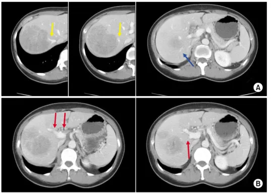
Fig. 1. Preoperative computed tomography scan shows a large mass abutting the right hepatic vein (yellow arrow) and the right posterior portal vein (blue arrow) (A) and type II portal vein (red arrow) anomaly (B).
caused by severe adhesions that will be encountered in the second stage.7 The laparoscopic intrahepatic glissonian ap- proach for right hepatectomy is a safe and feasible techni- que,8 but in ALPPS, which requires separate ligation of the portal vein, portal vein variations can pose significant technical demand.
We describe the technical aspects of a totally laparo- scopic ALPPS using the anterior approach in a HCC pa- tient with Type II portal vein anomaly.
CASE
A 44-year-old-woman with hepatitis B-related HCC was referred to our hospital. At admission, hepatitis B vi- rus-DNA level was 1.82×10⁶ IU/ml. But, she had a rela- tively well-preserved liver function according to the labo- ratory tests which showed a serum total bilirubin level of 0.53 mg/dl (normal range, <1.2 mg/dl), aspartate amino- transferase (AST) level of 67 U/L (normal range, <32 U/L), albumin level of 4.1 g/dl (normal range, 3.5-5.2 g/dl), and prothrombin time international normalized ratio (INR) of 0.99 (normal range, 0.85-1.5). Serum creatinine level was 0.71 mg/dl (normal range, 0.6-1.3 mg/dl) and the model for end-stage liver disease (MELD) score was 6. Computed tomography (CT) revealed an 8-cm hetero- geneous enhancing mass abutting the right hepatic vein
and wash out in the delayed venous phase in the right lobe of the liver (Fig. 1A). There were no evidences of ascites and portal hypertension. Alpha-fetoprotein (AFP) level and protein induced by vitamin K absence or antago- nist-II (PIVKA-II) level were markedly elevated to 8,423 ng/ml (normal range, <7 ng/ml) and 100,000 mAU/ml (normal range, <40 mAU/ml), respectively. Volumetric CT scan of the liver showed insufficient FLR (remnant left lobe with volume: 213 ml; FLR/total liver volume:
21.9%; FLR/standard liver volume: 20.5%) after right hepatectomy, which indicated that anatomical liver re- section with an adequate surgical resection margin was in- appropriate for the patient. The anatomic variation of the hepatic portal vein branching was identified as the tri- furcation type (Type II) (Fig. 1B).9
Surgical procedures
Laparoscopic ALPPS procedure using the anterior ap- proach was planned to induce rapid hypertrophy of the FLR.
In the first stage, the patient was placed in the left semi-lateral decubitus position with the right side elevated approximately 30 degrees. Five trocars were used. The first trocar (12 mm) was inserted in the upper umbilicus for placement of a 10-mm flexible 3-dimensional laparoscope. Four other trocars were placed 2 cm below
Fig. 2. The first stage operation findings. The right posterior portal vein and the right anterior portal vein were meticulously dissected and preservation of the right anterior and posterior hepatic arteries was also identified (A). After liver parenchymal transection using the anterior approach technique, the right anterior and posterior glissonian pedicles were separately isolated (B). A composite mesh graft was used to minimize adhesions between both transected cut surfaces (C). (Blue arrow, right posteri- or portal vein and hepatic artery; Yellow arrow, right anterior portal vein and hepatic artery).
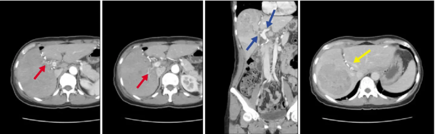
Fig. 3. Computed tomography scans 7 days after the first stage operation show intact right anterior and posterior hepatic arteries (red arrow), an adequately ligated right portal vein and a patent left portal vein (blue arrow), and a well-preserved middle hepatic vein (yellow arrow).
the costal margin on the right midclavicular line (12 mm), 2 cm below the costal margin on the right axillary line (5 mm), in the subxiphoid position (5 mm), and 2 cm be- low the costal margin on the left midclavicular line (5 mm). After exploration, a cholecystectomy was performed.
The right side of the hepatoduodenal ligament was dis- sected with lymphadenectomy for the exposure of the hi- lar structures. The right posterior portal vein and the right anterior portal vein were identified and isolated by metic- ulous dissection, and preservation of the right anterior and posterior hepatic arteries was also confirmed (Fig. 2A).
Without liver mobilization, parenchymal transection was performed along the line that was demarcated after liga- tion of the right anterior and posterior portal veins and temporary clamping of the hepatic artery. Liver trans- ection was performed using the Cavitron Ultrasonic
Surgical Aspiration (CUSA) and energy-source device (SonicisionⓇ; Covidien, Boulder, US) from the anterior surface of the liver to the anterior surface of the inferior vena cava, which was completely exposed. A composite mesh graft was attached to the transection surface of the right liver (Fig. 2C) and a closed suction drain was placed between the transection surfaces to prevent fluid collec- tion including biloma.
CT scan of the liver, which was performed 7 days after the first stage operation, demonstrated that the right ante- rior and posterior hepatic arteries were patent and the right anterior and portal veins were appropriately ligated.
The left portal vein, the left hepatic artery and the middle hepatic vein were well preserved for future liver remnant (Fig. 3).
The second stage operation (Fig. 4) was performed 9
Fig. 5. Postoperative liver function.
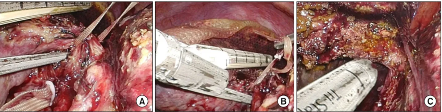
Fig. 4. The second stage operation findings. The right anterior glissonian pedicle (A) and the posterior glissonian pedicle (B) were sequentially transected with laparoscopic staplers (A). Finally, the right hepatic vein (C) was transected.
days after the first stage operation and the same trocar sites were used as in the first laparoscopic approach.
Initially, the right anterior and posterior glissonian pedi- cles, which had been isolated during the first stage oper- ation, were transected with laparoscopic staplers, respectively. Next, multiple right inferior hepatic veins were transected with Hem-O-lok clips. The transected right liver was fully mobilized and the right hepatic vein was finally transected with a laparoscopic stapler. The specimen was removed through the Pfannenstiel incision.
The time for the first-stage operation was 260 min and the time for the second-stage operation was 130 min.
During the two stage operation, blood transfusion was not necessary.
Changes in functional liver remnant volume and liver function
After the first stage operation, her liver function test gradually improved until reaching the normal range. CT scan 7 days after the first stage operation showed a sig-
nificant increase in FLR (remnant left lobe with volume:
339 ml; FLR/total liver volume: 30.2%; FLR/standard liv- er volume: 32.6%). FLR growth was 60% in 6 days. Liver function of the patient, including aspartate amino- transferase, alanine aminotransferase, total bilirubin and prothrombin time after the first stage operation, gradually improved (Fig. 5). The volume of the remaining left liver continuously increased after the second stage operation, and it was confirmed that the volume increased to 403 ml at 7 days and 604 ml at 2 months after the second stage operation. More stable liver function was obtained after the second stage operation (Fig. 5).
Postoperative course
Postoperative liver function of this patient steadily im- proved and complications did not occur. The patient was discharged 7 days after the second stage operation and she had recovered well. On follow-up at postoperative 15 months, imaging studies including a CT scan revealed no tumor in the remnant liver, and AFP and PIVKA II levels
were 7.2 ng/ml and 17.07 mAU/ml, respectively, which indicated an absence of tumor recurrence.
DISCUSSION
In patients who require an increase in volume of the planned future liver remnant to enable a potentially cura- tive resection, induction of hypertrophy of the tumor-free lobe prior to resection may mitigate morbidity and mortality.4 Preoperative portal vein embolization or liga- tion has been commonly used to achieve hypertrophy of remnant liver volume. But, these techniques are associated with some problems, including delay (between 3 and 8 weeks)10,11 or the absence of hypertrophy and the risk of tumor progression between the two stages. The delay or absence of sufficient hypertrophy may be related to the formation of intrahepatic vascular collaterals. Hence, a few reports have suggested that simultaneous occlusion of the intrahepatic collateral circulation and portal vein al- lows a rapid FLR growth of 40% to 160% in only 6 to 9 days.4,12-15 We were also able to complete this hep- atectomy with a much shorter interval of 9 days between the first and second stage operations.
ALPPS can achieve rapid FLR hypertrophy. However, the reported mortality in the initial series from Germany was 12% and 15% in a subsequent multi-centric analysis, reaching a rate of even 27% in experienced hepatobiliary centers.5,16,17 Extensive experience of laparoscopic liver resection suggests that laparoscopy may reduce operative severity and complications such as blood loss, adhesion, and bile leakage.18,19 Machado and colleagues believed that laparoscopic ALPPS, as an easy solution for adhe- sions and difficulties that may be encountered during the second stage, is feasible and does not appear to be inferior to the open approach in experienced hands.7,20 The treat- ment of the cut surface of the “deportalized” part of the liver and the use of devices to reduce adhesions for the second stage operation are significant points of concern.
The use of foreign bodies such as plastic bags or sheets around the liver, as a solution to avoid adhesions and fa- cilitate the second stage, requires a reoperation for their removal, even if the second stage operation cannot be per- formed because of clinical complications or insufficient hypertrophy of the remnant liver.7 Keeping this in mind, we used a composite mesh which offers a collagen barrier
on one side to limit visceral attachment and is able to completely avoid adherence to the cut surface of the rem- nant liver.
Laparoscopic ALPPS was introduced as the default pro- cedure for patients with very small FLR, and it is a very recent but a quite promising technique.20 Laparoscopy re- duces the extent of surgery and can minimize the risk of vascular or biliary injury because of the optimal visual- ization of the transection area.19 However, extensive dis- section of the hepatic hilum is essential for ligation of the portal vein and preservation of the hepatic artery.
Awareness regarding the variations in portal vein branch- ing patterns is very important in order to avoid major catastrophic events during laparoscopic hepatectomy. We believe that the magnification achievable via laparoscopy considerably facilitates separate dissection of the right hepatic pedicle with improvement in laparoscopic surgical skills and surgical instruments.
ALPPS was supposed to be an “all-touch” technique that would reduce the oncologic treatment efficacy for liv- er malignancy.16 But, totally laparoscopic associating liver tourniquet and portal ligation for staged hepatectomy via the anterior approach for cirrhotic HCC21 and totally lapa- roscopic ALPPS, conforming to the “No-Touch” princi- ple22 were reported. We experienced that laparoscopic ALPPS using the anterior approach technique could be a less invasive procedure that provides improved surgical and survival outcomes.
In conclusion, ALPPS has emerged as a new strategy to increase the resectability of hepatic malignancies.
Laparoscopic ALPPS using the anterior approach techni- que can provide a minimally invasive modification as a
“no-touch technique” that has greater oncologic efficacy.
The laparoscopic ALPPS technique seems to be feasible and safe in the hands of surgeons with expertise, even in patients with anatomical variation of the liver.
REFERENCES
1. Kishi Y, Abdalla EK, Chun YS, Zorzi D, Madoff DC, Wallace MJ, et al. Three hundred and one consecutive extended right hepatectomies: evaluation of outcome based on systematic liver volumetry. Ann Surg 2009;250:540-548.
2. Hemming AW, Reed AI, Howard RJ, Fujita S, Hochwald SN, Caridi JG, et al. Preoperative portal vein embolization for ex- tended hepatectomy. Ann Surg 2003;237:686-693.
3. Liu H, Zhu S. Present status and future perspectives of pre-
operative portal vein embolization. Am J Surg 2009;197:686-690.
4. Schadde E, Malagó M, Hernandez-Alejandro R, Li J, Abdalla E, Ardiles V, et al. Monosegment ALPPS hepatectomy: extending resectability by rapid hypertrophy. Surgery 2015;157:676-689.
5. Schnitzbauer AA, Lang SA, Goessmann H, Nadalin S, Baumgart J, Farkas SA, et al. Right portal vein ligation combined with in situ splitting induces rapid left lateral liver lobe hypertrophy en- abling 2-staged extended right hepatic resection in small-for-size settings. Ann Surg 2012;255:405-414.
6. Burpee SE, Kurian M, Murakame Y, Benevides S, Gagner M.
The metabolic and immune response to laparoscopic versus open liver resection. Surg Endosc 2002;16:899-904.
7. Machado MA, Makdissi FF, Surjan RC. Totally laparoscopic ALPPS is feasible and may be worthwhile. Ann Surg 2012;256:e13.
8. Cho A, Yamamoto H, Kainuma O, Ota T, Park S, Arimitsu H, et al. Extrahepatic Glissonean approach for laparoscopic major liver resection (with video). J Hepatobiliary Pancreat Sci 2013;
20:141-144.
9. Sureka B, Patidar Y, Bansal K, Rajesh S, Agrawal N, Arora A.
Portal vein variations in 1000 patients: surgical and radiological importance. Br J Radiol 2015;88:20150326.
10. Jaeck D, Oussoultzoglou E, Rosso E, Greget M, Weber JC, Bachellier P. A two-stage hepatectomy procedure combined with portal vein embolization to achieve curative resection for initially unresectable multiple and bilobar colorectal liver metastases.
Ann Surg 2004;240:1037-1049.
11. Broering DC, Hillert C, Krupski G, Fischer L, Mueller L, Achilles EG, et al. Portal vein embolization vs. portal vein liga- tion for induction of hypertrophy of the future liver remnant. J Gastrointest Surg 2002;6:905-913.
12. de Santibañes E, Clavien PA. Playing Play-Doh to prevent post- operative liver failure: the "ALPPS" approach. Ann Surg 2012;
255:415-417.
13. de Santibañes E, Alvarez FA, Ardiles V. How to avoid post-
operative liver failure: a novel method. World J Surg 2012;36:
125-128.
14. Adam R, Laurent A, Azoulay D, Castaing D, Bismuth H.
Two-stage hepatectomy: a planned strategy to treat irresectable liver tumors. Ann Surg 2000;232:777-785.
15. Alvarez FA, Iniesta J, Lastiri J, Ulla M, Bonadeo Lassalle F, de Santibañes E. New method of hepatic regeneration. Cir Esp 2011;89:645-649.
16. Aloia TA, Vauthey JN. Associating liver partition and portal vein ligation for staged hepatectomy (ALPPS): what is gained and what is lost? Ann Surg 2012;256:e9.
17. Dokmak S, Rasoaherinomenjanahary F, Cauchy F, Aussilhou B, Belghiti J. Does ALPPS regularly increase the future remnant liver and prevent postoperative liver failure? HPB (Oxford) 2014;16(Suppl 2):169.
18. Schadde E, Ardiles V, Robles-Campos R, Malago M, Machado M, Hernandez-Alejandro R, et al. Early survival and safety of ALPPS: first report of the International ALPPS Registry. Ann Surg 2014;260:829-838.
19. Wakabayashi G, Cherqui D, Geller DA, Buell JF, Kaneko H, Han HS, et al. Recommendations for laparoscopic liver re- section: a report from the second international consensus confer- ence held in Morioka. Ann Surg 2015;261:619-629.
20. Machado MA, Makdissi FF, Surjan RC, Basseres T, Schadde E.
Transition from open to laparoscopic ALPPS for patients with very small FLR: the initial experience. HPB (Oxford) 2017;19:
59-66.
21. Zhang Y, Yang H, Chen Y, Zhu S, Lu T, Jun X. Totally laparo- scopic associating liver tourniquet and portal ligation for staged hepatectomy via anterior approach for cirrhotic hepatocellular carcinoma. J Am Coll Surg 2015;221:e43-e48.
22. Xiao L, Li JW, Zheng SG. Totally laparoscopic ALPPS in the treatment of cirrhotic hepatocellular carcinoma. Surg Endosc 2015;29:2800-2801.