Abstract (J. Kor. Oral Maxillofac. Surg. 2009;35:304-309)
Ⅰ.
서 론섬유성 이형성증(Fibrous dysplasia)은 1938년 Lichtenstein 과 1942년 Lichtenstein과 Jaffe에 의해 기술된 골수 내에서 발견되는 양성의 섬유성 골 병소이다
1,2). 이는 골에 발생되 는 양성 병소 중에서 약 5~7% 빈도로 발견되는데 비록 발 병 빈도는 낮은 편이지만 골의 통증, 변형, 파절, 두개골 신 경마비, 청각장애, 두개골의 변형 등을 일으킬 수 있다고 보고되었다
3-7).
이 질병은 인간 염색체의 20q13.2-13.3 부위에 위치한
8)GNAS1 유전자의 stimulatory G 단백질의 α-subunit의 미스 센스 돌연변이(missense mutation)에 의해 일어나는 것으로 보 고 되 었 다
9-13). 이 러 한 돌 연 변 이 는 201번 째 코 돈 의 Arginine이 주로 Cysteine이나 Histidine으로 바뀌게 되면서 발생된다고 보고되고 있다
14,15). 통상의 G 단백질에서는 비 활성화 상태에서 α-subunit은 GDP와 결합하고 있으나 리간 드가 수용체와 결합되어 활성화되면 GDP 결합은 GTP 결 합으로 바뀌게 되고 βγ-subunit과 분리되게 된다. 하지만 돌 연변이에 의해 α-subunit과 결합된 GTP가 GTPase의 작용에 의하여 GDP로 다시 바뀌는 과정이 차단되고 결국 α-sub- unit의 계속적인 활성화로 adenylate cyclase를 자극하여 cAMP(cyclic adenosine monophosphate)의 생산을 증가시키 고 결과적으로 유전자를 과활성화시킨다고 알려져 있다.
c-fos 유전자에 의해 생산된 FOS 단백질은 activator protein-
서 병 무서울시 종로구 창경궁로
62-1
서울대학교 치의학대학원 구강악안면외과 Byoung Moo Seo
Department of Oral and Maxillofacial Surgery, Scchool of Dentistry, Seoul National University, 62-1 Changgyunggung-no Jongno-gu Seoul, Korea Tel: 82-2-2072-3369 Fax: 82-2-766-4948
E-mail: [email protected]
섬유성이형성증 유래세포의 특성연구
이찬희*, 한 인*, 서병무**
*서울대학교 치의학대학원, **서울대학교 치의학대학원 구강악안면외과, 치학연구소, BK21
CHARACTERISTICS OF FIBROUS DYSPLASIA DERIVED CELLS
Chanhee Lee*, Ihn Han*, Byoung Moo Seo**
*School of Dentistry, Seoul National University
**Department of Oral and Maxillofacial Surgery, School of Dentistry, Seoul National University, Dental Research Institute, BK 21
Purpose: Fibrous dysplasia (FD) is a fibro-osseous disease associated with activating missense mutations of the gene encoding the α-subunit of stimulatory G protein. FD may affect a single bone (called monostotic form) or multiple bones (called polyostotic form). The extent of lesions reflects the onset time of mutation. In this study, cells from monostotic FD in maxilla of a patient were isolated and cultured in vitro for characterization.
Materials and Methods: The single cells were released from FD lesion which was surgical specimen from 15 years-old boy. These isolated cells were cultured in vitro and tested their proliferation activity with MTT assay. In osteogenic media, these cells underwent differentiation process com- paring with its normal counterpart i.e. bone marrow stromal cells. The proliferated FD cells were detached and transplanted into the dordsal pocket of nude mouse and harvested in 6 weeks and 12 weeks.
Results and Summary: FD cells have an increased proliferation rate and poor differentiation. As a result, cells isolated from FD lesion decreased differentiation into osteoblast and increased proliferation capacity. MTT assay presented that proliferation rate of FD cells were higher than control.
However, the mineral induction capacity of FD was lesser than that of control. Monostotic FD cells make fewer amounts of bone ossicles and most of them are woven bone rather than lamellar bone in vivo transplantation. In transplanted FD cells, hematopoietic marrow were not seen in the marrow space and filled with the organized fibrous tissue. Therefore, they were recapitulated to the original histological features of FD lesion. Collectively, these results indicated that the FD cells were shown that the increased proliferation and decreased differentiation potential. These in vitro and in vivo system can be useful to test FD cell ’s fate and possible
Key words: fibrous dysplasia, in vivo transplantation, proliferation, differentiation
[원고접수일 2009. 9. 14 / 1차수정일 2009. 9. 21 / 2차수정일 2009. 9. 25 / 게재확정일 2009. 9. 30]
1(AP-1) 전사요소 복합체(transcription factor complex)의 구 성요소로써 조골세포의 증식기에는 발현이 많이 되나 분 화기에는 발현이 적다고 한다. 병소에서의 과발현된 cAMP 와 FOS 단백질은 직접적으로 interleukin-6(IL-6)를 과생산 시키고 이것에 의해 파골세포의 활동성을 증가시켜 섬유 성 조직의 발현 증가와 주변 골의 용해를 촉진시키는 결과 를 초래한다고 한다
16). 임상적으로 피질골이 흡수되는 용 해성 손상이 흔하고 과용해된 영역도 나타날 수 있다
17). 따 라서 병리조직학적 소견으로는 확장된 골수강 내에 성숙 된 골 대신에 미성숙한 편골(woven bone)이 나타나는 것이 특징이다.
섬유성 이형성증은 질환이 이환되는 범위에 따라 임상적 으로 크게 세 가지 형태를 보이는데
18), 단골성 섬유성 이형 성증(monostotic fibrous dysplasia)은 하나의 골에서만 발생 되고 간혹 피부 착색병소를 보이는 것 외에는 다른 골격이 상을 보이지 않는 경우이다
19). 가장 흔히 발생되는 부위는 대퇴골, 경골, 장골 등이며 단골성 병소의 25% 정도를 두개 악안면 영역에서 차지하고 있는데
5)그 중에서 하악골과 상 악골에서 84% 정도의 이환율을 보이고 있고
20), 하악골이나 전두골보다 상악골에서 두 배 이상 많이 발생하며 주로 편 측성으로 나타난다고 한다
21). 다골성 섬유성 이형성증 (polyostotic fibrous dysplasia)은 다수의 골에 이환되며 대퇴 골, 두개골 및 경골에 호발된다고 한다. 마지막으로 종종 커피색 반점(cafe-au-lait spot)이라고 불리는 피부 색소침착 을 동반하는 McCune-Albright 증후군은 이러한 골의 병변 외에 조발 사춘기, 갑상선종, 갑상선기능항진증, 부갑상선 기능항진증, Cushing 증후군과 말단비대증 등 여러 내분비 장애를 동반한다고 한다. 모든 경우의 McCune-Albright 증 후군에서 영향을 받은 내분비 기관의 생장이 증가하고, 뇌 하수체에서 분비되는 각각의 자극 호르몬(부신 피질 자극 호르몬[ACTH], 갑상선 자극 호르몬[TSH] 등)의 양이 적어 진 상태가 된다
22). 또한 내분비 기관뿐만 아니라 간, 이자, 심장 등의 기관에도 이환되는 경우가 발견되기도 한다.
이러한 병소의 이환 범위에 차이점이 존재하는 이유는 돌연변이가 일어나는 시기에 기인하는데 섬유성 이형성증 은 유전적인 질환이 아니라 출생 후의 돌연변이에 의한 질 환이라는 것이 지배적인 의견이다. 따라서 접합 후 낭배형 성기(gastrulation) 이전까지의 시기에 배반포(blastocyst) 상 태에서 돌연변이가 일어나면 이 시기의 세포들은 어느 기 관으로도 분화가 가능한 전분화능(pluripotency)을 지니고 있으므로 이후의 분화양상에 따라 피부나 안면골 등으로 분화하는 외배엽, 내분비 기관 등으로 분화하는 내배엽, 사 지의 골로 분화하는 중배엽 등에 영향을 끼치게 된다. 이로 인해 질환이 나타나는 범위가 다르게 되고 단순히 골에 국 한되지 않고 내분비 기관 등에도 영향을 미치게 되는 것이
라 한다
4). 이렇게 세포의 분화에 따라 몸의 각 기관으로 돌
연변이의 영향을 받을 수 있으므로 질병의 양상은 모자이 크와 같이 군데군데 흩어져 보일 수도 있다고 한다
10,23,24).
본 연구에서는 섬유성 이형성증 유래 세포를 in vitro상에 서 배양하여 조골세포로의 분화능과 세포의 증식능이 어 떠한 변화를 보이는지 알아보고, 이 세포를 전달체와 혼합 한 후 면역 억제된 쥐의 피하에 이식하여 병소가 생체 내에 서 과연 어떠한 양상으로 재현되는지를 관찰해 보고자 하 였다.
Ⅱ.
연구대상 및 방법1. 표본 세포의 분리와 배양
15세 남자 환자의 우측 상악골에서 발생한 단골성 섬유 성 이형성증 병소조직과 대조군으로 인체의 정상 상악골 표본을 서울대학교 치과병원의 구강악안면외과로부터 제 공받았다. 제공받은 샘플을 멸균된 phosphate-buffer saline (PBS)로 수세하고, 3 mg/ml 농도의 collagenase type 1(Gibco BRL, Carlsbad, CA)과 4 mg/ml 농도의dispase (Gibco BRL, Carlsbad, CA)를 이용하여 37℃, 5% CO
2의 조건에서 1시간 30분 동안 배양한 후 조직을 단일 세포들로 분리시킨다. 그 후 70 μm 크기의 격자를 갖고 있는 cell strainer (Falcon, BD labware, Franklin Lakes, NJ)를 이용하여 단일 세포를 얻고, 5분간 원심 분리하여 세포침전을 얻었다. 그 후 10% fetal bovine serum (Gibco BRL, Carlsbad, CA), 100 μM L-ascorbic acid 2-phosphate (Sigma, St. Louis, MO), 1% antibiotics (100 U/ml penicillin G sodium, Gibco BRL, Carlsbad, CA)가 첨가 된 α-modified Eagle's medium (α-MEM, Gibco BRL, Carlsbad, CA)을 배양배지로 이용하여 배양하였다. 본 실험 에 사용된 일차배양세포는 패세지 4~5사이의 세포들을 사 용하였다.
2. MTT assay를 이용한 증식능(Proliferation capacity)의 비교실험
섬유성 골이형성증 유래 세포들과 정상적인 골수 유래 세포들의 생존능과 증식력을 관찰하기 위하여 MTT assay 를 시행하였다. 96-well 배양접시에 실험군과 대조군의 세 포들을 하나의 well당 10,000개씩 분주하였다. 실험의 재현 성을 위하여 각각의 세포는 4개의 well에 반복되어 동시에 배양하였다. 5% CO
2, 37℃ 조건에서 24, 48, 72 시간 배양한 후 20 μl 의 MTT solution을 각각의 well에 처리하여 2시간 동안 37℃ incubator에 처리한다. 200 μl의 dimethyl sulfoxide solution을 각 well에 처리하여 formazan crystal을 용해시킨 후 multiwell 분광광도계(spectrophotometer)를 이용하여 540 nm 에서의 흡광도를 측정하였다.
실험 결과의 통계적인 분석을 위해 일원배치 분산분석법
(one-way ANOVA)으로 p<0.05를 기준으로 삼아 유의성 검
정을 시행하였다.
3. 무기질 유도 실험(mineral induction capacity)
각각의 병소에서 일차 배양 과정을 거친 일차배양세포를
60 mm 배양접시에 3×10
4개씩 분주하여 무기질 유도배지
(α-MEM 배지에 10% FBS, 50 μg/ml ascorbic acid, 5 mM β- glycerophosphate, 10 nM dexamethasone 첨가)를 이용하여 일주일에 3회 교체해가며 배양하였다. 섬유성 이형성증 병 소와 인체 골수 유래의 중간엽 줄기세포에서 무기질이 축 적된 정도를 파악하기 위하여 40 mM alizarin red S staining 을 이용하여 1주 간격으로 총 3주간 관찰하였다.
4. 생체 이식된 세포의 조직생성
인체의 골수 유래 세포와 섬유성 이형성증 유래 세포를 in vitro 상으로 배양한 뒤 각각 5×10
6개의 세포를 50 mg의 전달체인 hydroxyapatite/tricalcium phosphate (HA/TCP) 세 라믹 입자(Zimmer, Warsaw, IN)와 함께 1시간 반 동안 혼합 시키고, 면역 억제된 쥐(Hsd:NIHS-Lyst
bgFoxn1
nuBtk
xid, Harlan Sprague Dawley Inc., Indianapolis, IN)의 등쪽 피하에 이식 했다. 이식한지 6주와 12주가 경과한 후에 실험동물을 희 생시키고 조직을 채취하여 4% 파라포름 알데하이드 (Sigma, St. Louis, MO)용액으로 고정하고 pH 8.0의 10%
EDTA(ethylenediaminetetraacetic acid, Sigma, St. Louis, MO) 버퍼로 탈회하였다. 조직을 염색하기 위해 70%에서 100%
까지 순차적으로 에탄올 탈수하고 클로로포름 처리한 후 에 파라핀으로 블록을 만들고 5 μm 두께로 슬라이드 섹션 을 만들어 hematoxylin과 eosin (H&E staining)으로 염색한 후 광학현미경을 이용하여 절편을 관찰하였다.
Ⅲ.
연구 결과1. 방사선학적 소견
환자의 파노라마 방사선 사진에서 보듯이 우측 상악의 소구치부에서 상악결정에 이르는 부위에 간유리 모양 (ground glass appearance)의 방사선 불투과상을 보이고 있 다(Fig. 1A). 또한 우측 상악 제2소구치는 견치 및 제1소구 치의 구개측으로 전위되어 있었다.
컴퓨터 단층촬영 영상에서는(Fig. 1B) 피질골의 천공은 없으나 내부의 골수강 부위가 증식되어 좌측 상악골 부위 가 팽윤되어 있는 양상을 보여주고 있다.
2. MTT assay를 이용한 증식능의 비교실험
세포의 형태는 일차배양조건에서 골수유래세포와 섬유 성 이형성증 유래세포의 형태학적인 차이점은 발견할 수 없었다(Fig. 2). 섬유성 이형성증 유래 세포와 정상 골수 유 래 세포의 증식 능력을 비교하기 위하여 MTT assay를 실행 한 결과, 24, 48, 72 시간마다 흡광도를 측정하였을 때, 양측 의 세포군은 모두 시간이 지날수록 흡광도가 증가하는 양 상을 보였다(Fig. 3).
또한 각 시간대별로 증식양상을 비교한 결과에 따르면, 24시간째에 섬유성 이형성증 유래 세포의 증식능이 대조군 인 정상 골수 세포에 비해 통계적으로 유의하게 크다는 것 을 보여주고 있다(Table 1). 하지만 48시간, 72시간째에는 통계적으로는 유의한 결과를 보이지는 않으나 평균값이 모 두 섬유성 이형성증 유래 세포가 조금 더 높게 나타났다.
Fig. 1. Radiographic findings of fibrous dysplasia.
A panoramic radiograph (A) taken from the 15-year-old boy, shows the expansive lesion encroaching the right maxil-
lary sinus of fibrous dysplasia patient. The computed tomography (B) shows the expansion of the marrow cavity within
retained cortices of fibro-osseous tissue.
3. 무기질 침착 유도능
병적인 섬유성 이형성증 유래의 세포와 정상적인 골수 유래 세포 간에는 무기질 침착 유도능력에 차이를 보였다.
대조군인 정상적인 골수 유래 세포와 실험군인 섬유성 이 형성증 유래 세포를 무기질 유도 배지로 배양한 뒤 alizarin red S로 1, 2, 3주간 염색하였다(Fig. 4). Alizarin red S로 염 색하였더니 섬유성 이형성증 유래의 세포에서는 3주가 지 나도 뚜렷한 변화를 보이지 않고 일부에서만 무기질 결절 이 생성되어 산발적으로 붉은 염색상이 보이나 골수 유래
세포에서와 같은 높은 무기질 침착 유도능을 보여주지는 못하고 있다(Fig. 4C). 하지만 골수 유래 세포는 2주 때까지 는 뚜렷한 변화를 보이지 않았으나(Fig. 4E), 3주째에는 뚜 렷하게 무기질 결절을 형성하는 경향을 보였다(Fig. 4F).
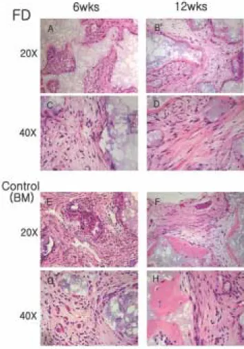
4. 생체 이식된 세포의 조직생성
섬유성 이형성증 유래 세포들을 배양하여 면역 억제된 쥐의 등쪽 피하에 이식한 뒤 6주(Fig. 5 A, C)뒤와 12주(Fig.
5 B, D) 뒤에 조직 슬라이드를 만들어 H&E 염색을 한 결과, 6주일 때는 HA/TCP 전달체 주변으로 별다른 골의 생성이 뚜렷이 관찰되지 않고 주변이 섬유성 조직으로만 둘러싸 인 모습을 보이고 있다. 12주가 경과하자 대조군인 정상 골 수 유래 세포를 배양한 것과 같이 조직화된 층판골의 형태 는 아니나(Fig. 5 F, H) 전달체를 감싸고 있는 형태로 골이 관찰되기 시작하고 있다. 이는 섬유성 이형성증에서 발견 되는 편골과 유사한 형태를 보여주는 것이다.
Fig. 2. Primary cell culture in regular media.
FD cells were cultured in the regular media for 1 week.
These cells are similar in shape with their normal bone marrow cells in this experiment.
Fig. 3. MTT assay for fibrous dysplasia(FD) cells and bone marrow(control) cells. Each cell group shows the increased proliferation over time. Comparing normal bone marrow stromal cells, fibrous dysplasia indicated high pro- liferation rate.
Fig. 4. Mineral induction capacity of comparing to the fibrous dysplasia (FD) cells and bone marrow (BM) cells. Alizarin red S stained bone marrow cells and fibrous dysplasia cells after 1, 2, 3 weeks cultured using mineral induction medium. In contrast to the nor- mal bone marrow cells, the FD originated cells produce less mineralized nodule until 3 weeks.
Table 1.
Result data by MTT assayFD Control (BM) p-value
24 Hours 0.487±0.0523 0.364±0.0323 0.0067 48 Hours 0.490±0.0499 0.409±0.0565 0.0738 72 Hours 0.794±0.0819 0.768±0.0990 0.7051 (average ± standard deviation)
*FD : fibrous dysplasia
*BM : bone marrow
Ⅳ.
고 찰본 연구에서는 인체의 골조직, 피부 및 내분비 기관에 이 환 가능한 양성의 종양성 병소인 섬유성 이형성증 유래의 세포를 in vitro 상에서 배양하여 면역 억제된 실험용 쥐의 피하에 이식함으로써 질환의 생체 내 이식실험에서 형성 된 조직의 형태를 관찰하였다. 이전에도 내분비 기관의 증 상을 동반하는 McCune-Albright 증후군 유래의 세포를 이 식하는 실험을 한 적은 있으나
23), 이번 실험에서는 두개악 안면 영역에서 호발하는 형태인 단골성 섬유이형성증 유 래의 세포를 이용하였다
20). McCune-Albright 증후군 유래의 세포에서와 유사하게 단골성 섬유이형성증 유래 세포에서 의 실험에서, 정상 골수 유래 이식에서 보이는 조직화된 층
판골(lamellar bone)의 형태는 보이지 않고 전달체인
HA/TCP 주위를 감싸고 있는 형태로 골이 관찰되었다. 이
는 섬유성 이형성증이 인체 내에서 발현될 때 나타나는 편 골(woven bone)과 유사한 형태를 보이고 있는 것이다. 그러 나 정상 골수 유래 세포의 이식 실험에서 볼 수 있는 조혈 세포 등은 관찰되지 않고 있다.
섬유성 이형성증으로부터 분리된 세포들은 증식능은 증 가되나 분화능은 감소하는 성질을 보인다고 보고되었다
16,25,26)
. 본 실험의 증식능을 관찰하기 위한 MTT assay 결과
에서는 섬유성 이형성증 유래 세포가 정상 골수 유래 세포 에 비해 관찰 기간 내내 흡광도가 더 높았고, 시간이 흐를 수록 두 세포군 모두에서 점차적으로 흡광도가 증가하는 모습을 보여줌으로써 세포의 증식능은 증가된다는 사실을 확인하였다. 또한 무기질 침착의 유도능 실험에서는 섬유 성 이형성증 유래 세포는 3주가 되어도 대조군인 정상적 골수 유래 세포에 비해 매우 적은 양의 무기질 침착을 보이 고 있다. 이는 곧 섬유성 이형성증 유래 세포가 정상 세포 에 비해 조골세포로의 분화능이 떨어짐을 보여주는 것이 라 할 수 있다.
섬유성 이형성증의 형질은 유전적으로 전달되지 않고, 접합 후 돌연변이에 의한다. 즉, 부모로부터의 유전적인 각 인 현상(imprinting)에 의하지 않고 무작위적이며 비대칭적 으로 유전형질을 전달받는다고 한다
27). 따라서 부계 혹은 모계로부터 특정적인 유전형을 전달받아 모든 세포에 이 환되지 않고, 인체 발생의 과정 중에서 GNAS1 유전자의 비특이적인 돌연변이가 발생하는 시기에 따라서 질환이 이환되는 범위의 차이가 생기게 된다. 각각의 분화 배엽에 따른 병소 분포상의 차이 이외에도, 같은 배엽 기원의 기관 이라 하더라도 특정 세포만이 돌연변이를 일으키고 자기 복제에 의해 증식하므로 정상적인 세포와 돌연변이를 가 진 세포가 혼재되어 mosaicism을 보이게 되는 것이다. 비록 이번 연구에서는 두개악안면 영역에서 호발하는 형태인 단골성 섬유성 이형성증 유래의 세포만을 이용하였지만, 다른 많은 연구들에서는 내분비 기관 이상을 동반하는 형 태인 McCune-Albright 증후군을 대상으로 삼고 있다. 이러 한 각기 다른 형태의 섬유성 이형성증은 비록 병소가 이환 되는 부위는 다르지만 돌연변이가 일어나는 장소나 양상, 돌연변이 이후의 분자적인 기전에는 커다란 차이가 없는 것으로 알려져 있다
28).
Ⅴ.
결 론결론적으로 섬유성 이형성증 유래 세포를 배양하여 생체 내에 이식하면 원래 병소의 형태와 유사한 발현 양상을 보 이게 된다. 즉, 조골세포의 분화능은 감소하여 정상 골수 유래 세포에 비하여 조혈세포, 적혈구세포 등의 발현은 줄 어들고, 증식능은 증가하여 전달체 사이의 대부분의 공간 을 섬유성 조직으로 채우고 있는 것이다. 따라서 본 연구에
Fig. 5. Representative histological section by in vivo trans-
planted fibrous dysplasia (FD) cells and normal bone mar-
row (BM) stromal cells in immunocompromised mice, har-
vested at 6, 12 weeks. Bone formation is limited to the thin
rim of osteoid tissue covering the carrier surfaces. In
experimental group, hematopoiesis is absent and the
spaces separating bone/carrier particles are occupied by
fibrous tissue. However, intercarrier spaces are occupied
by hematopoietic marrow (especially erythropoiesis, granu-
locytopoiesis etc.) in the control group.
서 적용된 in vivo 실험 방법은 환자의 병소 상태를 실험적 으로 재현할 수 있어 이에 대한 치료법의 개발 시 이를 토 대로 치료 방법의 유용성을 검증하기 위한 도구로 사용되 어 질 수 있으며 이와 함께 표준화된 치료를 사용함에 있어 예후를 판단하는 방법으로도 이용될 수 있으리라 판단된 다.
참고문헌