hPDX1 유전자의 삽입에 의한 직접 췌도세포 분화
옥선아†․오건봉․황성수․김영임․권대진․임기순
농촌진흥청 국립축산과학원 동물바이오공학과
Transdifferentiation of α-1,3-Galactosyltransferase Knock Out (GalT KO) Pig Derived Bone Marrow Mesenchymal Stromal Cells (BM-MSCs) into
Pancreatic Cells by Transfection of hPDX1
Sun A Ock†, Keon Bong Oh, Seongsoo Hwang, Youngim Kim, Dae-Jin Kwon and Gi-Sun Im Animal Biotechnology Division, National Institute of Animal Science, Rural Development Administration,
Wanju 565-851, Republic of Korea
ABSTRACT
Diabetes mellitus, the most common metabolic disorder, is divided into two types: type 1 and type 2. The essential treatment of type 1 diabetes, caused by immune-mediated destruction of β-cells, is transplantation of the pancreas;
however, this treatment is limited by issues such as the lack of donors for islet transplantation and immune rejection.
As an alternative approach, stem cell therapy has been used as a new tool. The present study revealed that bone marrow- derived mesenchymal stromal cells (BM-MSCs) could be transdifferentiated into pancreatic cells by the insertion of a key gene for embryonic development of the pancreas, the pancreatic and duodenal homeobox factor 1 (PDX1). To avoid immune rejection associated with xenotransplantation and to develop a new cell-based treatment, BM-MSCs from α-1,3-galactosyltransferase knockout (GalT KO) pigs were used as the source of the cells. Transfection of the EGFP-hPDX1 gene into GalT KO pig-derived BM-MSCs was performed by electroporation. Cells were evaluated for hPDX1 expression by immunofluorescence and RT-PCR. Transdifferentiation into pancreatic cells was confirmed by morphological transformation, immunofluorescence, and endogenous pPDX1 gene expression. At 3∼4 weeks after transduction, cell morphology changed from spindle-like shape to round shape, similar to that observed in cuboidal epithelium expressing EGFP. Results of RT-PCR confirmed the expression of both exogenous hPDX1 and endogenous pPDX1. Therefore, GalT KO pig-derived BM-MSCs transdifferentiated into pancreatic cells by transfection of hPDX1.
The present results are indicative of the therapeutic potential of PDX1-expressing GalT KO pig-derived BM-MSCs in β-cell replacement. This potential needs to be explored further by using in vivo studies to confirm these findings.
(Key words: diabetes, pig, PDX1, GalT KO derived BM-MSCs)
* 이 연구는 차세대 바이오그린 21 사업(PJ011311022015)의 지원에 의하여 수행되었다.
†Correspondence : [email protected], [email protected]
서 론
전 세계적으로 당뇨병 환자는 3억 명이 넘는 것으로 알려져 있으며, type 1과 type 2로 나눠진다. Type 1 당뇨의 경우, 전체 당뇨의 5∼10% 정도를 차지하고, 원인은 인슐린 생산을 책임 지는 췌장 내β세포를 면역 체계가 공격하여 파괴하는 대사성 질환으로 인슐린 주사의 지속적인 투여에 의해서만 치료가 가 능한 것으로 알려져 있다(Kim과 MacDonal 2002; Associattion 2013). 따라서 1형 당뇨를 치료하기 위한 근본적인 방안으로 는 사체로부터 췌장을 이식 받거나 췌장으로부터 췌도를 분리
하여 이식 받는 방법만이 유일하지만, 췌도 이식을 위한 공여 장 기의 부족과 면역 거부 반응으로 현실적인 대안이 필요한 상황 이다(Miyazaki 등, 2004; Allahyerdi 등, 2015).
최근에는 이상의 문제를 해결하기 위해 면역 거부반응을 최 소화할 수 있는 줄기세포를 이용한 세포 치료제 연구가 활발 히 이루어지고 있으며, 이것은 배아줄기세포와 유사한 induced pluripotent stem cell(iPSCs)의 개발과 동시에 가속화되고 있으 며, 다른 한편으로는 성체줄기세포 중 대표적인 중간엽 줄기세 포(Mesenchymal stem cell)를 이용하는 두 가지 방향으로 연구 가 진행되고 있다(Zhang 등, 2009; Kunisada 등 2012; Allahyerdi
등, 2015).
줄기세포를 이용한 췌도 세포나 인슐린 분비세포의 분화는 크게 small과 large molecules(Activin A, Fibroblast Growth Factor(FGF), Sodium Butyrate, Retinoic acid, Betacellulin, 등) (Kumer 등, 2014)을 이용하는 방법, epigenetic을 조절하는 방법 (Pennarossa 등, 2012), 췌장 분화의 근원이 되는 유전자를 중 간엽 줄기세포에 삽입하여 insulin producing cells(IPCs)로 분 화 유도를 하는 방법(Karnieli 등, 2007; Xu 등, 2012; Yuan 등, 2012; Allahyerdi 등, 2015) 등이 알려져 있다.
췌도에서 인슐린을 분하는 β-cell로 분화가 이루어지기 위 해서는 췌장의 배아발달 단계 중 내배엽에서 췌장세포로 가기 위해서 중요한 transcriptional factor로 Pancreatic and duodenal homeobox factor 1(PDX1)임이 밝혀졌다(Allahyerdi 등, 2015).
선행 연구결과에 따르면 사람의 중간엽 줄기세포에서 이 유전 자의 삽입으로 IPCs를 유도하였다고 보고하였다(Yuan 등, 2012;
Allahyerdi 등, 2015). 이러한 싱글 유전자의 과발현이 췌도 세 포로 분화하는데 중요한 key의 역할을 한다면 상대적으로 많 은 유전자와 복잡한 배양 방법과 시간을 필요로 하는 iPSCs 만 드는 것이 불필요해질 것이다.
과거에는 당뇨병을 치료를 위한 인슐린은 돼지로부터 분리 정제하여 사용하여 왔다. 그 이유는 사람과 비교하여 돼지 인 슐린의 경우, 단지 염기서열 1개만이 달라 상당히 유사하기 때 문이다. 그래서 돼지의 췌장으로부터 췌도를 분리․정제하여 사람에 주입하여 1형 당뇨를 근본적으로 치료하는 연구들이 많 이 진행되고 있으며, 이 단계를 넘어 면역 거부 반응을 최소화하 기 위해 이종 간 이식을 위해 사람에는 존재하지 않는 α-1,3- galactosyltransferase 제거된 돼지(GalT KO 돼지)를 이용하여 세포 치료제로 활용하려고 노력하고 있다(Thompson 등, 2011).
이 연구는 α-1,3-galactosyltransferase knock out된 돼지의 중간엽 줄기세포를 이용하여 hPDX1 유전자의 삽입을 통하여 췌도 세포의 전환이 가능한지를 형태학적, 면역 형광염색, 유 전자 증폭에 의한 exogenous와 endogenous PDX1 유전자 발현 증가를 조사하였다.
재료 및 방법
1. 시약 및 배양액
이 실험에 사용된 모든 시약은 특별한 언급이 없는 경우, Sigma Chemical Company(St Louis, MO, USA)에서 구입하였 고, 배양액은 Gibco(Life Technologies, Rockville, MS, USA) 에서 구입하였다. 모든 배양액은 pH 7.2, 삼투압 280 mOsm/kg 으로 조정하여 사용하였다.
2. 중간엽 줄기세포(Mesenchymal Stromal Cells, MSCs)의 추출 및 배양
이 실험은 국립축산과학원 동물윤리위원회의 승인을 받아 수행되었다. GalT KO 돼지는 Ahn 등(2011)의 방법에 의해 생 산되었고, 교배에 의해 호모개체를 생산하여 실험에 사용되었 다. 골수유래 중간엽 줄기세포(BM-MSCs)는 5주령 돼지의 대 퇴부 골수로부터 외과적으로 회수되어 Faast 등(2006)의 방법 에 의해 Ficoll-Paque Plus(Amersham Science, USA)를 사용하여 제조사의 분리방법에 따라 분리되어졌다. GalT KO BM-MSCs 를 위한 기본 배양액으로는 Advanced Dulbecco’s modified Eagle medium(ADMEM)에 10% fetal bovine serum, 1 mM Napyruvate, 100 U/ml penicillin G, 100 μg/ml streptomycin sulfate를 첨가 하여 사용하였고, 세포들은 모두 38.5℃ 인큐베이터 속에서 5%
CO2 농도를 유지하면서 배양되었고, 중간엽 줄기세포인지의 확인은 옥 등(2013)의 방법에 의해 미리 확인되었다. 이 실험에 사용된 세포는 모두 5 passages까지만 사용되었다.
3. pEGFP-hPDX1 Vector 백터 구축
hPDX1 mRNA은 NCBI GeneBank의 accession #:BC111592 를 기반으로 cDNA를 합성하였고, pEGFP-C2 백터의 multi- cloning site의 EcorR I과 BamH I 제한 효소에 삽입하였다.
Vector의 선별마크로 EGFP와 Neo 유전자가 발현되도록 설계 되었다(Fig. 1A).
4. hPDX1 Vector가 삽입된 세포 생산 및 검정
선형의 hPDX1 vector의 세포내 도입은 p1 primary cell 4d- Nucleofector®X kit(Lonza, USA)와 4d-Nucleofector-system (Lonza, Germany) 기계를 이용하여 수행되었다. 2×10⁵개의 GalT KO BM-MSCs는 100 μl p1 primary cell 4d-Nucleofector®X 용 액과 선형의 hPDX1 vector 2 μg을 섞어 4d-Nucleofector-syste 을 이용하여 제조사의 매뉴얼에 따라 전기 천공법(electropo- ration)에 의해 transfection을 유도 후 신속하게 35 mm dish에 옮겨 기본배양액으로 배양하였다.
세포의 외부 유전자 삽입 효율을 조사하기 위하여 Nucleo- Counter NC-3000을 이용하여 분석하였다. 배양된 세포는 single cells로 분리 후에 1 μg/ml Hoechst와 Propidiumiodide(PI)이 함 유된 4℃ PBS를 이용하여 염색을 실시하였고, slide에 seeding 후 분석하였다. 모든 분석은 Hoechst가 발현되는 세포(유핵세 포의 기준)만을 기준으로 다음과 같이 분류하였다. EGFP가 발 현되는 경우는 외부유전자가 도입된 살아 있는 세포로 PI와 EGFP가 발현되는 경우는 죽은 세포에서 외부유전자가 도입된 것으로 판단하였다(Fig. 1B).
5. 면역 형광염색에 의한 PDX1 발현 확인
3주 동안 배양된 세포는 형태학적 변화를 도립현미경을 통 해 확인하였고, 세포들은 4% formalin을 이용하여 1시간 동안
hPDX1vector
(A)
(a) (b)
(B)
(C)
Fig. 1. Construction of hPDX1 vectors and their transfection into α-1,3-galactosyltransferase knockout (GalT KO) pig-derived bone marrow- derived mesenchymal stromal cells (BM-MSCs). (A) structure of the hPDX1 vector, (B) analysis of the transfection rate by Nucleo- Counter NC-3000, (C) transfection ratio of hPDX1 in GalT KO pig-derived BM-MSCs. Ba and Bb indicate the scatterplot and immu- nofluorescence analyzed with NucleoCounter NC-3000, respectively.
고정하였으며, 0.1% triton X-100, 1% BSA가 첨가된 PBS를 사용하여 3번 수세를 하였다. 최종적으로 30분 동안 이 용액 에 담가 세포의 투과성을 증가시키고, blocking을 유도한다.
RNase 첨가된 1 μg/ml PI 용액을 사용하여 핵산에 대한 대조 염색을 30분간 실시하였다. PBS를 사용하여 3번 수세 후 Vec- tashield antifade mounting medium(Vector Laboratories Inc., CA USA)을 사용하여 슬라이드에 한 방울 떨어드리고, 커브 글라스를 덮어 형광현미경 아래에서 green과 red 형광의 발현 을 관찰하였다.
6. Reverse Transcription Polymerase Chain Reaction(RT-PCR)에 의 한 Exogenous와 Endogenous PDX1 mRNA 발현 확인 유전자 삽입 4주후 세포는 회수되었고, RNeasy® Mini kit (Qiagen, UAS)를 사용하여 total RNA를 추출하였다. cDNA 합 성을 위해 1 μg/ml total RNA를 사용하여 Omniscript® reverse transcription kit(Qiagen)를 사용하여 cDNA를 합성하였다. 특 이 유전자의 발현을 확인하기 primer로 hPDX1는 F-AAGTCC- GCCCCATHCCCGAAGGCTAC’와 R-ACGCGCATGGGTCC- TTGTAAAGCTG, pPDX1은 F-AAGTCTACCAAGGCTCAC-
(A)
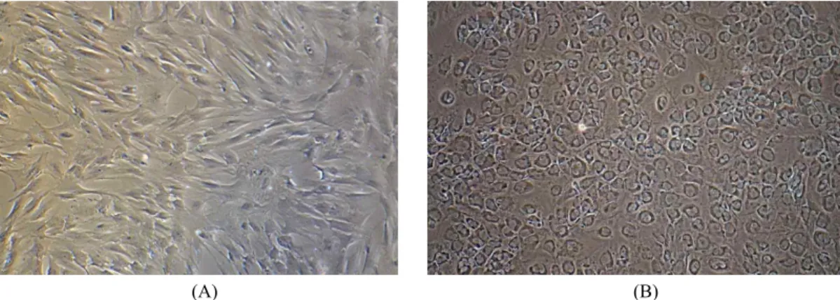
(B)
Fig. 2. Morphological changes in α-1,3-galactosyltransferase knockout pig-derived bone marrow-derived mesenchymal stromal cells trans- fected with the hPDX1 gene. (A) cells without genetic modification, (B) cells transduced with the hPDX1 vector were cultured for 3 weeks (×100).
GC’와 R-GCGCGGCCTAGAGATGTATT’, β-actin은 F-ATC- ATGTTTGAGACCTTCAACACCC와 R-CATGGTGGTGCCG- CCAGACAG를 사용하였다. PCR은 premix(Maxime PCR Pre- Mix kit(Intron biotechnology))를 사용하여 증폭하여 1% agarose gel에 loading하여 전기영동 후 확인하였다. Housekeeping gene 으로는 β-actin을 사용하여 Internal control로 사용하였다.
결 과
1. hPDX1 Vector가 삽입된 세포 구축
hPDX1 vector와 같이 CMV promoter에 EGFP와 hPDX1이 발현되도록 선형으로 제작되었다(Fig. 1). 기본배양액에서 3주 간 배양된 세포에서 형질전환율을 Fig. 1과 같이 조사하였고, 다음과 같이 분류되었다. 살아있으며 EGFP가 발현된 세포는 green으로 발현되었고, 유전자가 삽입되지 않아 죽은 세포는 red로, 유전자가 삽입된 상태로 죽은 세포는 yellow로 분류됨 을 확인할 수 있었다. 정확한 비율은 Fig. 1B와 같이 생존세포 에서 transfection 비율은 50%였고, 죽은 세포에서는 transfection 비율이 25%, 전체 세포에서는 41%의 transfection율을 보였다.
2. hPDX1 Vector가 삽입된 세포의 분화
hPDX1 vector가 삽입된 세포는 3주간 기본 배양액에서 배 양되었다. 유전자가 삽입되기 전에는 중간엽 줄기세포의 전형 적인 형태인 빗살 무늬형태(Fig. 2A)를 보였지만, 배양이 진행 되고 유전자의 전사가 시작됨으로 자갈돌 모양의 입방형 형태 로(Fig. 2B) 형태학적 변화를 보였다.
3. 면역 형광염색에 의한 hPDX1 발현 확인
3주에서 4주간 분화된 세포에서 면역 형광염색을 통하여
EGFP 발현을 대조염색인 PI와 함께 관찰하였다. 자갈돌 모양
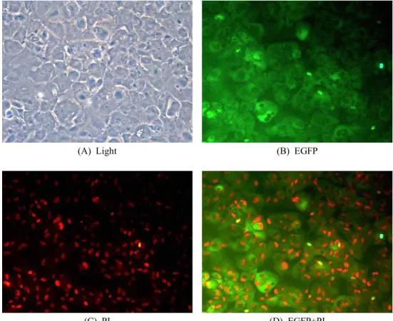
의 형태학적 변화를 보인 세포에서 EGFP의 경우, 핵이 아닌 세 포의 세포질에서 강하게 발현됨을 확인할 수 있었다(Fig. 3). 또 한 대부분의 세포에서 EGFP가 발현됨을 확인할 수 있었다.
4. RT-PCR에 의한 Exogenous와 Endogenous PDX1 mRNA 발현 확인
hPDX1 vector가 transfection 4주간 배양된 세포에서 exoge- nous hPDX1의 발현을 관찰하였다(Fig. 4B). Positive control로 사용된 hPDX1 vector와 같이 hPDX1이 transfection된 세포들에 서는 hPDX1 유전자의 삽입이 확인되었다. Endogenous pPDX1의 경우, transfection 전에는 확인되지 않았지만, transfection 후에 는 이 유전자의 활성이 진행되어 발현됨을 확인할 수 있었다 (Fig. 4A). β-actin의 경우, 내부 유전자량의 control로 사용되었 고, transfection된 세포와 되지 않은 세포 간에 유전자의 양이 같음을 보여주었다(Fig. 4C).
고 찰
이 연구는 사람의 당뇨병 세포치료제를 개발하기 위해 면 역학적 거부 반응이 최소화된 GalT KO 돼지의 중간엽 줄기세 포를 사용하여 췌도 분화를 위한 초기 전사인자인 hPDX1의 단일 유전자 삽입만으로 특수한 배양액이 아닌 기본 배양액 만을 목적으로 하는 내배엽성 췌도세포로 분화를 유도할 수 있음을 형태학적 변화, 면역 형광염색, RT-PCR에 의해 증명 하였다. 특히 외부에서 도입된 hPDX1은 돼지 세포내 잠자고 있던 pPDX1을 깨워 활성화 하도록 하는 역할을 충분이 할 수 있음을 보여주었다.
선행 연구 결과에 따르면 내배엽성 유래가 아닌 중배엽성 유래인 Bone marrow는 체내에 이식되었을 때 자발적으로 내 분비 췌장 세포로 분화할 수 있음이 보고되었고(Ianus 등, 2003;
(A) Light
(B) EGFP
(C) PI
(D) EGFP+PI
Fig. 3. Expression of hPDX1 in α-1,3-galactosyltransferase knockout pig-derived bone marrow-derived mesenchymal stromal cells trans- fected with the hPDX1 vector. (A) light source, (B) GFP (green) fluorescence, (C) PI (red) fluorescence, (D) Merged. Nuclei evidenced by PI staining.
Fig. 4. Evaluation of PDX1 expression using RT-PCR. Two types of bone marrow-derived mesenchymal stromal cells from the α-1,3-galactosyltransferase knockout pig were used:
cells with and without genetic modification. A: expression of porcine PDX1, B: expression of human PDX1, C: β-actin was used as the positive control gene.
Choi 등, 2003), 이 연구는 이 자료를 바탕으로 형질 전환 돼지 의 골수 중간엽 줄기세포를 이용하여 진행되었다.
PDX1은 내분비 췌장의 발달과β-cell 분화에 핵심적인 전사 인자로 알려져 있다(Zalzman 등, 2003). 이 연구에서는 hPDX1 의 세포내 전사인자의 발현에 췌도세포로 분화를 유도하고자 전기 천공법을 사용하여 세포를 유전자 조작하였지만, 전체 세 포에서 발현 효율은 50% 수준에 머물렀다. 전기 천공법의 장
점은 쉬운 조작이 가능하다는 점과 viral transfection을 하지 않 음으로써 안전성을 높일 수 있는 장점이 있지만, 상대적으로 낮 은 세포생존율과 viral transfection에 비해 낮은 transfection 효 율이 문제가 된다(Xu 등, 2012; Yuan 등, 2012). 우리의 결과도 일치하였다. 따라서 FACS를 통한 cell sorting을 통하여 EGFP 가 삽입된 세포만을 선별하여 배양하는 과정이 추가로 이루어 져야 할 것으로 생각된다.
hPDX1 삽입된 중간엽 줄기세포는 형태학적으로 fibroblast like cell로 spindle-shape 형태에서 배양 후 7일경부터 round- shape한 모양으로 바뀌기 시작했으며, 3주 배양 시에는 전형적 인 cuboidal epithelial 형태로 변화되어 내분비세포의 형태로 변화하였음을 확인할 수 있었다(Pokrywczynska 등, 2015). 우 리의 결과는 사람에서 lentiviral vector를 이용하여 수행한 연 구 결과와도 일치하였다(Allahyerdi 등, 2015). 그러나 3주 이 상 배양하였을 때 Allahyerdi 등(2015)의 보고와 달리 전형적 인 islet cluster 형태의 colonies은 극히 낮은 빈도로 관찰되었 다. 다른 결과를 보인 이유는 유전자 삽입을 위하여 lentiviral vector가 아닌 전기 천공법을 사용함으로써 유전자 도입율이 상대적으로 낮은데서 기인한 것으로 추정된다.
3주에서 4주간 배양된 중간엽 줄기세포에서 hPDX1의 발 현을 확인하였고, EGFP는 세포질에서 강하게 발현되는 것을 확인할 수 있었다. 선행 연구 결과들에서는 viral vector를 사 용하여 PDX1 유전자의 발현을 유도함으로써, 인체에 사용 시 안전성을 보장할 수 없는 단점이 있지만 우리의 결과는 2가지 선별마크의 사용이 가능함과 동시에 형광현미경 아래에서 유 전자의 삽입을 실시간 확인할 수 있어 기존의 연구 결과보다 한 단계 진보된 결과라 할 수 있다.
일반적으로 iPSCs를 만들기 위해 삽입된 외부 유전자는 내 부에 잠자고 있던 내부 유전자를 활성화를 유도하여 세포가 줄기세포능을 가지도록 하는 것으로 알려져 있다. 그래서 이 연구에서는 외부 유전자의 삽입을 확인할 뿐만 아니라, 잠자 고 있던 돼지 PDX1 유전자의 활성을 RT-PCR을 통하여 분석 하였고, 외부 유전자의 삽입이 돼지 내부 유전자의 활성을 유 도할 수 있음을 동시에 확인할 수 있었다. 따라서 내부 유전자 의 활성과 동시에 이 세포는 췌도 세포로 분화가 유도되는 경 로 신호가 발생되었음을 확인할 수 있었다.
결 론
이 연구는 이종 간 세포 치료제 개발을 위해 사람과 돼지 사 이에 세포 이식 시 발생될 수 있는 초급성 거부 반응을 제어하 기 위해 GalT KO 돼지의 골수 유래 중간엽 줄기세포를 사용 하여, 췌장 분화 시작에 중요한 전사인자인 PDX1 유전자를 삽입함으로써 추가적인 약물의 처치나 메틸화의 조절 없이도 췌도 세포로 분화할 수 있음을 증명하였다. 따라서 이 연구 결 과는 환자 맞춤형 iPSCs를 만들기 위해 많은 시간과 노동력의 소비 없이도, 쉽고 안전하면서도 효율적으로 당뇨치료용 세포 를 대량 생산할 수 있는 기반을 확립하는데 이용될 수 있을 것이다.
REFERENCES
Ahn KS, Kim YJ, Kim M, Lee BH, Heo SY, Kang MJ, Kang YK, Lee JW, Lee KK, Kim JH, Nho WG, Hwang SS, Woo JS, Park JK, Park SB and Shim H. 2011. Resurrection of an alpha-1,3-galactosyltransferase gene-targeted miniature pig by recloning using postmortem ear skin fibroblasts. The- riogenology. 75:933-999.
Allahverdi A, Abroun S, Jafarian A, Soleimani M, Taghikhani M and Eskandari F. 2015. Differentiation of human mesen- chymal stem cells into insulin producing cells by using a lentiviral vector carrying PDX1. Cell J. 17:231-242.
Association AD. 2013. Standards of medical care in diabetes-
2013. Diabetes Care. 36(Suppl 1):S11-66.
Choi JB, Uchino H, Azuma K, Iwashita N, Tanaka Y, Mochi- zuki H, Migita M, Shimada T, Kawamori R and Watada H.
2003. Little evidence of transdifferentiation of bone marrow- derived cells into pancreatic beta cells. Diabetologia. 46:
1366-1374.
Ianus A1, Holz GG, Theise ND and Hussain MA. 2003. In vivo derivation of glucose-competent pancreatic endocrine cells from bone marrow without evidence of cell fusion. J. Clin.
Invest. 111:843-850.
Karnieli O, Izhar-Prato Y, Bulvik S and Efrat S. 2007. Gene- ration of insulin-producing cells from human bone marrow mesenchymal stem cells by genetic manipulation. Stem Cells.
25:2837-2844.
Kim SK and MacDonald RJ. 2002. Signaling and transcriptional control of pancreatic organogenesis. Curr. Opin. GenetDev.
12:540-547.
Kumar SS, Alarfaj AA, Munusamy MA, Singh AJ, Peng IC, Priya SP, Hamat RA and Higuchi A. 2014. Recent develop- ments in β-cell differentiation of pluripotent stem cells induced by small and large molecules. Int. J. Mol. Sci. 15:
23418-23447.
Kunisada Y, Tsubooka-Yamazoe N, Shoji M and Hosoya M.
2012. Small molecules induce efficient differentiation into insulin-producing cells from human induced pluripotent stem cells. Stem. Cell. Res. 8:274-284.
Miyazaki S, Yamato E and Miyazaki J. 2004. Regulated ex- pression of pdx-1 promotes in vitro differentiation of insulin- producing cells from embryonic stem cells. Diabetes. 53:
1030-1037.
Pellegrini S, Ungaro F, Mercalli A, Melzi R, Sebastiani G, Dotta F, Broccoli V, Piemonti L and Sordi V. 2015. Human in- duced pluripotent stem cells differentiate into insulin-pro- ducing cells able to engraft in vivo. Acta. Diabetol. [Epub ahead of print]
Pennarossa G, Maffei S, Campagnol M, Tarantini L, Gandolfi F and Brevini TA. 2013. Brief demethylation step allows the conversion of adult human skin fibroblasts into insulin-se- creting cells. Proc. Natl. Acad. Sci. USA. 110:8948-8953.
Pokrywczynska M1, Lewandowska MA, Krzyzanowska S, Jund- zill A, Rasmus M, Warda K, Gagat M, Deptula A, Helmin- Basa A, Holysz M, Nowacki M, Buchholz L, Bodnar M, Marszalek A, Grzanka A, Jozwicki W, Michalkiewicz J and Drewa 2015. Transdifferentiation of bone marrow mesen-
chymal stem cells into the islet-like cells: The role of ex- tracellular matrix proteins. T. Arch. Immunol. Ther. Exp.
(Warsz). [Epub ahead of print]
Thompson P, Badell IR, Lowe M, Cano J, Song M, Leopardi F, Avila J, Ruhil R, Strobert E, Korbutt G, Rayat G, Ra- jotte R, Iwakoshi N, Larsen CP and Kirk AD. 2011. Islet xenotransplantation using gal-deficient neonatal donors im- proves engraftment and function. Am. J. Transplant. 11:
2593-602.
Xu ZF, Pan AZ, Yong F, Shen CY, Chen YW and Wu RH.
2012. Human umbilical mesenchymal stem cell and its adi- pogenic differentiation: Profiling by nuclear magnetic reso- nance spectroscopy. World J. Stem. Cells. 4: 21-27.
Yuan H, Liu H, Tian R, Li J and Zhao Z. 2012. Regulation of mesenchymal stem cell differentiation and insulin secretion by differential expression of Pdx-1. Mol. Biol. Rep. 39:
7777-7783.
Zalzman M1, Gupta S, Giri RK, Berkovich I, Sappal BS, Karnieli O, Zern MA, Fleischer N and Efrat S. 2003. Re- versal of hyperglycemia in mice by using human expandable insulin-producing cells differentiated from fetal liver pro- genitor cells. Proc. Natl. Acad. Sci. USA. 100(12):7253- 7258.
Zhang D, Jiang W, Liu M, Sui X, Yin X, Chen S, Shi Y and Deng H. 2009. Highly efficient differentiation of human ES cells and iPS cells into mature pancreatic insulin-producing cells. Cell Res. 19:429-438.
옥선아, 오건봉, 황성수, 임석기, 김영임, 박진기. 2013. α-1,3- Galactosyltransferase Knock Out(GalT KO) 돼지유래 골수 중간엽 줄기세포의 특성 규명. 한국수정란이식학회지. 28:
281-287.
Received September 1, 2015, Revised September 23, 2015, Accepted September 23, 2015