• 검색 결과가 없습니다.

Biology and Potential Use of Chicken Bone Marrow-derived Cells

N/A
N/A
Protected

Academic year: 2021

Share "Biology and Potential Use of Chicken Bone Marrow-derived Cells"

Copied!
10
0
0

로드 중.... (전체 텍스트 보기)

전체 글

(1)

Difference in Embryogenesis between Aves and Mammals

The aves such as chickens, quails and pheasants have unique characteristics, which is based on its evolutional position between mammals and other species. Ex vivo development of the aves is the most prominent difference from the mammalian species which undertake in vivo organogenesis and relevant or addition difference are found during embyogenesis and organogenesis. Differing from mammals, physiological polyspermy is usually induced at the time of fertilization and numerous sperms are visible, regardless of embryonic nuclei formation. Consequently, asymmetric cleavage yielding pre-blastodermal cells is observed throughout early embryogenesis and the intrauterine eggshell formation consisting of three stages is underwent before laying. According to developmental stage, the eggs consisting of 1 to 3,000 cells form yellowish soft eggshell membrane at the phase I. Phase II eggs consisting of 3,000 to 30,000 cells have light yellowish, flexible eggshell and phase III eggs consisting of 30,000 to 60,000 cells have milky white stiffened eggshell. Such difference from the mammals may induce unique cell fate determination in various systems, which may be prominent in mesenchyme-derivative tissues and organs.

Anatomic Feature of Chicken Skeletal System

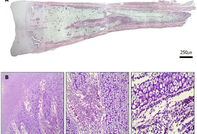
Endochondral ossification is responsible for the formation of the long bones such as the femurs and humeruses. Unlikely to intramembranous ossification, cartilage tissue is formed prior to ossification, and subsequent ossification acquires the activities of rudimentary long bone formation, extension of its length and natural healing against various damages. Intramembranous ossification induce bone formation without cartilage development. In both process, rudimentary bone formation is completed before birth, while

Biology and Potential Use of Chicken Bone Marrow-derived Cells

Dongwoo Ko

1,†

and Jeong Mook Lim

1,2,†

1

Department of Agricultural Biotechnology, Seoul National University, Seoul 151-921, Korea

2

Research Institute of Agriculture and Life Sciences, Seoul National University, Seoul 151-921, Korea

Abstract

Developmental aspects of chicken embryos showed dramatic difference compared with those of mammals and consequently, such difference in various developmental events leads to different feasibility in both clinical and industrial application. We have concentrated on the studies for using of chicken bone marrow cells and currently we found number of unique cellular properties. Through this article, we reviewed characteristics and cell signaling of osteogenic cells during endochondral ossification in chicken long bone.

Received :22 December 2017 Revised :28 February 2018

Accepted:27 March 2018 Key Words : Bone marrow cell, Sox9, Chicken, Biological therapies

(2)

the extension of bone length continues until puberty. In mammals, endochondral ossification was completed until the birth and primitive cells for ossification usually disappeared in neonatal offsprings.

The long bones are the source of bone marrow cells, of which endochondral ossification following hypertrophic proliferation and calcium deposition occurs. There has been no report on the details of long bone development in chicken. Consequently, we collected the femurs from 4-day-old neonatal chicks and examined their histological characteristics. As shown in figure 1, no growth plate (hypophyseal line) is detected and proliferate chondrocytes are dominant on the middle of long bone. Probably, cell niche and microenvironment is unique in the long bones collected from neonatal chicks.

Bone Formation (Osteogenesis)

Two different processes, endochondral and intramembraneous ossifications, are involved in bone formation and mesenchymal stem cells differentiating into osteoblasts and chondrocytes play a pivotal role in inducing bone morphogenesis (Shapiro, 2008;

Zhang et al., 2011a). A variety of signaling pathways are involved in mesenchymal stem cells differentiation and of those, BMP, Wnt, Notch signaling are important as extracellular mediators of differentiation (Lin and Hankenson, 2011).

The bone marrow cells retrieved from chicks may contain various undifferentiated progenitors and precursors cells and those cells can provide enormous information on cell differentiation when they can be manipulated under a laboratory condition (Csaki, Matis, Mobasheri, Ye, & Shakibaei, 2007; Dai et al., 2014; Friedenstein, Chailakhyan, & Gerasimov, 1987; Hudson et al., 2011;

Pittenger et al., 1999). The lineaged cells of bone morphogenetic cells that derives from mesenchymal stem cells includes osteochondro progenitor cells, preosteoblasts, osteoblasts, osteocytes, chondrocytes, proliferate chondrocytes and hypertrophic chondrocytes (Aubin, 2001; Hofstetter et al., 1991). Osteochondro progenitor cells have the ability to become osteoblasts or chondroblasts. Osteochondral progenitor cells are located in the inner layer of the perichondrium and the periosteum, and in the endosteum. From these locations, they either derive new osteoblasts or chondroblasts or differentiate into osteoblastic or chondroblast lineaged cells (Friedenstein, Piatetzky, & Petrakova, 1966; Morikawa et al., 2009; Orlic et al., 2001; Toma, Pittenger, Cahill, Byrne, & Kessler, 2002). Osteochondro progenitor cells having the activity to differentiate into osteoblasts and chondrocytes and continuously find inner surface of the long bones. Isolation of the progenitor cells may be helpful to develop novel therapy for tissue regeneration. For clinical application, optimized and standardized process of cell isolation, purification and amplication for tissue regeneration is essential.

As stated, the osteochondro progenitor cells have the potential to develop into specific lineaged cells related bone morphogenesis when they respond to appropriate each lineage related signals (Garside et al., 2015; Langhans, Yu, & Tuan, 2016; Visvader &

Stingl, 2014). Multiple gene and surface protein markers of osteochondro progenitor cells are used for detection. Of those, Sox9 is a transcription factor, which regulates chondrocyte differentiation and anti-Mullrian hormone (AMH) gene transcription (Garside et al., 2015). Sox9 is key regulatory signal for the osteochondro progenitor cells that undergo osteogenic and chondrogenic differentiation and it can control lots of signal pathways. Sox9 is also expressed in osteochondro progenitor cells, which may demonstrate its importance for early signal pathways of bone formation (Pan et al., 2008). At the end of each pathway, osteoblasts form a bone and chondroblasts make a cartilage. Runx2 is known as a core-binding factor subunit a-1 (CBF-a-1) and Osx (a zinc finger containing transcription factor) is necessary for lineage differentiation of osteochondro progenitor cells (S. Chen et al., 2009;

Shahi, Peymani, & Sahmani, 2017). Runx2 regulates the expression of major bone matrix genes on differentiation (Chung, 2004;

Fujita et al., 2004) and acts as a factor to mature hypertrophic chondrocyte. Wnt/β-catenin signaling influences Runx2 function.

A β-catenin signal pathway regulates cell fate determination and WNT/β-catenin signal affects Runx2 expression (Dong, Soung

do, Schwarz, O'Keefe, & Drissi, 2006; Hill, Spater, Taketo, Birchmeier, & Hartmann, 2005). In the presence of Wnt3a, an activator

of the canonical WNT signaling pathway, β- catenin transport to the nucleus, where it binds TCF/LEF-1 and leads to the

transcription of Runx2 (Komori, 2011; Voronkov & Krauss, 2013).

(3)

Through Sox9 as a transcriptional mediator, transforming growth factor (TGF)-β and bone morphogenetic protein (BMP) signaling are responsible for initiating expression of cartilaginous extracellular matrix such as aggrecan, collagen types II and XI, fibronectin and tenascin as shown in in vitro cultures (Hatakeyama et al., 2004; Chimal-Monroy and Diaz de Leon, 1999). TGF-β is a representative marker for cell proliferation and differentiation during the stage of bone and cartilage development (Sakaki-Yumoto, Katsuno, & Derynck, 2013). TGF-β signal relates with Sox9 and Runx2 signalling pathways and regulates cell lineages during endochondral ossification (G. Chen, Deng, & Li, 2012; Yang et al., 2001). TGF-β signal stimulates differentiation potential into chondrocytes and osteoblasts with multiple signals like FGF, Msx1, and Ctgf signalling pathways (Guo & Wang, 2009; Parada, Li, Iwata, Suzuki, & Chai, 2013). Alkaline phosphatase (ALP) is a functional gene expressed early in the process of calcification and cartilage formation (Amano et al., 2009). Regulatory signaling pathways involve osteogenic and chondrogenic differentiations, and alkaline phosphatase expression is controlled with WNT/beta-catenin, BMP, Runx2 signaling cascades.

Collagen type I is abundant in extracellular matrix component and its gene is listed as a marker for the end product of bone and tendons (Liu, Yang, al-Shaikh, & Lane, 1995). Fully differentiated cells reduce expression of collagen type I and II and increase collagen type X and Aggrecan (Aigner, Dietz, Stoss, & von der Mark, 1995; Haaijman, D'Souza, Bronckers, Goei, &

Burger, 1997).

A

B

Figure 1. Morphology of the femur retrieved from 4-day-old chicks. (A) The femur has not formed epiphyseal line, which the stromal

tissue of diaphysis and epiphysis are mixed. Hematoxylin and eosin staining demonstrated numerous positive cells spreaded

into both the epiphysis and the diaphysis regions in the femur. (B) The picture of each site of femur.

(4)

Chicken Bone Marrow Cells

Bone marrow tissue is soft and flexible, which finds in interior hollow spaces of bones. Bone marrow consists of red and yellow marrow, which is determined by the predominance of hematopoietic tissue or fat composition (Malkiewicz & Dziedzic, 2012). It is well known that red marrow tissue contains the hematopoietic cells that produces red blood cells, white blood cells and platelets.

Hematopoietic cells matured in bone marrow subsequently migrate into capillary sinusoids for entering blood stream (Lang et al., 1992). In adult tissue, red bone marrow is found on the flat bones such as the pelvic girdle and the sternum. In children, red bone marrow is found in the medullary cavity of the long bones, such as the femur (Gurevitch, Slavin, Resnick, Khitrin, &

Feldman, 2009; Moore & Dawson, 1990). Yellow marrow acts as a stromal cell producing tissue that derives subcomponents of cartilage and bone (Gurevitch, Slavin, & Feldman, 2007; Zakaria & Shafrir, 1967). As the age is increased, bone marrow becomes yellow tissue and main function of bone marrow changes to store the adipocytes that serves as an energy source (Ortiz-Nieto, Johansson, Ahlstrom, & Weis, 2010; Tavassoli, Houchin, & Jacobs, 1977).

In addition to osteoblasts and osteoclasts, cellular components of bone marrow tissue contain fibroblasts and macrophages, which contribute to producing red blood cells by delivering iron for hemoglobin production. Also, fat cells and sinusoid-constituring endothelial cells are included in bone marrow tissue (Gordon, Pluddemann, & Martinez Estrada, 2014; Mansour et al., 2012; Yin

& Li, 2006). Most cells derive from the endothelial stem cells that are also present in the bone marrow. The mesenchyme of embryonic connective tissue, which is derived from the mesoderm, differentiates into hematopoietic and connective tissues, whereas mesenchymal stem cells do not differentiate into hematopoietic cell (Phinney & Prockop, 2007). Mesenchymal stem cells are multipotent stem cells that have various abilities to differentiate into multi-lineage cells (Pittenger et al., 1999) such as osteoblasts (Heino & Hentunen, 2008), adipocytes, chondrocytes (Mackay et al., 1998), myocytes (Xie, Wang, Cao, & Zhang, 2006).

Mesenchymal-derived, multipotent cells are valuable resources for cell-to-tissue regeneration and experimental modelling for differentiation and reprogramming (Anbari et al., 2014). Also, Mesenchymal-derived cells are crucial for developing novel stem cell biotechnologies. Various characteristics of bone marrow cells are considered as the basic material for clinical application for treating traumatic injuries and degenerative diseases (Krebsbach, Kuznetsov, Bianco, & Robey, 1999). Stable maintenance of these cells is important for optimizing manipulation protocol and cell amplication.

Bone marrow-derived mesenchymal stem cells can differentiate into multi lineages of mesodermal, ectodermal and endodermal (Woodbury, Reynolds, & Black, 2002). They can differentiate into bone, fat, chondrocyte, muscle, neuron and liver cells under a specific cellular environment (Campagnoli et al., 2001; Pittenger et al., 1999). Differentiation is controlled by the specific genetic regulation pathway that can lead to specific lineage cells. Induction chemicals and growth factors are further accelerate for differentiation and proliferation (Indrawattana et al., 2004; Kim et al., 2005; Li et al., 2007). However, osteochondro progenitors are different from stem cells. In osteogenic differentiation, differentiation medium usually contain dexamethasone, ß- glycerophosphate, ascorbic-2-phosphate was conducted to induce differentiation into osteoblast (Eslaminejad, Fani, & Shahhoseini, 2013). Alizarin red can be used for detecting differentiation with accumulating of calcium (Gregory, Gunn, Peister, & Prockop, 2004). In chonrogenic differentiation, differentiation medium usually contains insulin-transferrin-selenium, bovine serum album, linoleic, ascorbic acid-2-phosphate, TGF-β1 (Day et al., 2005; Joyce, Roberts, Sporn, & Bolander, 1990; Kolambkar, Peister, Soker, Atala, & Guldberg, 2007; Roark & Greer, 1994). As a differentiation marker, alcian blue staining to detect the formation of proteoglycan is used (H. Akiyama, Chaboissier, Martin, Schedl, & de Crombrugghe, 2002; Day et al., 2005).

Gene expression regulates lineage repression. PPAR-γ and C/EBP are involved in adipogenesis and SOX9 induce chondrogenesis (Hu, Tontonoz, & Spiegelman, 1995; Wu, Xie, Bucher, & Farmer, 1995). There is a correlation between PPAR-γ and Cbfa-1 genes.

Overexpression of the PPAR-γ gene in adipogenesis represses Cbfa-1 gene expression in osteogenic cells (Heim et al., 2004; Lian

et al., 2004).

(5)

Possible Application

Limitation of stem cell therapy

Stem cell therapy majorly targets autologous and allogenic cell transplantation into patients by local transplantation or systemic infusion. Regarding bone-derived, cell therapy, mesenchymal stem cells may be available for various clinical applications, which has been continuously studied in regards to stem cell transplantation and autoimmunity (Alexander et al., 2013; Daikeler &

Tyndall, 2007; LoCascio, Spinelli, & Kurtz, 2011). Mesenchymal stem cells have low levels of major histocompatibility complex (MHC) class I antigens and do not express MHC class II and other stimulatory molecules (Koch, Lemke, & Lange, 2015).

However, different expression pattern is detected, which is influenced by cell type and the cell characteristics and culture condition may effect on mesenchymal stem cells immunogenicity. Based on stem cells characteristic, immunosuppressive properties of transformed (differentiated) cells can be imaged as a potential risk (Flores-Figueroa, Montesinos, & Mayani, 2006; Merino- Gonzalez et al., 2016). Recent study on mesenchymal stem cell injection in the rats showed bone-like tissue differentiation and triggering into calcification of heart tissue (Lv, Liu, Wang, & Zhang, 2016). To avoid potential risk of cell therapy, good manufacturing practices (GMP), standard operating procedures (SOP) for stem cell manipulation is essential to be established.

Clinical therapeutics of Bone marrow derived cells

Mesenchymal stem cells can differentiate into osteoblasts and chondrocytes. The osteochondro-progenitor cells are developed into osteoblast and chondroblast then deposit into outer layer of bones and cartilages during intramembranous and endochondral ossification (Shapiro, 2008). In current clinical therapeutics for cartilage and joint injuries, bone marrow-derived, mesenchymal stem cell directly injects into articular space. (Jin et al., 2007; Frisbie et al., 2009). To advance cell transplantation technology, isolation of osteochondro progenitor cells and optimization of a culture technique are required. Advanced regenerative medicine field is currently using a cartilage autograft implantation system. The current trend leads to the short-term recovery of bone- and cartilage-related symptoms using progenitor cells of osteogenesis and chondrogenesis (Ankrum et al., 2010).

CONCLUSION

Bone marrow cells including bone marrow-derived, multipotent cells are crucial for the development of novel biotechnologies for cell therapy. The bone marrow cells will become a resource of cell-to-cell tissue regeneration and experimental modelling for differentiation and reprogramming (Wagers & Weissman, 2004). Also, they are the critical for the therapy of bone injuries and degenerative diseases (Lu, Shen, & Broxmeyer, 1996). For further clinical application of bone marrow-derived cells, stable maintenance of the cells is important and the optimal culture system for large-scale expansion is necessary and to obtain clinical feasibility, basic researches on identifying target cells and their controlling signals are required.

REFERENCES

Aigner, T., Dietz, U., Stoss, H., & von der Mark, K. (1995). Differential expression of collagen types I, II, III, and X in human osteophytes. Lab Invest, 73(2), 236-243.

Akiyama, H., Chaboissier, M. C., Martin, J. F., Schedl, A., & de Crombrugghe, B. (2002). The transcription factor Sox9 has essential roles in successive steps of the chondrocyte differentiation pathway and is required for expression of Sox5 and Sox6.

Genes Dev, 16(21), 2813-2828. doi:10.1101/gad.1017802

Akiyama, K., You, Y. O., Yamaza, T., Chen, C., Tang, L., Jin, Y., . . . Shi, S. (2012). Characterization of bone marrow derived

mesenchymal stem cells in suspension. Stem Cell Res Ther, 3(5), 40. doi:10.1186/scrt131

(6)

Alexander, T., Schneider, S., Hoyer, B., Cheng, Q., Thiel, A., Ziemer, S., . . . Hiepe, F. (2013). Development and resolution of secondary autoimmunity after autologous haematopoietic stem cell transplantation for systemic lupus erythematosus: competition of plasma cells for survival niches? Ann Rheum Dis, 72(6), 1102-1104. doi:10.1136/annrheumdis-2012-202729

Amano, K., Hata, K., Sugita, A., Takigawa, Y., Ono, K., Wakabayashi, M., . . . Yoneda, T. (2009). Sox9 family members negatively regulate maturation and calcification of chondrocytes through up-regulation of parathyroid hormone-related protein.

Mol Biol Cell, 20(21), 4541-4551. doi:10.1091/mbc.E09-03-0227

Anbari, F., Khalili, M. A., Bahrami, A. R., Khoradmehr, A., Sadeghian, F., Fesahat, F., & Nabi, A. (2014). Intravenous transplantation of bone marrow mesenchymal stem cells promotes neural regeneration after traumatic brain injury. Neural Regen Res, 9(9), 919-923. doi:10.4103/1673-5374.133133

Aubin, J. E. (2001). Regulation of osteoblast formation and function. Rev Endocr Metab Disord, 2(1), 81-94.

Campagnoli, C., Roberts, I. A., Kumar, S., Bennett, P. R., Bellantuono, I., & Fisk, N. M. (2001). Identification of mesenchymal stem/progenitor cells in human first-trimester fetal blood, liver, and bone marrow. Blood, 98(8), 2396-2402.

Chanda, D., Kumar, S., & Ponnazhagan, S. (2010). Therapeutic potential of adult bone marrow-derived mesenchymal stem cells in diseases of the skeleton. J Cell Biochem, 111(2), 249-257. doi:10.1002/jcb.22701

Chen, G., Deng, C., & Li, Y. P. (2012). TGF-beta and BMP signaling in osteoblast differentiation and bone formation. Int J Biol Sci, 8(2), 272-288. doi:10.7150/ijbs.2929

Chen, H., Ghori-Javed, F. Y., Rashid, H., Adhami, M. D., Serra, R., Gutierrez, S. E., & Javed, A. (2014). Runx2 regulates endochondral ossification through control of chondrocyte proliferation and differentiation. J Bone Miner Res, 29(12), 2653-2665.

doi:10.1002/jbmr.2287

Chen, S., Gluhak-Heinrich, J., Wang, Y. H., Wu, Y. M., Chuang, H. H., Chen, L., . . . MacDougall, M. (2009). Runx2, osx, and dspp in tooth development. J Dent Res, 88(10), 904-909. doi:10.1177/0022034509342873

Chu, C. R. (2015). The Challenge and the Promise of Bone Marrow Cells for Human Cartilage Repair. Cartilage, 6(2 Suppl), 36S-45S. doi:10.1177/1947603515574839

Chung, U. I. (2004). Essential role of hypertrophic chondrocytes in endochondral bone development. Endocr J, 51(1), 19-24.

Csaki, C., Matis, U., Mobasheri, A., Ye, H., & Shakibaei, M. (2007). Chondrogenesis, osteogenesis and adipogenesis of canine mesenchymal stem cells: a biochemical, morphological and ultrastructural study. Histochem Cell Biol, 128(6), 507-520.

doi:10.1007/s00418-007-0337-z

Dai, R., Rossello, R., Chen, C. C., Kessler, J., Davison, I., Hochgeschwender, U., & Jarvis, E. D. (2014). Maintenance and neuronal differentiation of chicken induced pluripotent stem-like cells. Stem Cells Int, 2014, 182737. doi:10.1155/2014/182737 Daikeler, T., & Tyndall, A. (2007). Autoimmunity following haematopoietic stem-cell transplantation. Best Pract Res Clin

Haematol, 20(2), 349-360. doi:10.1016/j.beha.2006.09.008

Day, T. F., Guo, X., Garrett-Beal, L., & Yang, Y. (2005). Wnt/beta-catenin signaling in mesenchymal progenitors controls osteoblast and chondrocyte differentiation during vertebrate skeletogenesis. Dev Cell, 8(5), 739-750. doi:10.1016/j.devcel.

2005.03.016

Dong, Y. F., Soung do, Y., Schwarz, E. M., O'Keefe, R. J., & Drissi, H. (2006). Wnt induction of chondrocyte hypertrophy through the Runx2 transcription factor. J Cell Physiol, 208(1), 77-86. doi:10.1002/jcp.20656

Eslaminejad, M. B., Fani, N., & Shahhoseini, M. (2013). Epigenetic regulation of osteogenic and chondrogenic differentiation of mesenchymal stem cells in culture. Cell J, 15(1), 1-10.

Flores-Figueroa, E., Montesinos, J. J., & Mayani, H. (2006). [Mesenchymal stem cell; history, biology and clinical application].

Rev Invest Clin, 58(5), 498-511.

Friedenstein, A. J., Chailakhyan, R. K., & Gerasimov, U. V. (1987). Bone marrow osteogenic stem cells: in vitro cultivation and transplantation in diffusion chambers. Cell Tissue Kinet, 20(3), 263-272.

Friedenstein, A. J., Piatetzky, S., II, & Petrakova, K. V. (1966). Osteogenesis in transplants of bone marrow cells. J Embryol Exp Morphol, 16(3), 381-390.

Frisbie, D. D., Lu, Y., Kawcak, C. E., DiCarlo, E. F., Binette, F., & McIlwraith, C. W. (2009). In vivo evaluation of autologous

(7)

cartilage fragment-loaded scaffolds implanted into equine articular defects and compared with autologous chondrocyte implantation. Am J Sports Med, 37 Suppl 1, 71S-80S. doi:10.1177/0363546509348478

Fujita, T., Azuma, Y., Fukuyama, R., Hattori, Y., Yoshida, C., Koida, M., . . . Komori, T. (2004). Runx2 induces osteoblast and chondrocyte differentiation and enhances their migration by coupling with PI3K-Akt signaling. J Cell Biol, 166(1), 85-95.

doi:10.1083/jcb.200401138

Garside, V. C., Cullum, R., Alder, O., Lu, D. Y., Vander Werff, R., Bilenky, M., . . . Hoodless, P. A. (2015). SOX9 modulates the expression of key transcription factors required for heart valve development. Development, 142(24), 4340-4350. doi:10.1242/

dev.125252

Goldberg, A., Mitchell, K., Soans, J., Kim, L., & Zaidi, R. (2017). The use of mesenchymal stem cells for cartilage repair and regeneration: a systematic review. J Orthop Surg Res, 12(1), 39. doi:10.1186/s13018-017-0534-y

Gordon, S., Pluddemann, A., & Martinez Estrada, F. (2014). Macrophage heterogeneity in tissues: phenotypic diversity and functions. Immunol Rev, 262(1), 36-55. doi:10.1111/imr.12223

Gregory, C. A., Gunn, W. G., Peister, A., & Prockop, D. J. (2004). An Alizarin red-based assay of mineralization by adherent cells in culture: comparison with cetylpyridinium chloride extraction. Anal Biochem, 329(1), 77-84. doi:10.1016/j.ab.2004.02.002 Guo, X., & Wang, X. F. (2009). Signaling cross-talk between TGF-beta/BMP and other pathways. Cell Res, 19(1), 71-88.

doi:10.1038/cr.2008.302

Gurevitch, O., Slavin, S., & Feldman, A. G. (2007). Conversion of red bone marrow into yellow - Cause and mechanisms. Med Hypotheses, 69(3), 531-536. doi:10.1016/j.mehy.2007.01.052

Gurevitch, O., Slavin, S., Resnick, I., Khitrin, S., & Feldman, A. (2009). Mesenchymal progenitor cells in red and yellow bone marrow. Folia Biol (Praha), 55(1), 27-34.

Haaijman, A., D'Souza, R. N., Bronckers, A. L., Goei, S. W., & Burger, E. H. (1997). OP-1 (BMP-7) affects mRNA expression of type I, II, X collagen, and matrix Gla protein in ossifying long bones in vitro. J Bone Miner Res, 12(11), 1815-1823.

doi:10.1359/jbmr.1997.12.11.1815

Heim, M., Frank, O., Kampmann, G., Sochocky, N., Pennimpede, T., Fuchs, P., . . . Bendik, I. (2004). The phytoestrogen genistein enhances osteogenesis and represses adipogenic differentiation of human primary bone marrow stromal cells. Endocrinology, 145(2), 848-859. doi:10.1210/en.2003-1014

Heino, T. J., & Hentunen, T. A. (2008). Differentiation of osteoblasts and osteocytes from mesenchymal stem cells. Curr Stem Cell Res Ther, 3(2), 131-145.

Hill, T. P., Spater, D., Taketo, M. M., Birchmeier, W., & Hartmann, C. (2005). Canonical Wnt/beta-catenin signaling prevents osteoblasts from differentiating into chondrocytes. Dev Cell, 8(5), 727-738. doi:10.1016/j.devcel.2005.02.013

Hofstetter, W., Guenther, H. L., Stutzer, A., Schenk, R., Fleisch, H., & Friis, R. (1991). Establishment and characterization of two immortalized cell lines of the osteoblastic lineage. J Bone Miner Res, 6(6), 609-622. doi:10.1002/jbmr.5650060612 Hu, E., Tontonoz, P., & Spiegelman, B. M. (1995). Transdifferentiation of myoblasts by the adipogenic transcription factors PPAR

gamma and C/EBP alpha. Proc Natl Acad Sci U S A, 92(21), 9856-9860.

Hudson, J. E., Mills, R. J., Frith, J. E., Brooke, G., Jaramillo-Ferrada, P., Wolvetang, E. J., & Cooper-White, J. J. (2011). A defined medium and substrate for expansion of human mesenchymal stromal cell progenitors that enriches for osteo- and chondrogenic precursors. Stem Cells Dev, 20(1), 77-87. doi:10.1089/scd.2009.0497

Indrawattana, N., Chen, G., Tadokoro, M., Shann, L. H., Ohgushi, H., Tateishi, T., . . . Bunyaratvej, A. (2004). Growth factor combination for chondrogenic induction from human mesenchymal stem cell. Biochem Biophys Res Commun, 320(3), 914-919.

doi:10.1016/j.bbrc.2004.06.029

Jin, X. H., Yang, L., Duan, X. J., Xie, B., Li, Z., & Tan, H. B. (2007). [In vivo MR imaging tracking of supermagnetic iron-oxide nanoparticle-labeled bone marrow mesenchymal stem cells injected into intra-articular space of knee joints: experiment with rabbit]. Zhonghua Yi Xue Za Zhi, 87(45), 3213-3218.

Joyce, M. E., Roberts, A. B., Sporn, M. B., & Bolander, M. E. (1990). Transforming growth factor-beta and the initiation of

chondrogenesis and osteogenesis in the rat femur. J Cell Biol, 110(6), 2195-2207.

(8)

Kim, D. H., Yoo, K. H., Choi, K. S., Choi, J., Choi, S. Y., Yang, S. E., . . . Koo, H. H. (2005). Gene expression profile of cytokine and growth factor during differentiation of bone marrow-derived mesenchymal stem cell. Cytokine, 31(2), 119-126.

doi:10.1016/j.cyto.2005.04.004

Koch, M., Lemke, A., & Lange, C. (2015). Extracellular Vesicles from MSC Modulate the Immune Response to Renal Allografts in a MHC Disparate Rat Model. Stem Cells Int, 2015, 486141. doi:10.1155/2015/486141

Kolambkar, Y. M., Peister, A., Soker, S., Atala, A., & Guldberg, R. E. (2007). Chondrogenic differentiation of amniotic fluid-derived stem cells. J Mol Histol, 38(5), 405-413. doi:10.1007/s10735-007-9118-1

Komori, T. (2011). Signaling networks in RUNX2-dependent bone development. J Cell Biochem, 112(3), 750-755. doi:10.1002/

jcb.22994

Krebsbach, P. H., Kuznetsov, S. A., Bianco, P., & Robey, P. G. (1999). Bone marrow stromal cells: characterization and clinical application. Crit Rev Oral Biol Med, 10(2), 165-181.

Lang, P., Fritz, R., Vahlensieck, M., Majumdar, S., Berthezene, Y., Grampp, S., & Genant, H. K. (1992). [Residual and reconverted hematopoietic bone marrow in the distal femur. Spin-echo and opposed-phase gradient-echo MRT]. Rofo, 156(1), 89-95.

doi:10.1055/s-2008-1032842

Langhans, M. T., Yu, S., & Tuan, R. S. (2016). Stem Cells in Skeletal Tissue Engineering: Technologies and Models. Curr Stem Cell Res Ther, 11(6), 453-474.

Leung, V. Y., Gao, B., Leung, K. K., Melhado, I. G., Wynn, S. L., Au, T. Y., . . . Cheah, K. S. (2011). SOX9 governs differentiation stage-specific gene expression in growth plate chondrocytes via direct concomitant transactivation and repression.

PLoS Genet, 7(11), e1002356. doi:10.1371/journal.pgen.1002356

Li, Y., Yu, X., Lin, S., Li, X., Zhang, S., & Song, Y. H. (2007). Insulin-like growth factor 1 enhances the migratory capacity of mesenchymal stem cells. Biochem Biophys Res Commun, 356(3), 780-784. doi:10.1016/j.bbrc.2007.03.049

Lian, J. B., Javed, A., Zaidi, S. K., Lengner, C., Montecino, M., van Wijnen, A. J., . . . Stein, G. S. (2004). Regulatory controls for osteoblast growth and differentiation: role of Runx/Cbfa/AML factors. Crit Rev Eukaryot Gene Expr, 14(1-2), 1-41.

Liu, S. H., Yang, R. S., al-Shaikh, R., & Lane, J. M. (1995). Collagen in tendon, ligament, and bone healing. A current review.

Clin Orthop Relat Res(318), 265-278.

LoCascio, S. A., Spinelli, J., & Kurtz, J. (2011). Hematopoietic stem cell transplantation for the treatment of autoimmunity in type 1 diabetes. Curr Stem Cell Res Ther, 6(1), 29-37.

Loebel, C., Czekanska, E. M., Bruderer, M., Salzmann, G., Alini, M., & Stoddart, M. J. (2015). In vitro osteogenic potential of human mesenchymal stem cells is predicted by Runx2/Sox9 ratio. Tissue Eng Part A, 21(1-2), 115-123. doi:10.1089/ten.TEA.

2014.0096

Lu, L., Shen, R. N., & Broxmeyer, H. E. (1996). Stem cells from bone marrow, umbilical cord blood and peripheral blood for clinical application: current status and future application. Crit Rev Oncol Hematol, 22(2), 61-78.

Lv, Y., Liu, B., Wang, H. P., & Zhang, L. (2016). Intramyocardial implantation of differentiated rat bone marrow mesenchymal stem cells enhanced by TGF-beta1 improves cardiac function in heart failure rats. Braz J Med Biol Res, 49(6), e5273.

doi:10.1590/1414-431X20165273

Mackay, A. M., Beck, S. C., Murphy, J. M., Barry, F. P., Chichester, C. O., & Pittenger, M. F. (1998). Chondrogenic differentiation of cultured human mesenchymal stem cells from marrow. Tissue Eng, 4(4), 415-428.

Malkiewicz, A., & Dziedzic, M. (2012). Bone marrow reconversion - imaging of physiological changes in bone marrow. Pol J Radiol, 77(4), 45-50.

Mansour, A., Abou-Ezzi, G., Sitnicka, E., Jacobsen, S. E., Wakkach, A., & Blin-Wakkach, C. (2012). Osteoclasts promote the formation of hematopoietic stem cell niches in the bone marrow. J Exp Med, 209(3), 537-549. doi:10.1084/jem.20110994 Merino-Gonzalez, C., Zuniga, F. A., Escudero, C., Ormazabal, V., Reyes, C., Nova-Lamperti, E., . . . Aguayo, C. (2016).

Mesenchymal Stem Cell-Derived Extracellular Vesicles Promote Angiogenesis: Potencial Clinical Application. Front Physiol, 7, 24. doi:10.3389/fphys.2016.00024

Moore, S. G., & Dawson, K. L. (1990). Red and yellow marrow in the femur: age-related changes in appearance at MR imaging.

(9)

Radiology, 175(1), 219-223. doi:10.1148/radiology.175.1.2315484

Morikawa, S., Mabuchi, Y., Kubota, Y., Nagai, Y., Niibe, K., Hiratsu, E., . . . Matsuzaki, Y. (2009). Prospective identification, isolation, and systemic transplantation of multipotent mesenchymal stem cells in murine bone marrow. J Exp Med, 206(11), 2483-2496. doi:10.1084/jem.20091046

Nooeaid, P., Salih, V., Beier, J. P., & Boccaccini, A. R. (2012). Osteochondral tissue engineering: scaffolds, stem cells and applications. J Cell Mol Med, 16(10), 2247-2270. doi:10.1111/j.1582-4934.2012.01571.x

Orlic, D., Kajstura, J., Chimenti, S., Limana, F., Jakoniuk, I., Quaini, F., . . . Anversa, P. (2001). Mobilized bone marrow cells repair the infarcted heart, improving function and survival. Proc Natl Acad Sci U S A, 98(18), 10344-10349. doi:10.1073/

pnas.181177898

Ortiz-Nieto, F., Johansson, L., Ahlstrom, H., & Weis, J. (2010). Quantification of lipids in human lower limbs using yellow bone marrow as the internal reference: gender-related effects. Magn Reson Imaging, 28(5), 676-682. doi:10.1016/j.mri.2010.03.014 Pal, B., & Das, B. (2017). In vitro Culture of Naive Human Bone Marrow Mesenchymal Stem Cells: A Stemness Based Approach.

Front Cell Dev Biol, 5, 69. doi:10.3389/fcell.2017.00069

Pan, Q., Yu, Y., Chen, Q., Li, C., Wu, H., Wan, Y., . . . Sun, F. (2008). Sox9, a key transcription factor of bone morphogenetic protein-2-induced chondrogenesis, is activated through BMP pathway and a CCAAT box in the proximal promoter. J Cell Physiol, 217(1), 228-241. doi:10.1002/jcp.21496

Parada, C., Li, J., Iwata, J., Suzuki, A., & Chai, Y. (2013). CTGF mediates Smad-dependent transforming growth factor beta signaling to regulate mesenchymal cell proliferation during palate development. Mol Cell Biol, 33(17), 3482-3493. doi:10.1128/

MCB.00615-13

Phinney, D. G., & Prockop, D. J. (2007). Concise review: mesenchymal stem/multipotent stromal cells: the state of transdifferentiation and modes of tissue repair--current views. Stem Cells, 25(11), 2896-2902. doi:10.1634/stemcells.2007-0637 Pittenger, M. F., Mackay, A. M., Beck, S. C., Jaiswal, R. K., Douglas, R., Mosca, J. D., . . . Marshak, D. R. (1999). Multilineage

potential of adult human mesenchymal stem cells. Science, 284(5411), 143-147.

Roark, E. F., & Greer, K. (1994). Transforming growth factor-beta and bone morphogenetic protein-2 act by distinct mechanisms to promote chick limb cartilage differentiation in vitro. Dev Dyn, 200(2), 103-116. doi:10.1002/aja.1002000203

Sakaki-Yumoto, M., Katsuno, Y., & Derynck, R. (2013). TGF-beta family signaling in stem cells. Biochim Biophys Acta, 1830(2), 2280-2296. doi:10.1016/j.bbagen.2012.08.008

Shahi, M., Peymani, A., & Sahmani, M. (2017). Regulation of Bone Metabolism. Rep Biochem Mol Biol, 5(2), 73-82.

Shapiro, F. (2008). Bone development and its relation to fracture repair. The role of mesenchymal osteoblasts and surface osteoblasts. Eur Cell Mater, 15, 53-76.

Tavassoli, M., Houchin, D. N., & Jacobs, P. (1977). Fatty acid composition of adipose cells in red and yellow marrow: A possible determinant of haematopoietic potential. Scand J Haematol, 18(1), 47-53.

Tevlin, R., Walmsley, G. G., Marecic, O., Hu, M. S., Wan, D. C., & Longaker, M. T. (2016). Stem and progenitor cells: advancing bone tissue engineering. Drug Deliv Transl Res, 6(2), 159-173. doi:10.1007/s13346-015-0235-1

Toma, C., Pittenger, M. F., Cahill, K. S., Byrne, B. J., & Kessler, P. D. (2002). Human mesenchymal stem cells differentiate to a cardiomyocyte phenotype in the adult murine heart. Circulation, 105(1), 93-98.

Visvader, J. E., & Stingl, J. (2014). Mammary stem cells and the differentiation hierarchy: current status and perspectives. Genes Dev, 28(11), 1143-1158. doi:10.1101/gad.242511.114

Voronkov, A., & Krauss, S. (2013). Wnt/beta-catenin signaling and small molecule inhibitors. Curr Pharm Des, 19(4), 634-664.

Wagers, A. J., & Weissman, I. L. (2004). Plasticity of adult stem cells. Cell, 116(5), 639-648.

Wang, C., Meng, H., Wang, X., Zhao, C., Peng, J., & Wang, Y. (2016). Differentiation of Bone Marrow Mesenchymal Stem Cells in Osteoblasts and Adipocytes and its Role in Treatment of Osteoporosis. Med Sci Monit, 22, 226-233.

Woodbury, D., Reynolds, K., & Black, I. B. (2002). Adult bone marrow stromal stem cells express germline, ectodermal, endodermal, and mesodermal genes prior to neurogenesis. J Neurosci Res, 69(6), 908-917. doi:10.1002/jnr.10365

Wu, Z., Xie, Y., Bucher, N. L., & Farmer, S. R. (1995). Conditional ectopic expression of C/EBP beta in NIH-3T3 cells induces

(10)

PPAR gamma and stimulates adipogenesis. Genes Dev, 9(19), 2350-2363.

Xie, X. J., Wang, J. A., Cao, J., & Zhang, X. (2006). Differentiation of bone marrow mesenchymal stem cells induced by myocardial medium under hypoxic conditions. Acta Pharmacol Sin, 27(9), 1153-1158. doi:10.1111/j.1745-7254.2006.00436.x Yang, X., Chen, L., Xu, X., Li, C., Huang, C., & Deng, C. X. (2001). TGF-beta/Smad3 signals repress chondrocyte hypertrophic

differentiation and are required for maintaining articular cartilage. J Cell Biol, 153(1), 35-46.

Yin, T., & Li, L. (2006). The stem cell niches in bone. J Clin Invest, 116(5), 1195-1201. doi:10.1172/JCI28568 Zakaria, E., & Shafrir, E. (1967). Yellow bone marrow as adipose tissue. Proc Soc Exp Biol Med, 124(4), 1265-1268.

Zhang, X., Siclari, V. A., Lan, S., Zhu, J., Koyama, E., Dupuis, H. L., . . . Qin, L. (2011). The critical role of the epidermal

growth factor receptor in endochondral ossification. J Bone Miner Res, 26(11), 2622-2633. doi:10.1002/jbmr.502

수치

Figure 1. Morphology of the femur retrieved from 4-day-old chicks. (A) The femur has not formed epiphyseal line, which the stromal  tissue of diaphysis and epiphysis are mixed

참조

관련 문서

Effects of pulse frequency of low-level laser thrapy (LLLT)on bone nodule formation in rat calvarial cells.. Low-level laser therapy stimulats

LIPUS therapy is a recently developed method for application of mechanical stress, and used clinically to promote bone healing. Author evaluated the effect

Determining optimal surface roughness of TiO₂blasted titanium implant material for attachment, proliferation and differentiation of cells derived from

Approved clinical use of bone marrow stem cells for myocardial infarction treatment... Cardiac

Higher power view of the box area of the left figure showed poor new-bone formation around the implant (asterisk) and low BIC. Villanueva

of mineralized cancellous bone allograft(Puros) and anorganic bovine bone matrix(Bio-oss) for sinus augmentation: Histomorphometry at 26 to 32 weeks after

Graph showing improving Constant Shoulder Score in both groups but more improvement in autologous bone marrow injection group which is statistically

Meterials and Methods: Our study involved three stem cell sources-subacromial bursal tissue, bone marrow, and umbilical cord-derived stem cells. Under consent we