Comparison of MicroRNA Expression in Placenta-derived Mesenchymal Stem Cells and Bone Marrow-derived Stem Cells
Soo Hwan Kim*
Department of Integrated Biomedical and Life Science, Graduate School, Korea University, 1 JeongReung-Dong, Sungbuk-Gu, Seoul 136-703, Korea
Received August 11, 2014 /Revised October 6, 2014 /Accepted November 11, 2014
Mesenchymal stem cells (MSCs) have been widely used as cellular therapeutic agents. They have their own characteristic stemness, and thus, they can be used in the treatment of many chronic diseases and in anticancer therapy. MSC therapy has many advantages over chemical therapy. MSC therapy is based on self or homogeneous origin; as such, it is expected to be effective in the treatment of vari- ous diseases. In addition, microRNAs in particular have been studied for their structure and function, and they are also expected to prove effective for use as therapeutic agents in cancer or chronic diseases. MicroRNAs are largely associated with metabolism and homeostasis. Therefore, over- or un- der-expression of microRNAs leads to chronic diseases. Conversely, effective control of the expression of specific microRNAs reduces the risk of many chronic diseases. However, there have been no re- ports thus far on the synergistic effects of MSCs and microRNAs. Therefore, in this study, we exam- ined the relationship between MSCs and microRNAs using placenta-derived MSCs (PDSCs), bone marrow-derived MSCs (BM-MSCs), and fibroblast (WI-38) cells. We studied the expression of some microRNAs in MSCs and compared the expression in each cell line and cell passage. As a result, we found that the expression of microRNA-34a was higher in PDSCs than in BM-MSCs and that the ex- pression of microRNA-27a, 33a, 33b, and 211 was higher in BM-MSCs than in PDSCs. Therefore, we expect that each MSC line will be used as cell therapy, considering its expressed functional microRNA.
Key words : BM-MSC (bone marrow-derived MSC), cell therapy, microRNA, MSC (Mesenchymal stem cell), PDSC (placenta-derived stem cell)
*Corresponding author
*Tel : +82-2-940-2810, Fax : +82-2-940-2819
*E-mail : [email protected]
This is an Open-Access article distributed under the terms of the Creative Commons Attribution Non-Commercial License (http://creativecommons.org/licenses/by-nc/3.0) which permits unrestricted non-commercial use, distribution, and reproduction in any medium, provided the original work is properly cited.
Journal of Life Science 2014 Vol. 24. No. 11. 1238~1243 DOI : http://dx.doi.org/10.5352/JLS.2014.24.11.1238
서 론
줄기세포(Stem cell)는 분화되지 않은 미성숙한 체내 세포 로써, 특정 미세환경하에서 특정한 조직의 세포로 분화할 수 있는 능력과 자기복제능력을 가진 세포이다[22]. 이러한 특성 때문에 최근 줄기세포는 여러 가지 만성질환이나 난치병에 적용되어 세포치료제로 널리 활용될 것으로 예상된다[17, 20].
기본적으로 줄기세포는 연구실에서 세포배양기법을 통하여 배양한 후, 질병이 발병한 환자의 조직이나 기관에 선택적으 로 투여된다. 그리고 투여된 세포는 해당 조직이나 기관의 세 포로 분화되어 손상된 조직, 기관의 재생을 유도하여 근본적 인 질병의 치료 방법이 될 것으로 여겨지고 있다. 줄기세포는 기본적으로 배아 초기에서 발견되는 배아줄기세포(Embryonic stem cell)과 성인의 조직에 존재하는 성체줄기세포(adult stem cell)로 크게 구분 되어진다. 배아줄기세포는 수정란의
초기발달 단계에서 형성되며, 신체의 모든 세포유형으로 분화 가능하다[20]. 따라서 배아줄기세포는 세포치료에 있어서 아 주 높은 효용성을 갖고 있으며, 난치병이나 만성질환치료에 활용될 수 있다고 여겨지고 있지만, 배아줄기세포는 생명윤리 문제를 가지고 있어 신중한 접근이 요구된다. 반면에 성체줄 기세포는 제대혈(umbilical cord blood)이나 성인의 골수와 혈 액 등과 같이 이미 분화된 조직으로부터 추출한 것으로 장기 를 구성하는 특정의 세포로 분화되기 전의 조직 특이적인 (tissue-specific) 줄기세포로 조혈모세포(hematopoietic stem cell)와 중간엽 줄기세포(mesenchymal stem cell), 신경줄기세 포(neural stem cell) 등을 그 예로 들 수 있다[4, 18]. 이들 성체 줄기세포는 특정 조직에 분포하고 있다가 생명이 끝난 세포를 대체하거나, 조직의 손상이나 질병과 같은 특정한 상황에서는 분화를 유도하여 조직을 재생시켜 항상성을 유지한다. 일반적 으로 배아줄기세포와는 다르게 이들 성체줄기세포는 특정 범 위내의 세포 유형으로만 분화가 가능하다[15]. 성체줄기세포 는 이미 분화된 신체조직에서 추출할 수 있기 때문에 배아의 파괴라는 윤리적인 비판을 피할 수 있고 특히 자기세포를 이 용하는 자가치료의 경우에는 면역 거부반응을 해결할 수 있 다. 특히 태반유래 줄기세포(Placenta-derived stem cell, PDSC) 는 성체줄기세포의 일종으로 태반이라는 특수한 조직에서 유 래한 덕분에 분화 또는 증식능력이 뛰어나 다른 종류의 성체
줄기세포보다 더욱 강력한 세포치료 효과를 가진다[13, 14].
마이크로RNA(microRNA, miRNA)는 작은 비암호화(non- coding) RNA로, 최근 그들의 다양한 역할이 밝혀지고 있고, 이에 많은 연구가 진행되고 있다. 마이크로RNA는 기존의 유 전자 발현조절의 중심인 전사조절인자(Transcription factor) 와 유사하게 유전자 발현을 조절하는 주요한 Switch로 작용한 다고 알려졌으며[5], 유전자 발현 조절 외에 발생과정의 세포 분화에서 많은 영향을 미치는 인자의 하나로 작용한다고 알려 져 있다. 인간계에서는 4,500여개 이상의 마이크로RNA가 규 명 되었으며[12], 전사인자와 유사하게 단일 마이크로RNA가 여러 개의 유전자 발현을 조절할 수 있는 다면 발형성(pleio- trophy)과 함께, 다양한 마이크로RNA를 하나의 특정 유전자 를 공동의 목표물로 삼는 중복성(redundancy)을 갖고 있는 것으로 알려져 있다[1, 3]. 본 연구에서 대상으로 하는 마이크 로 RNA는 각각 마이크로RNA-15, 27a, 33a, 33b, 34a, 106b, 211이며 이들은 체내에서 다양한 생리활성기능을 가지고 있 다. 마이크로RNA-15b의 경우는 세포증식과 세포대사, 세포스 트레스조절기능을 가지고 있으며[10], 마이크로RNA-27는 세 포분화, 혈관신생, 세포자멸사조절의 기능을 가지고 있다[7, 19]. 마이크로RNA-33a와 33b family는 골수에서의 조혈작용 을 촉진하나 콜레스테롤 수송막 단백질(ABCA1)의 생성을 억 제하여 혈관에 지질축적으로 인한 죽상동맥경화를 유발할 수 있기 때문에 지질대사 질환의 세포치료에 있어서 줄기세포를 이용한 세포치료 시에는 이를 충분히 검토하고 선별할 필요가 있다[5, 11, 16]. 마이크로RNA-34a는 SIRT1 (SIRT1 silent in- formation regulator 1)의 억제를 통하여 p53과 p21의 발현을 높여, 세포주기를 늦추고 세포 자멸사를 촉진하는 것으로 알 려져 있다[24]. 마이크로RNA-106b는 마이크로RNA-17 fam- ily로 구조적으로 비슷한 서열을 가지고 있으며[21] 세포자멸 사를 유도한다[9]. 마지막으로 마이크로RNA-211은 항암활성 을 가지고 있기 때문에 많은 항암치료에 적극 활용될 것으로 기대된다[23]. 또한 마이크로 RNA-211은 세포자멸사를 유도 하는 전사개시인자인 CHOP의 발현을 억제하여 세포내 망상 구조물인 소포체(Endoplasmic reticulum)의 스트레스로 인한 세포자멸사를 조절하는 기능을 가지고 있다[6].
본 연구에서는 세포치료에 사용되는 성체줄기세포에서 발 현되는 마이크로RNA를 측정하여, 세포치료가 적용됨에 나타 날 수 있는 마이크로RNA에 의한 부작용이나 혹은 보조효과 를 나타낼 수 있는 마이크로RNA 항목을 측정하였다. 특히 줄기세포 이식을 통해 나타날 수 있는 세포 스트레스, 세포자 멸사, 종양세포의 조절기능등연관된 마이크로RNA에 중점을 두어 연구를 수행하였다.
재료 및 방법 줄기세포주 배양
골수유래 중간엽 줄기세포(Bone marrow mesenchymal
stem cells, BM-MSCs)와 정상 섬유아세포(WI-38)세포는 각각 Cambrex Bioscience (Cambrex Bioscience Walkersville, East Rutherford, NJ, USA)와 ATCC (ATCC; Manassas, VA, USA) 로 부터 구입하였으며, 이들 세포를 alpha-MEM 배지(Invitro- gen, Carlsbad, CA, USA)에 10% FBS (Gibco, Uxbridge, UK), penicillin-streptomycin (Gibco), 2 mM L-glutamine (Gibco) 가 첨가된 상태에서 배양하였다. 태반유래 줄기세포는 이전연 구와 같이 획득하여[13], alpha-MEM 배지에 10%의 FBS, 25 ng/ml의 FGF-4 (Sigma-Aldrich, St Louis, Mo, USA), 1 μg/ml 의 Heparin (Sigma-Aldrich)를 첨가하여 배양하였다. 각각의 배양세포는 배양 플라스크를 70% 이상 덮기 전에 계대배양 하였으며, 마이크로RNA측정을 위하여 6세대(Passage) 및 12 세대의 세포를 수획하였다.
마이크로RNA분리
분리된 줄기세포는 1 ml의 Trizol-LS (Invitrogen)에 5분간 실온에서 처리 후, 200 μl의 Chloroform (Sigma-Aldrich)을 추 가하여 잘 섞은 뒤, 10분간 실온에서 더 반응하였다. 그리고 15,000 g에서 15분간 원심 분리하여 맑은 상층을 분리하였다.
분리된 상층은 Isopropyl alcohol 500 μl와 섞어준 뒤, 4℃에서 15분간 반응시키고, 15,000 g에서 15분간 원심분리 하였다. 원 심분리 후, 상층은 모두 버리고 1 ml의 에탄올을 넣은 뒤, 15,000 g에서 10분간 원심 분리하여 잔존한 RNA를 세척하였 다. 원심분리 후, 상층은 모두 버리고 RNA침전을 모두 실온에 서 말린 뒤, DEPC treated water에 녹여서 Nanodrop (Thermo Fisher Scientific, San Jose, CA, USA)을 사용하여 RNA농도를 측정하였다.
마이크로RNA 발현 측정
분리된 RNA샘플 1 μg을 DNase I Buffer (Clontech, Mountain View, USA) 와 0.5 M EDTA (Sigma-Aldrich) 수용 액을 처리하여 샘플에 잔존한 DNA의 활성을 제거하였다.
Mir-XTM miRNA First-Strand Synthesis Kit (Clontech)를 사용 하여 역전사를 실시하여 마이크로RNA에 대해서만 선별적으 로 cDNA합성이 이루어 졌다. cDNA합성과정은 제조사가 제 공한 프로토콜에 따라 진행되었다. 합성된 cDNA는 SYBR® Advantage® qPCR Premix (Clontech)를 사용하여 Real-time PCR을 통해 각각의 마이크로RNA발현이 분석되었다. 마이크 로RNA PCR primer는 Tabe 1과 같으며, 마이크로RNA re- verse primer는 mRQ 3’ Primer (Clontech)를 사용하였다.
RG-6000 (Corbett Life Science, Concorde, NSW, Australia)을 사용하여 PCR이 수행되었고, PCR 내부 대조군으로 U6 snRNA가 측정되었다. 이때 사용된 마이크로RNA 측정용 Primer는 Table 1과 같다. PCR후, 각각의 마이크로RNA 항목 의 Ct값과 U6의 Ct값을 얻은 뒤, 마이크로RNA Ct값에서 U6 의 Ct값을 뺀 delta-Ct (ΔCt)값을 계산하였다. 그리고 그로부터 대조군인 WI-38그룹의 평균 ΔCt값을 빼준 delta delta-Ct (ΔΔ
Table 1. Sequence of microRNA primer
miRNA Sequence
15b 27a 33a 33b 34a 106b
211 U6 snRNA Forward
U6 snRNA Reverse
5‘-TAG CAG CAC ATC ATG GTT TAC A-3‘
5‘-AGG GCT TAG CTG CTT GTG AGC A-3‘
5‘-GTG CAT TGT AGT TGC ATT GCA-3‘
5‘-GTG CAT TGC TGT TGC ATT GC-3‘
5‘-TGG CAG TGT CTT AGC TGG TTG T-3‘
5‘-TAA AGT GCT GAC AGT GCA GAT-3‘
5‘-TTC CCT TTG TCA TCC TTC GCC T-3‘
5‘-GCT TCG GCA GCA CAT ATA CTA AAA T-3‘
5‘-CGC TTC ACG AAT TTG CGT GTC AT-3‘
A
B
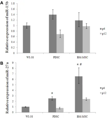
Fig. 1. Relative expression of miR-15b (A) and 27a (B). p6 and p12 means passage 6 and 12. Data are given as means of values ± S.D. from three independent experiments.
Level of signifcance was identifed statisticaly (*, # p<0.05) using Student's t-test (*: significance with WI-38,
#: significance with PDSC).
Ct)값을 구하고, 2의 지수로 ΔΔCt를 넣어 계산하여, 상대적인 마이크로RNA 발현량을 측정하였다.
통계분석
각 실험은 3회 이상 반복실험을 통하여 그 결과를 얻어 각각 의 마이크로RNA 발현량에 대하여 평균±표준편차로 나타내 었다. 각 마이크로RNA 발현량은 세포주간 Student’s t test를 실시한 후, p<0.05값을 통계적으로 유의성 있는 결과로 간주하 였다.
결과 및 고찰 마이크로RNA-15b, 27a의 발현
PDSC와 BM-MSC모두 WI-38과 비교해 보았을 때 세포분 열 및 스트레스 조절기능을 가지는 마이크로RNA-15b의 발현 량에 있어서 유의적인 차이를 보이지 않는 것을 볼 수 있다.
하지만 PDSC의 경우는 6세대에 비하여 12세대에서 현격하게 발현이 떨어지는 것을 확인 할 수 있다(Fig. 1A). 이는 줄기세 포가 세대가 지속될수록 줄기세포의 고유성질인 줄기성 (stemness)를 잃어감에 따라 마이크로RNA의 발현수준이 떨 어지는 것으로 추정된다. 마이크로RNA 27a의 경우에는 PDSC 와 BM-MSC모두 WI-38에 비하여 발현량이 크게 유의적으로 높은 것을 확인할 수 있다(Fig. 1B). 마이크로 RNA-27a는 분화 능력 외에도 혈관신생이나 세포자멸사를 조절하는 기능을 가 지고 있기 때문에[7, 19] 이식된 줄기세포의 조직분화에도 상 당히 도움이 될 것으로 생각되며, 이와 관련된 만성질환인 종 양치료에 있어서 줄기세포주와 마이크로RNA의 효과에 의해 높은 세포치료효과를 볼 수 있을 것으로 예상되며 12세대에서 는 6세대에 비해 발현수준이 감소하므로 치료효과를 높이기 위하여 세대수가 낮은 세포를 활용하는 것이 종양치료에 효과 적인 것으로 보인다. 여기서 흥미로운 것은 PDSC가 BM- MSC 보다 세포치료효과가 더 크게 나타난다고 알려진 게 일반적인 사실인데[2], 마이크로RNA-27a의 경우에는 오히려 BM- MSC 가 더 발현량이 PDSC에 비해 유의적으로 높은 것을 볼 수 있다.
마이크로RNA-33a, 33b의 발현
마이크로RNA-33a발현을 비교해보면 WI-38과 PDSC에 비 하여 BM-MSC에서의 발현이 유의적으로 상당히 높은 것을 확인할 수 있다(Fig. 2A). 이는 이들 줄기세포의 유래를 생각하 면 이해할 수 있다. BM-MSC의 경우에는 성인골수에서 채취 한 중간엽 줄기세포로 PDSC에 비해 상대적으로 골수의 주요 기능인 조혈작용에 더 크게 관련이 되어있기 때문에 더 높은 마이크로RNA-33a발현이 나타났을 것으로 사료된다. 따라서 BM-MSC의 체내 이식을 통한 세포치료를 통하여 조혈기능의 개선이 기대된다. 마이크로RNA-33b역시 WI-38이나 BM-MSC 에 비해 PDSC의 발현이 낮은 것을 볼 수 있다. 하지만 BM-
A
B
Fig. 2. Relative expression of miR-33a (A) and 33b (B). p6 and p12 means passage 6 and 12. Data are given as means of values ± S.D. from three independent experiments.
Level of signifcance was identifed statisticaly (*, # p<0.05) using Student's t-test (*: significance with WI-38,
#: significance with PDSC).
A
B
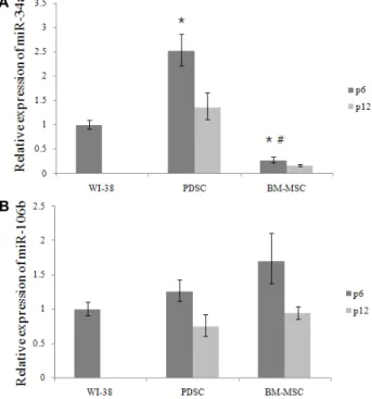
Fig. 3. Relative expression of miR-34a (A) and 106b (B). p6 and p12 means passage 6 and 12. Data are given as means of values ± S.D. from three independent experiments.
Level of signifcance was identifed statisticaly (*, # p<0.05) using Student's t-test (*: significance with WI-38, #: sig- nificance with PDSC).
Fig. 4. Relative expression of miR-211. p6 and p12 means pas- sage 6 and 12. Data are given as means of values ± S.D.
from three independent experiments. Level of signi- fcance was identifed statisticaly (*, # p<0.05) using Student's t-test (*: significance with WI-38, #: significance with PDSC).
MSC의 경우 6세대에서 12세대로 넘어갈수록 마이크로RNA 33b의 발현이 낮아지는 것에 비하여, PDSC의 경우에는 높아 지는 것을 볼 수 있다(Fig. 2A).
마이크로RNA-34a, 106b의 발현
마이크로RNA-34a의 발현이 WI-38이나 BM-MSC에 비하 여 PDSC에서 유의적으로 크게 높은 것을 확인할 수 있다 (Fig.
3A). PDSC 12세대의 경우에도 6세대에 비하여 발현은 떨어지 지만 이는 세포노화에 따른 발현저하로 보여지며, 그래도 BM-MSC에 비해서는 현저하게 높은 것을 확인할 수 있다. 따 라서 항암치료에 있어서 암세포의 세포자멸사를 유도할 수 있을 것으로 예상된다. 마이크로RNA-106b는 다소간의 차이 는 보이나 각 그룹간 유의적인 차이는 보이지 않는 것을 볼 수 있다(Fig. 3B).
마이크로RNA-211의 발현
마이크로RNA-211은 Fig. 4를 보면 PDSC의 경우엔 오히려 WI-38보다 발현이 떨어지는 것으로 나타났으나 BM의 경우에 는 WI-38이나 PDSC에 비해 최대 8배 이상 높은 발현이 나타 나는 것을 볼 수 있다. 12세대의 경우에도 6세대보다는 떨어지 지만 여전히 높은 마이크로RNA-211발현을 보이고 있는 것을 확인할 수 있다. 마이크로RNA-211의 경우에는 특히 흑색종과
같은 피부암에 큰 효과를 보이는 것으로 알려져 있어 이들 피부암종의 세포치료에 BM을 사용할 경우 높은 효과를 얻을 수 있을 것으로 예상된다[23].
References
1. Ambros, V. 2004. The functions of animal microRNAs.
Nature 431, 350-355.
2. Barlow, S., Brooke, G., Chatterjee, K., Price, G., Pelekanos, Rebecca., Rossetti, T., Doody, M., Venter, D., Pain, S., Gilshenan, K. and Atkinson, K. 2008. Comparison of human placenta- and bone marrow-derived multipotent mesen- chymal stem cells. Stem Cells Dev 17, 1095-1108.
3. Bartel, D. P. 2009. MicroRNAs: Target recognition and regu- latory functions. Cell 136, 215-233.
4. Bhatia, M., Wang, J. C., Kapp, U., Bonnet, D. and Dick, J.
E. 1997. Purification of primitive human hematopoietic cells capable of repopulating immune-deficient mice. Proc Natl Acad Sci USA 94, 5320-5325.
5. Cerlos, F., Yajaira, S., Katey, R. and Kathryn, M. 2011.
MicroRNAs in lipid metabolism. Curr Opin Lipidol 22, 86-92.
6. Chitnis, N. S., Pytel, D., Bonrovnikiva-Marjon, E., Pant, D., Zheng, H., Maas, N. L., Frederick, B., Kushner, J. A., Chodosh, L. A., Koumenis, C., Fuchs, S. Y. and Diehl, J.
A. 2012. miR-211 Is a prosurvival microRNA that regulates chop expression in a PERK-dependent manner. Mol Cell 48, 353-364.
7. Gang, L., Peng, C., Huajing, C., Wen, Y., Jianxi, W. and Xianye, T. 2013. MiR-27a regulates apoptosis in nucleus pul- posus cells by targeting PI3K. PLoS One 8, e75251.
8. Heo, I., Joo, C., Kim, Y. K., Ha, M., Yoon, M. J., Cho, J., Teom, K. H., Han, J. and Kim, V. N. 2009. TUT4 in concert with Lin28 suppresses microRNA biogenesis through pre-microRNA uridylation. Cell 138, 696-708.
9. Ivanovska, I., Ball, S., Diaz, L., Magnus, F., Kibukawa, M., Schelter, M., Kobayashi, V., Lim, L., Burchard, J., Jackson, L., Linsley, S. and Cleary, A. 2008. MicroRNAs in the miR-106b family regulate p21/CDKN1A and promote cell cycle progression. Mol Cell Biol 28, 2167-2174.
10. John, R., Wang-Xia, W., Sebastien, S., Bernard, R., Guogen, M. and Peter, T. 2010. The miR-15/107 group of microRNA genes: evolutionary biology, cellular functions, and roles in human diseases. J Mol Biol 402, 491-509.
11. Jose, J. and Vicente, A. 2010. A role for miR-33 in p53 regu- lation: New perspectives for hematopoietic stem cell research. Cell Cycle 9, 3397-3398.
12. Kozomara, A. and Griffiths-Jones, S. 2013. miRBase : Anno- tating high confidence microRNAs using deep sequencing data. Nucleic Acids Res 2013, 1-6.
13. Lee, M. J., Jung, J., Na, K. H., Moon, J. S., Lee, H. J., Kim, J. H., Kim, G. I., Kwon, S. W., Hwang, S. G. and Kim, G.
J. 2010. Anti-fibrotic effect of chorionic plate-derived mes enchymal stem cells isolated from human placenta in a rat nodel of CCl4-injured liver: potential application to the
treatment of hepatic diseases. J Cell Biochem 111, 1453- 1463.
14. Parolini, O. and Caruso, M. 2011. Review: Preclinical studies on placenta-derived cells and amniotic membrane: an update. Placenta 32(Suppl 2), S186-195.
15. Pittenger, M. F., Mackat, A. M, Stephen, C. B., Jaiswal, R.
K., Dauglas, R., Masca, J. D., Moorman, M. A., Simonetti, D. W., Craig, S. and Marshak, D. R. 1999. Multilineage po- tential of adult human mesenchymal stem cells. Science 284, 143-147.
16. Ramirez, C., Goedeke, L., Rotllan, N., Yoon, J. H., Cirera- Salinas, D., Mattison, J., Suarez, Y., Cabo, R., Gorospe, M.
and Fernandez-Hernando, C. 2013. MicroRNA 33 regulates glucose metabolism. Mol Cell Biol 33, 2891-2902.
17. Rubinoff, B. E., Pera, M. F., Fong, C. Y., Trounson, A. and Bongso, A. 2000. Embryonic stem cell lines from human blastocysts: somatic differentiation in vitro. Nat Biotechnol 18, 399-404.
18. Spangrude, G. J., Heimfeld, S. and Weissman, I. L. 1998.
Purification and characterication of mouse hematopoietic stem cells. Science 241, 58-62.
19. Tang, W., Yu, F., Yao, H., Cui, X., Jiao, Y., Lin, L., Chen, J., Yin, D., Song, E. and Liu, Q. 2014. miR-27a regulates en- dothelial differentiation of breast cancer stem like cells.
Oncogene 33, 2629-2638.
20. Thomson, J. A., Itskovitz-Eldor, J., Shapiro, S. S., Waknitz, M. A., Swiergiel, J. J., Mardchall, V. S. and Jones, J. M. 1998.
Embryonic stem cell lines derived from human blastocysts.
Science 282, 1145-1147.
21. Trompeter, H. I., Abbad, H., Iwaniuk, K. M., Hafner, M., Renwick, N., Tuschl, T., Schira, J. Muller, H. W. and Werner, P. 2011. MicroRNAs MiR-17, MiR-20a, and Mir-106b act in concert to modulate E2F activity on cell cycle arrest during neuronal lineage differentiation of USSC. PLoS One 6, e16138.
22. Weissman, I. L. 2000. Translating stem and progenitor cell biology to the clinic: barriers and opportunities. Science 287, 1442-1446.
23. Xu, Y., Brenn, T., Brown, R., Doherty, V. and Melton, W.
2012. Differential expression of microRNAs during melano- ma progression: miR-200c, miR-205 and miR-211 are down- regulated in melanoma and act as tumour suppressors. Br J Cancer 106, 553-561.
24. Yamakuchi, M., Ferlito, M., Lowenstein, C. J., Yamakuchi M., Ferlito, M. and Lowenstein, C. J. 2008. miR-34a re- pression of SIRT1 regulates apoptosis. Proc Natl Acad Sci USA 105, 13421-13426.
초록:태반유래 줄기세포와 골수유래 줄기세포에서의 마이크로RNA 발현비교 김수환*
(고려대학교 일반대학원 의생명융합과학과)
중간엽줄기세포(mesenchymal stem cell, MSC)은 세포치료로 각광받아 널리 사용되고 있다. 이들은 줄기세포의 분화성을 이용하여 많은 만성질환에 연관되어 치료제로 사용되고 있다. 줄기세포는 다른 화학적 치료법에 비해 많은 장점을 가지고 있다. 왜냐하면 줄기세포치료는 자기자신, 혹은 동종의 세포를 이용한 치료이기 때문에 화학 치료에 비해 부작용이나 치료의 위험성이 덜하다. 그리고 마이크로RNA또한 최근 기 존재와 기능이 밝혀져서 연 구되고 있는데 특히 항암, 세포생장촉진 등의 기능을 이용해 항암, 만성질환 치료에 접목되어 치료제로의 역할이 기대된다. 마이크로RNA는 대부분의 대사과정이나 항상성조절에 관여되어있다. 따라서 마이크로RNA가 저 발현 혹은 과 발현하게 되면 만성질환으로 이어지게 된다. 하지만 줄기세포와 마이크로RNA의 상호간 보조효과는 잘 연구되어 있지 않다. 따라서 이들 간의 상관관계를 확인하기 위하여 태반유래 줄기세포(PDSC)와 골수줄기세포 (BM-MSC), 대조군으로 섬유아세포(Fibroblast, WI-38)을 사용하여 이들이 발현하는 마이크로RNA 발현을 확인해 보았다. 각각의 MSC 세포주에 대하여 특정 마이크로RNA의 발현량을 확인해 보았다. 결과 PDSC의 경우엔 마이 크로RNA-34a의 발현이 높았고 BM-MSC의 경우에는 마이크로RNA-27a, 33a, 33b, 211의 발현이 높은 것을 확인 할 수 있었다. 따라서 우리는 각각의 MSC세포주와 그들이 발현하는 기능성 마이크로RNA을 연관지어 효과적인 세포치료에 활용될 수 있을 것을 기대한다.