• 검색 결과가 없습니다.

A Case of Polyostotic Fibrous Dysplasia Masquerading as a Multiple Bone Metastases

N/A
N/A
Protected

Academic year: 2021

Share "A Case of Polyostotic Fibrous Dysplasia Masquerading as a Multiple Bone Metastases"

Copied!
6
0
0

로드 중.... (전체 텍스트 보기)

전체 글

(1)

Introduction

Fibrous dysplasia (FD) is a rare, sporadically occurring bone disease, characterized by abnormal bone growth where normal bone is replaced by fibrous skeletal tissue composed of bone-forming mesenchyme [1]. FD represents abnormal growth and swelling of bone results poor mechanical

strength which can lead to pathologic fractures.

Most patients have lesions involved in one bone(monostotic) and others have them in multiple bone(polyostotic) [2].

The majority of monostotic lesions are a s y m p t o m a t i c a n d a r e d i s c o v e r e d w h e n radiographs of the involved region are obtained incidentally or during other intentions [3]. On the

Abstract

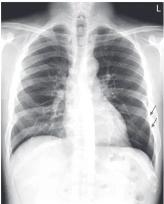
Fibrous dysplasia is a benign, bony abnormality that is usually asymptomatic. A 41-year-old male with minimal symptoms presented at this hospital with abnormal findings incidentally seen in his ribs on the chest radiograph.

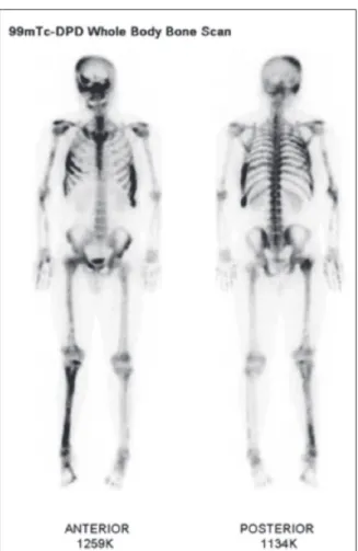
A skeletal survey showed numerous, osteolytic lesions throughout multiple bones. Diagnostic processes for malignancy of undefined primary origin (MUO) were performed in order to identify the underlying primary neoplasm, although abnormal findings were not seen except for multiple bone lesions. A computed tomography guided bone biopsy was performed on his left rib. The final diagnosis was fibrous dysplasia. This case demonstrates that fibrous dysplasia should be considered in the differential diagnosis in young patients with multiple, osteolytic lesions and without a prior history suggesting malignancy.

Key Words : Bone neoplasm, Fibrous dysplasia, Metastasis

Corresponding Author:

Jae Lyun Lee, M.D., Department of Oncology, Asan Medical Center, University of Ulsan College of Medicine, 88 Olympic-ro 43-gil, Songpa-gu, Seoul 138-736, Korea

Tel : +82-2-3010-5977 E-mail : jaelyun@amc.seoul.kr

Departments of Medicine, Radiology

1

, Pathology

2

, and Oncology

3

, Asan Medical Center, University of Ulsan College of Medicine, Seoul, Korea

Jaekyung Cheon, M.D., Yumun Jeong, M.D., Ji Young Yang, M.D., Sunpyo Lee, M.D., Misun Chung

1

, M.D., Eun Mi Son

2

, M.D., Jae Lyun Lee

3

, M.D.

A Case of Polyostotic Fibrous Dysplasia Masquerading as a Multiple

Bone Metastases

(2)

other hand, polyostotic forms may sometimes be accompanied by bone pain and deformities. The typical radiologic description is the “ground-glass”

pattern of the bone, and which is helpful in a correct diagnosis. However, in some patients, it is difficult to differentiate the radiologic diagnosis between FD and malignant bone diseases.

We report a case of polyostotic FD with minimal symptoms masquerading as multiple bone metastases.

Case Report

In February 2013, a 41-year-old male visited to this hospital with abnormal findings on his chest radiograph taken during his annual health screening. He had suffered from fatigue and mild low back pain for one year. He was otherwise in good health and no specific past medical or family history. His physical examination results were unremarkable. His chest radiograph revealed several, osteolytic bulging masses in his left ribs (Fig. 1). His full blood count and biochemistry showed almost normal except for a mild elevation in alkaline phosphatase (228 IU/L). He was admitted for further evaluation and got an examination processes which usually used for diagnosing malignancies of unknown origin (MUO). The level of serum alpha-fetoprotein (AFP) and β-subunit of human chorionic gonadotropin (β-hCG) were normal. A skeletal survey showed multiple, osteosclerotic, bulging lesions in his skull, both ribs, both femurs, the right tibia, both humeri, a n d t h e r i g h t r a d i u s. S e r u m a n d u r i n e electrophoresis were done and there were no abnormal peaks. The esophagogastroduodeno- scopy showed short segment of Barrett’s esophagus without any other abnormalities. Computed tomography (CT) of his chest, abdomen, and pelvis

revealed multiple skeletal lesions in the right ischium, both femurs, cervical spines, and both side of ribs. In his abdominopelvic CT showed a 2.7-cm nodule in the right adrenal gland, but there were no other abnormal findings. On the delayed phase of enhanced CT, the washout ratio was 71%, and which met the radiologic criteria for the diagnosis of a lipid-poor, adrenal adenoma [4]. The bone scan showed multiple abnormal areas of radio-uptakes, which suggested the lesion might be metastasis. But, the scan showed diffusely increased uptakes in his right tibia, including the metaphysis and diaphysis, these findings were somewhat different from that of metastasis. The skull base was also involved, but the spine was spared in the scan, which is also unusual in malignant bone metastasis (Fig. 2). We could not find any evidence of malignancy. All results

Fig. 1. Initial chest radiograph.

(3)

suggested a benign bone disease, such as fibrous dysplasia or Paget’s disease, rather than metastatic bone tumor. The fluorodeoxyglucose-positron emission (FDG-PET)/CT scan showed increased FDG uptake in multiple, skeletal lesions [maximum standardized uptake value (SUVmax)=12.2 g/mL in the left, 11

th

rib], thus favoring metastatic lesions rather than benign bone tumors (Fig. 3). However, patient’s FDG-PET/CT showed only bony lesion and no other evidence of the primary site of solid malignancies. The patient underwent percutaneous, CT-guided, needle biopsy of the left, 11

th

rib, and three cores were obtained. Microscopically, the pathology specimens revealed a benign, fibro- osseous lesion, thus confirming the diagnosis of fibrous dysplasia. Cellular atypia, mitotic figures, or necrosis were not observed (Fig. 4).

The patient then received 60 mg of intravenous pamidronate every three months and regularly followed in the outpatient clinic.

Fig. 2. Initial bone scan.

Fig. 3. FDG-PET/CT scan.

(4)

Discussion

FD was described by Lichtenstein [5] in 1938, and the incidence and prevalence of FD are difficult to estimate, these lesions are not so rare.

FD accounts for about 5% to 7% of benign bone tumors and consists nearly 1% of primary bone tumors [6].

Most lesions are monostotic, and approximately 20% to 30% of FD cases are polyostotic. The majority of monostotic lesions are asymptomatic, although polyostotic cases may be accompanied by bone pain or deformities, and two thirds of these patients are symptomatic before the 10 years of age [2].

Sometimes it is difficult to differentiate polyostotic FD from disseminated bone metastasis.

The clinical and radiologic characteristics of polyostotic FD and bone metastasis are very similar in the occurrence of multiple lesions and location of, anatomic sites, but the prognosis and methods of treatment are totally different.

CT is the most successful technique for demonstrating the typical radiographic findings of FD. The CT findings are intramedullary, expansile, well-defined lesions with intact cortex and a characteristic ground-glass appearance, although some cases may appear almost radiolucent or sclerotic [7]. In this patient, the radiologic findings showed multiple, bulging lesions, and which suggested bone metastasis or multiple myeloma.

However, his CT images did not show a periosteal reaction, cortical destruction or a soft-tissue mass lesions. Therefore, the CT images suggested a benign condition rather than malignancy.

Although radionucleotide bone scintigraphy is

non-specific, these scans are helpful in making a

more reliable diagnosis. Many benign bone lesions,

such as eosinophilic granulomas, disseminated

tuberculosis, fibrous dysplasia, and enchondroma,

appear similar to bone metastasis on bone scans

[8,9]. However, there are some useful factors

which differentiate FD from metastasis. First, the

involved FD sites are usually extremities and ribs,

Fig. 4. Microscopic pathology.

(5)

unlike metastatic bone tumors which favor the axial skeleton, especially the spine and pelvis. Second, the hot uptakes usually appear in the entire parts of the involved bone in the FD [10].

With regard to FDG-PET/CT, it is known that there is a large variability in the FDG avidity of FD.

The SUVmax values of the FD have ranged from 3.73 to 9.64 in previous reports [11]. But in this case, the FDG-PET/CT scan showed higher FDG uptake in the left 11

th

rib. (SUVmax = 12.2 g/mL), therefore the possibility of FD should not be excluded on the basis of FDG avidity.

Radiologic findings are usually sufficient for making a diagnosis of FD [10], and a bone biopsy is not necessary. The bone biopsy leads likely to result in a fracture which can be difficult to heal [12]. Therefore, bone biopsy should be avoided whenever possible, and the diagnosis of FD should be based on the clinical and radiological findings.

In this patienst, objective evidence of malignancy based on the clinical, laboratory, and radiologic findings were insufficient. A potentially harmful diagnostic approach, such as bone biopsy, had been chosen due to the patient or clinician's over concern, was inappropriate.

Conclusion

Polyostotic FD may be misdiagnosed as metastatic bone disease, and FD should be included in the differential diagnosis in young patients with multiple, osteolytic lesions but who do not have a prior history of malignancy.

Conflict of Interest

The authors report no conflict of interest in this work.

References

1. Keskin M, Karabekmez FE, Ozkan BT, Tosun Z, Avunduk MC, Savaci N. Simultaneous occurrence of facial fibrous dysplasia and ameloblastoma. J Craniomaxillofac Surg 2009;37:102-5.

2. Harsh M, Preeti M, Isneet M, Sudhir K. Fibrous dysplasia of bone: a clinicopathologic review. Patholo Lab Med Int 2011:3:31-42

3. Ippolito E, Bray EW, Corsi A, De Maio F, Exner UG, Robey PG, et al. Natural history and treatment of f i b r o u s d y s p l a s i a o f b o n e : a m u l t i c e n t e r clinicopathologic study promoted by the European Pediatric Orthopaedic Society. J Pediatr Orthop B 2003;12:155-77.

4. Caoili EM, Korobkin M, Francis IR, Cohan RH, Dunnick NR. Delayed enhanced CT of lipid-poor adrenal adenomas. AJR Am J Roentgenol 2000;175:1411-5.

5. Lichtenstein L. Polyostotic fibrous dysplasia. Arch Surg 1938;36:874-98.

6. Singer FR. Fibrous dysplasia of bone: the bone lesion unmasked. Am J Pathol 1997;151:1511-5.

7. Fitzpatrick KA, Taljanovic MS, Speer DP, Graham AR, Jacobson JA, Barnes GR, et al. Imaging findings of fibrous dysplasia with histopathologic and intraoperative correlation. AJR Am J Roentgenol 2004;182:1389-98.

8. Wang K, Allen L, Fung E, Chan CC, Chan JC, Griffith JF. Bone scintigraphy in common tumors with osteolytic components. Clin Nucl Med 2005;30:655-71.

9. Zhang Y, Zhao C, Liu H, Hou H, Zhang H. Multiple metastasis-like bone lesions in scintigraphic imaging. J Biomed Biotechnol 2012;2012:957364.

10. Zhibin Y, Quanyong L, Libo C, Jun Z, Hankui L, Jifang Z, et al. The role of radionuclide bone scintigraphy in fibrous dysplasia of bone. Clin Nucl Med 2004;29:177-80.

11. Su MG, Tian R, Fan QP, Tian Y, Li FL, Li L, et al.

Recognition of fibrous dysplasia of bone mimicking

(6)

skeletal metastasis on 18F-FDG PET/CT imaging.

Skeletal Radiol 2011;40:295-302.

12. Chapurlat RD, Meunier PJ. Fibrous dysplasia of bone.

Clin Rheumatol 2000;14:385-98.

수치

Fig. 1. Initial chest radiograph.
Fig. 2. Initial bone scan.

참조

관련 문서

: Bone response to unloaded titanium implants with a sandblasted and acid-etched surface : A histomorphometric study in the canine mandible.. : Early

Expanded foams, gas-filled powders, fibrous materials, vacuum alone, evacuated powders and fibrous materials, opacified powders 그리고 multilayer insulations 등이

Bone transplants, autogenous bones, allogenic bones, xenogenic bones, and synthetic bone substitutes have all been used as bone graft materials.. However,

Histopathologic findings of control group at 4 weeks show little bone- implant contact (BIC) around the implant (asterisks) and new-bone formation in the defect

Purpose: Calcaneal fracture is a rare fracture, which accounts for about 2% of all fractures, but is one of the most common fractures in the ankle bone.. There is

Alveolar ridge augmentation with titanium mesh and a combination of autogenous bone and anorganic bovine bone: a 2-year prospective study.. Corinaldesi G, Pieri F, Sapigni

LIPUS therapy is a recently developed method for application of mechanical stress, and used clinically to promote bone healing. Author evaluated the effect

It might also act in conjunction with a growth factor, such as bone morphogenic protein (BMP), insulin-like growth factor, or platelet-derived growth factor.