대구외지 2009;35:372-375
372
Abstract (J. Kor. Oral Maxillofac. Surg. 2009;35:372-375)
Ⅰ.
서 론중심성 치성 섬유종은 극히 드문 양성 종양으로서, 전체 치성 종양의 0.1% 미만을 차지한다
1). 가장 최근의 문헌 보 고에 의하면, 오직 69 증례만이 발표 되었다
2). 이 종양은 서 서히 자라고, 무통성의 피질골 팽창을 하는 임상적 특징을 나타내며, 단방성 혹은 다방성의 경계가 잘 형성된 방사선 투과성 병소를 보인다
1). 종종 인접치의 치근 흡수와 치근 이개를 일으킨다. 남성에 비해 여성에게 더 호발하는 경향 이 있고, 상악골과 하악골의 발생 비율은 비슷하다. 수술적 절제로 치료하며, 악성으로 변화하는 경향은 거의 없다.
저자 등은 27세 남자 환자의 상악 전치부에 발생한 중심 성 치성 섬유종 증례를 경험하였기에 문헌 고찰과 함께 이 를 보고하는 바이다.
Ⅱ.
증례보고본 27세 남환은 상악 전치부 상실된 부위의 임플란트 치 료를 위해 개인치과에 방문하여 촬영한 파노라마 영상에 서 상악 전치부의 치근 흡수 양상 및 방사선 투과성 병소가 보여 2008년 10월 13일 본원에 의뢰 되었다. 2~3년 전 해당치 아(#21)가 자연 탈락 하였다고 하며, 특이할 통증이나 출혈 소견은 없었으며, 그 외 병력상 전신 질환은 없었다(Fig. 1).
내원 당시의 임상 소견은 #21 치아를 상실한 상태였으며,
#22 치아의 Mob(++), Per(-)였으며, #11치아와 #23치아는 특별한 흔들림은 없었고, 해당 부위의 촉진 시 압통도 없었 다. 파노라마 영상(Fig. 2)에서 #11 치아에서 #23 치아에 걸 친 방사선 투과성 병소가 있었으나, 구강내로 누관 형성은 없었다. 치아 생활력 검사에서 #11, 22, 23치아 모두 cold 및 EPT 음성 반응이 나타났다. 술 전 촬영한 routine Maxilla CT상 좌측 상악동의 외측 경계 및 협측 부의 피질골 흡수 양상이 나타났으며, 방사선 불투과성 병소는 보이지 않았 다(Fig. 3).
두 번째 내원 당시 incisional biopsy후 중심성 치성 섬유종 으로 진단되어 해당치아(#11, 22, 23)의 근관치료 후 종양 절제술 및 유리 장골 이식술을 통한 골결손부의 재건을 계 획하였다. 2008년 11월 18일 전신 마취하에 계획된 수술을
남 웅120-752
서울시 서대문구 성산로250
연세대학교치과대학치의학전문대학원구강악안면외과학교실, 구강종양연구소
Woong NamDepartment of Oral and Maxillofacial Surgery, Oral Cancer Research Institute, Yonsei University College of Dentistry, 250 Sungsan-no, Seodaemun-gu, Seoul, Korea, 120-752 Tel: 82-2-2228-2971 Fax: 82-2-364-0992
E-mail: [email protected]
상악 전치부에 발생한 중심성 치성 섬유종 : 증례 보고
함태훈
1∙김학진
1,2∙김형준
1,2∙차인호
1,2∙남 웅
1,2연세대학교 치과대학
1구강악안면외과학교실,
2구강종양연구소
CENTRAL ODONTOGENIC FIBROMA IN ANTERIOR MAXILLA - A CASE REPORT
Tae-Hoon Hahm
1, Hak-Jin Kim
1,2, Hyung Jun Kim
1,2, In-Ho Cha
1,2, Woong Nam
1,21
Department of Oral and Maxillofacial Surgery,
2Oral Cancer Research Institute, College of Dentistry, Yonsei University, Seoul, Korea
Central odontogenic fibroma is an extremely rare benign tumor, accounting for less than 0.1% of all odontogenic tumors. The most recent literature review that only 69 cases have so far been reported. This tumor has a slow persistent growth that results in painless cortical expansion clinically, and well defined unilocular or multilocular radiolucent lesion. Root resorption of associated teeth is common, and lesions located between the teeth often cause root divergence. There is occurring tendency to female more than male, and occurring in the mandible and in the maxilla with equal frequency.
The treatment is surgical excision with no tendency to undergo malignant transformation. We report a case of central odontogenic fibroma in the max- illa of a 27-year male with literatures review.
Key words: Central odontogenic fibroma, Maxilla, Iliac bone graft
[원고접수일 2009. 8. 28 / 1차수정일 2009. 9. 2 / 2차수정일 2009. 9. 10 / 게재확정일 2009. 9. 22]
373 하였으며, 수술 당시 palatal approach후 #22 치아와 함께 섬
유성 종물을 주변 조직과 분리하였으며, 해당 부위 치아 (#11, 23)의 치근단 절제술을 하였다(Fig. 4). 종물은 주변 조직과 부착 소견이 없었고, 종물외 정상 조직은 잘 보존되
었다. 골 결손부의 재건을 위하여 좌측 장골에서 2×2 cm 크기의 피질골과 수질골의 채취하였다. 수질골과 beta-TCP 성분인 alloplastic bone(PolyBone
�)을 혼합하여 골 결손부 을 채운 후 피질골을 협측 barrier로 사용하였다(Fig. 5).
Fig. 1. Intraoral view: #21 was missed 2~3 years ago.
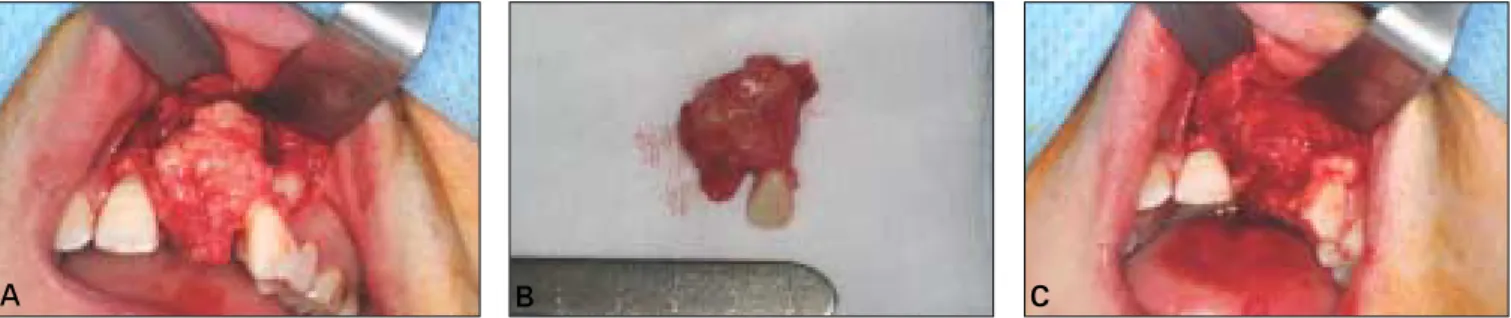
Fig. 4. operative findings.
A. Well-defined margin, easily enucleated mass. B. Removal of mass and no oronasal communication. C. mass involved #22 tooth, sized 2×3 cm.
Fig. 3. Coronal & axial CT scan showing cortical bone thin- ning & resorption.
Fig. 2. Panoramic view showing radiolucency on
#11,21,22,23.
Fig. 5. Immediate reconstruction with autogenous bone graft and allograft.
A. Grafted Bone blending with iliac cancellous bone and PolyBone
�. B. Bone barrier on buccal aspect, arrow : iliac inner cortex (2×2cm). C. Flap was closed interrupted suture.
A B C
A B C
상악 전치부에 발생한 중심성 치성 섬유종 : 증례 보고
대구외지 2009;35:372-375
374
Ⅲ.
고 찰중심성 치성 섬유종은 악골에 매우 드물게 발생하는 종 양으로, 남성에 비해 여성에 2.2배로, 상악골에 발생할 경 우 전치부(29%)쪽에 하악골에 발생할 경우 구치부(29%)쪽 에 주로 호발한다
1). 중심성 치성 섬유종 병소는 종종 10~20 대에 발생한다.
가장 빈번하게 관찰할 수 있는 증상은 종창이며, 몇몇 경 우에 있어서 치근 이개를 야기하며 서서히 병소가 자라는 것이다. 통증이나 감각이상과 같은 임상적 양상은 드물다
3). 치성 섬유종의 많은 경우에서 인접치의 위치 이상이 보고 되며, 종종 치근 흡수를 야기 한다
4). 또한, 그 크기가 작을 경우 주로 (2cm정도) 단방성이며, 크기가 클 경우 다방성
(4cm이상)을 나타낸다
4). 이 증례의 경우 치근흡수를 야기하
였으며, 인접치의 치근이개와 단방성 병소를 나타내었다.
중심성 치성 섬유종의 방사선학적 특징은 외상성 골 낭, 단방성의 법랑아세포종, 치성낭, 중심성 거대세포 육아종 과 비슷한 양상을 나타내며, 감별을 요한다
5). 주로 방사선 투과성의 병소를 나타내며, 드물게 이형성의 상아질 또는 백악질 유사 물질이 있을 경우 병소내 방사선 불투과성이 있을 수 있다
6).
조직학적 양상을 통해 분류 할 수 있는데, 1980년대
Gardner
7)에 의한 분류에 의하면 3가지 종류로 묘사될 수 있
다. 첫째, 과증식성 치성 낭포 둘째, Simple type-치성 상피
섬을 담고 있는 다양한 섬유성 결합 조직을 가지고 있는 섬 유성 종양 셋쩨, WHO type-다양한 양의 치성 상피와 이형 성 상아질 또는 백악질 유사 물질을 가지고 있는 복합적 병 소이다
2). 이 증례의 조직학적 특징을 볼 때(Fig. 6) Gardner 분류 중 simple type에 속하며, 200배 확대에서 치성 상피가 편평상피보다 입방 상피의 소견을 보인다.
치료는 수술적 절제를 하는 것이며, 재발은 드물다.
Barker와 Dunlap
8)은 2 증례의 상악 치성 섬유종을 소파술을 통해 치료한 후 각각 9년, 10년 동안 경과 관찰한 결과 재발 하지 않았다. 하악골에 발생한 중심성 치성 섬유종을 적출 한 Daniel 등의 보고에 의하면 5년 경과 관찰 결과 재발하 지 않았다
2). 하지만, 몇몇 재발 증례가 보고된 바도 있다.
Heimdal등
9)은 술 후 9년째 재발 소견을 보였으며, Svirsky 등
10)은 13%(15증례 중 2증례)의 재발율을 보고하였다. 본 증례의 경우 술 후 9개월째 재발 소견은 없다.
중심성 치성 섬유종은 최근까지 보고에 의하면 69증례만 이 발표되었으며, 임상적 양상이 초기부터 특징적으로 나 타나지 않고 주로 방사선 검사를 받다 우연히 발견되기 때 문에 초기 진단 및 치료가 어렵다. 본 증례 역시 병소 발병 후 3~4년 뒤 치료가 된 것으로 추정되며, 중심성 치성 섬유 종의 정확한 기전을 밝히기 어렵다. 본 증례를 통해 중심성 치성 섬유종의 임상적, 방사선학적, 조직학적 특징을 확인 할 수 있었다.
Fig. 6. Histologic features : The tumor is composed of bundles of interlacing col- lagenous fibers. (A: H & E stain ×100) Inactive odontogenic epithelium was cuboidal form more than squamous form in this case. (B: H & E stain ×200)
Fig. 7. Panoramic view at 1week after
operation. #22 tooth was extracted, and
bone defect was reconstructed iliac bone
properly. #11, 23 teeth were treated
endodontically.
375
참고문헌1. Neville BW, Damm DD, Allen CM, Bouquet JE. Oral and Maxillofacial Pathology. 2nd ed. Philadelphia, WB Saunders 2002.
2. Daniels JS : Central odontogenic fibroma of mandible: a case re- port and review of the literature. Oral surg Oral Med Oral Pathol Oral Radiol Endod 2004; 98: 295-300.
3. Covani U, Crespi R, Perrini N, Barone A : Central odontogenic fibroma- a case report. Med Oral Patol Oral Cir Bucal 10 Suppl 2005; 2: 154-7.
4. Regezi JA : Odontogenic cysts, odontogenic tumors, fi- broosseous, and giant cell lesions of the jaws. Modern Pathology 2002; 15: 331-41.
5. Ramer M, Buonocore P, Krost B : Central odontogenic fibroma-
report of a case and review of the literature. Periodontal Clin Investig 2002;24: 27-30.
6. Gardner DG : Central odontogenic fibroma current concepts. J Oral Pathol Med 1996; 25: 556-61.
7. Gardner DG : The central odontogenic fibroma - an attempt at clarification. Oral surg Oral Med Oral Pathol Oral Radiol Endod 1980; 5: 425-32.
8. Barker BF, Dunlap CL : Central odontogenic fibroma of the WHO type. Oral surg Oral Med Oral Pathol Oral Radiol Endod 1984; 57: 390-4.
9. Heimdal A, Isacsson G, Nilsson L : Recurrent central odonto- genic fibroma. Oral Surg Oral Med Oral Pathol Oral Radiol Endod. 1980;50:140-5.
10. Svirsky JA, Abbey LM, Kaugars GE : A clinical review of cen- tral odontogenic fibroma: with the addition of three new cases. J Oral Med 1986;41: 51-4.
상악 전치부에 발생한 중심성 치성 섬유종 : 증례 보고