Ⅰ. 서 론
치성유령세포암종 (Odontogenic ghost cell carcinoma)은 1985년 Ikemura 등
1)이 처음 보고한 이래로, 영자 문헌에 16예만이 보고 된 드문 암종으로 석회화치성낭종 (Calcifying odontogenic cyst)의 악성으로 알려져 있다. 이는 석회화치성낭종에서 유래한 치성유 령세포암종이 보고되었을 뿐 아니라 조직학적으로 석회화치성 낭종에서 특징적으로 나타나는 유령세포 (Ghost cell)가 그 병명 에도 나타나 있듯이 치성유령세포암종에서도 발견되기 때문이 다. 치성유령세포암종은 조직학적으로 무핵의 유령세포의 증식 과 함께 세포의 다형성 (pleomorphism), 비정상적인 유사분열 (mitosis), 괴사 (necrosis) 등의 악성종양의 특징을 보이는 경우 해 당된다.
본 증례는 초진 시 상악골 낭종으로 진단되어 적출술 및 이종 골 이식 (allograft)을 시행받은 후 재발하여 조직 생검 결과 치성 유령세포암종으로 진단받은 25세 남환의 것으로 문헌고찰과 함 께 이를 보고한다.
Ⅱ. 증례보고
25세 남자 환자가 국군 수도 병원으로부터 조직생검 결과 치성 유령세포암종으로 진단받고 외과적 처치를 위해 대진의뢰 되었 다. 환자는 2002년 낭종적출술을 시행받을 때까지는 특이할 만한 과거력이나 가족력은 없었다.
환자는 2001년 8월 처음으로 좌측상악전치부에 통증을 느꼈으 며, 동년 12월 동부위에 종창이 발생하여 2002년 1월 개인의원에 서 치근단 낭종 진단받고 22, 23번 치아에 대한 근관치료 후 치근 단 절제술을 동반한 낭종적출술과 함께 이종골 이식(bovine bone)을 시행받았다. 당시 조직검사는 시행하지 않았다.
2002년 7월 동부위에 다시 종창이 발생하여 동병원에서 다시 적출술 및 이종골 이식(bovine bone) 시행받았다. 당시 광학 현미 경 소견에서 균질의 호산성 세포질을 갖는 무핵의 유령세포가 다형성 핵을 포함한 상피세포 군과 함께 한 군데에서 관찰되었
치성상피종양으로부터 상악에 발생한 치성유령세포암종의 치험례
김진학∙김문기∙차인호∙김 진*∙김현실*∙최희수**∙김형준 연세대학교 치과대학 구강악안면외과학교실, 구강병리학교실*, 구강종양연구소*,
국군수도병원 구강악안면외과**
Abstract (J. Kor. Oral Maxillofac. Surg. 2004;30:218-222)
김 형 준
120-752 서울특별시 서대문구 신촌동 134번지 연세대학교 치과대학병원 구강악안면외과교실 Hyung-Jun Kim
Dept. of OMFS, School of Dentistry, Yonsei University 134 Shinchon-Dong Seodaemoon-Gu, Seoul, 120-752 Korea Tel : 82-2-361-8759
E-mail : [email protected]
ODONTOGENIC GHOST CELL CARCINOMA ARISING FROM ODONTOGENIC EPITHELIAL TUMOR IN MAXILLA - A CASE REPORT
Jin-Hak Kim, Moon-Key Kim, In-Ho Cha, Jin Kim * , Hyun-Sil Kim * , Hee-Soo Choi ** , Hyung-Jun Kim
Dept. of Oral and Maxillofacial Surgery,
Dept. of Oral Pathology*, Oral Cancer Research Institute*, College of Dentistry, Yonsei University Dept. of Oral and Maxillofacial Surgery, The Armed Forced Capital Hospital**
The neoplastic variant of calcifying odontogenic cyst has various designation, and its malignant counterpart has been reported as aggressive epithelial ghost cell tumor or odontogenic ghost cell carcinoma. Odontogenic ghost cell carcinoma(OGCC) is a rare carcino- ma first documented in 1985. It is composed of varying sized islands of anucleated cells with homogenous, pale eosinophilic cyto- plasm, so called ghost cells, were admixed with nucleated cells.
We report a case of maxillary OGCC developed from odontogenic epithelial tumor in a 25-year-old man with literature review.
Key words : Calcifying odontogenic cyst, Odontogenic ghost cell carcinoma, Ghost cell
으며 치성상피종양으로 진단 되었다(Fig. 1).
그 후 동년 10월에 다시 종창이 발생하여 동년 11월 국군수도 병원 구강악안면외과로 내원하였고, 전신마취 하에 적출술을 시 행하였다. 당시 수술 소견에서 이전의 수술로 인하여 박리가 힘 들었다고 하며, 진홍색의 젤과 같은 액체가 약 1cc 정도 배출되었 다. 22번 치근 부위에 예전의 이종골 이식 물질 관찰되어 소파술 을 시행하였으며 상악동과도 일부분 개통되어 있는 소견이 보였 다. 당시 광학 현미경 저배율 소견에서 피막이 없으며, 부분적으 로 종양이 주위의 정상 골로 침윤되는 양상을 보였고(Fig. 2), 또 한 이물질로 사료되는 부분이 관찰되었다(Fig. 3). 고배율 소견에 서 유령세포와 비정형 세포, 즉 다형성 핵의 분열이 관찰되는 악 성 세포들이 혼재되어 있는 소견이 보였고(Fig. 4), 병리학적 진단 은 치성유령세포암종이었다.
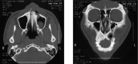
술후 연세대학교 치과병원 구강악안면외과로 의뢰되었고, 내 원 당시 좌측 중안면 부위에 약간의 종창이 관찰되었으며, 23번 협측 치근단 부위에 누공이 관찰되었고, 22, 23번 치아에 약간의 동요도와 타진시의 통증이 있었다. 파노라마 방사선 사진 소견 에서 22, 23번 치근 부위로부터 상방으로 좌측의 상악동 하방 벽 과, nasal floor의 하연까지 연장되어 있는 방사선 투과성 병소가 관찰되었다. 하방의 경계는 비교적 명확하였으나 상방의 경계는 명확하지 않은 것으로 관찰되었다(Fig. 5). 전산화 컴퓨터 단층 촬 영 결과 좌측 전방 상악골에 이전의 병소와 적출술로 인한 골파 괴 양상이 관찰되었고, 경계는 비교적 불명확하고 상악동과 개 통되어 있는 소견이 보였다(Fig. 6). 전신 마취하에 11번 치아에서 부터 25번 치아를 포함하여 전방부 부분 상악골 절제술 시행하 였는데, 상방으로는 좌측 상악동 하방벽의 일부와 하비갑개
Fig. 1. Photomicrograph showing epithelial nests composed of closely packed polyhedral epithelial cells showing nuclear pleomorphism, and spherical spaces filled with eosinophilic homogenous material are also seen. (H-E stain, ×100)
Fig. 2. Photomicrograph showing no encapsulation and partially infiltrative growth into surrounding normal bone. (H-E stain, ×40)
Fig. 3. Photomicrograph showing foreign material. (H- E stain, ×40)
Fig. 4. Photomicrograph showing nucleated cells
adjacent to ghost cells. (H-E stain, ×400)
(inferior concha)를 포함하고 외측으로는 근육의 일부를 포함하 여 수술 경계를 설정하였다. 절제 후 결손부는 obturator로 재건 하였으며, 1년 동안 주기적인 경과 관찰하였으나 재발 및 전이 소견은 관찰되지 않았다.
Ⅲ. 고 찰
치성암종 (Odontogenic carninoma)에 대한 분류는 그 증례가 적 다는 한계로 인해 분류의 어려움이 있었는데, 1971년 세계보건기 구(WHO)에서는 치성암종을 세 가지 형태로 분류하였으며, 악성 법랑아세포종 (Malignant ameloblastoma), 일차성 골내 암종 (Primary intraosseous carcinoma), 그리고 치성낭종을 포함하여 치 성 상피에서 기원한 암종으로 분류하였다. 그 후 1982년 Elzay 등
2)은 12 증례를 고찰하며 치성암종에 대해 수정 보완하였는데 치 성낭종으로부터 발생하는 암종, 법랑아세포종으로부터 발생한 암종, 그리고 새로이 (de novo) 발생한 암종으로 분류하였다. 또
한 1992년 세계보건기구(WHO) 분류에서는 석회화치성낭종을 종양으로 분류하면서 치성낭종으로부터 악성변화를 일으키는 암종 (Malignant changes in odontogenic cysts)을 치성암종 (Odontogenic carcinomas)의 한 분류에 포함하였다
3).
석회화치성낭종은 1962년 Gorlin 등에 의해 처음 기술되었고, 1972년 Fejerskov와 Krogh
4)는 석회화유령세포치성종양 (Calcifying ghost cell odontogenic tumor)라는 용어를 사용하였다. 1981년 Praetorius 등
5)은 석회화치성낭종을 세 종류의 낭종과 한 종류의 신생물로 분류하고 Dentinogenic ghost cell tumor라 명명한 바 있 다. 석회화치성낭종에서 발생한 혹은 석회화치성낭종의 한 분류 로 치성유령세포암종이란 용어를 1983년 Pinborg 등이 처음 언급 하였는데, 정식으로 문헌화되어 발표된 최초의 증례는 1985년 Ikemura 등이 발표한 증례라 할 수 있다. Ikemura 등의 증례보고 이후에 1986년 Ellis와 Shmookler 등
6)은 세 증례를 보고하며, 각화 된 유령세포 (ghost-nucleated keratinizing cells)와 연관시켜 상피성 치성유령세포 종양 (Epithelial odontogenic ghost cell tumor,
Fig. 5. Panoramic view showing relative well-defined cystic lesion on apex of #22, 23 teeth with extension to the inferior border of maxillary sinus and nasal floor.
Fig. 6. CT scans showing bony defect on left anterior maxilla with infiltrative growth
tendency to the surrounding structures.
EOGCT)이라 명명하였고, 1987년 Grodjesk 등
7)은 46세 남환의 상 악골 암종이 국소 재발과 폐로 원격전이된 증례를 보고하였는 데, 1983년 Pinborg 등이 처음 사용하였던 치성유령암종이란 용 어를 사용할 것을 주장하며, 각화석회화치성낭종 (Keratinizing and calcifying odontogenic cyst, KCOC)의 악성 부분으로 정의하였 다. 이 논문에서는 유령세포를 세포질 내 각화의 특징적인 형태 로 정의하였으며, 다양한 형태의 기질 반응 (stromal reaction)을 유발한다고 언급하였다. 1989년 Scott 등
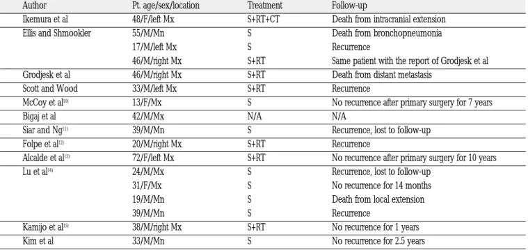
8)은 법랑아세포종으로 처 음 진단되었던 33세 남환의 증례를 보고하였는데, 조직학적 소 견의 특징 때문에 dentinogenic ghost cell ameloblastoma라 명명하 였다. 현재까지 영자문헌에 보고되었던 치성유령세포암종은 모 두 16예로 남자 대 여자의 비율은 3:1이었고, 상악골에 11예가 발 생하였으며, 평균 연령은 35.5세(13세-72세)이었다. 국소재발이나 원격 전이 된 증례는 11례로 초기에 낭종으로 진단받고 보존적 처치를 받았기 때문에 국소 재발한 증례가 많았다. 치료는 모든 증례에서 외과적 처치가 시행되었고, 방사선 치료가 행해진 증 례는 모두 6예가 있었다(Table 1). 치성유령세포암종의 조직학적 소견은 다형성 상피 세포와 유령세포가 nucleated cell들과 혼재 된 침윤성 종양의 형태로 나타나는데, 2000년 김 등
9)은 33세 남환 의 하악골에 발생한 치성유령세포암종에 대한 증례보고와 함께 유령세포와 세포소멸 과정에 대한 연관성을 언급하며, 유령세포 주위의 다형성 상피 세포와 nucleated cell들이 TUNEL assay에 양 성 반응이 나타남(nucleolar reactivity가 없을 경우에 양성 반응을 나타냄)을 관찰하고 유령세포는 분화가 덜 된 치성 세포의 세포
소멸 과정 또는 각질세포 (Keratinocyte)로의 비정상적인 terminal differentiation의 결과로 나타난 것이라 제안하였다. 본 증례는 초 기에 치성낭종으로 진단되어 적출술 및 이종골 이식을 시행받았 으나 재발하여 치성유령세포암종으로 진단된 증례로 첫 번 째 적출술 시 조직검사가 시행되지 않았기 때문에 두 번째 적출술 시의 조직검사 결과인 치성종양이 암종의 초기 기원인지 혹은 다른 병소가 초기의 기원인지에 대한 정보를 알기 어렵다는 한 계가 있다. 또한, 지금까지 이종골 이식 후의 암종으로의 진행에 대해 보고된 바는 없으나 치성유령세포암종으로 최종 진단되기 전에 두 번의 이종골 이식을 시행하였기 때문에 이러한 술식이 암종의 발생에 어떤 영향을 주었을 가능성을 배제하기 힘들다고 사료된다.
Ⅳ. 요 약
치성유령세포암종은 석회화치성낭종의 악성 부분으로 치성낭 종, 치성종양 모두에서 기인할 수 있다. 다형성 상피 세포와 유령 세포가 nucleated cell들과 혼재된 침윤성 종양의 형태로 나타나 는 것이 특징이고, 국소 재발 및 원격 전이된 증례도 보고된 바 있다. 저자들은 드문 치성종양인 치성유령세포암종을 상악골에 서 경험하였기에 문헌 고찰과 함께 보고하였으며, 본 증례의 경 우 향후 재발 및 전이 여부 파악을 위해 주기적인 경과 관찰이 필 요하리라 사료된다. 또한 악성 변화의 요인으로 이종골 이식의 영향에 대한 연구도 필요하리라 사료된다.
Table 1. Previously reported cases of odontogenic ghost cell carcinoma
Author Pt. age/sex/location Treatment Follow-up
Ikemura et al 48/F/left Mx S+RT+CT Death from intracranial extension
Ellis and Shmookler 55/M/Mn S Death from bronchopneumonia
17/M/left Mx S Recurrence
46/M/right Mx S+RT Same patient with the report of Grodjesk et al
Grodjesk et al 46/M/right Mx S+RT Death from distant metastasis
Scott and Wood 33/M/left Mx S+RT Recurrence
McCoy et al
10)13/F/Mx S No recurrence after primary surgery for 7 years
Bigaj et al 42/M/Mx N/A N/A
Siar and Ng
11)39/M/Mn S Recurrence, lost to follow-up
Folpe et al
12)20/M/right Mx S+RT Recurrence
Alcalde et al
13)72/F/left Mx S+RT No recurrence after primary surgery for 10 years
Lu et al
14)24/M/Mx S Recurrence, lost to follow-up
31/F/Mx S No recurrence for 14 months
19/M/Mn S Death from local extension
39/M/Mn S Recurrence
Kamijo et al
15)38/M/right Mx S+RT No recurrence for 1 years
Kim et al 33/M/Mn S No recurrence for 2.5 years
M=male; F=female, Mx=maxilla, Mn=mandible, S=surgery, RT=radiotherapy, CT=chemotherapy, N/A=not available
참고문헌