Ⅰ. 서 론
CGCOT는 호산성 육아세포들과 비활성의 치성상피세포들 로 이루어진 매우 드문 양성치성종양이다. Werthemann(1950) 에 의해 sponginocytic adamantinoma1)라는 이름으로 처음 보고되 었다. 그 이후 현재까지 31개의 증례보고가 있었다. CGCOT는 granulacell ameloblastic fibroma(Cough, 1962), Central granular cell tumor(White, 1978), central granular cell odontogenic fibroma(Vincent, 1987), central granular cell tumor(Chen, 1991)2)의 다 양한 이름으로 불려왔으나 1995년 이후 WHO3)에서 CGCOT로 명명되었다4).
CGCOT의 임상적 특징을 살펴보면, 남성에서보다 여성에서 호발하며(여성 75%) 평균발생연령은 46세로 주로 40대에서 70 대에서 호발한다. 악골부위별 호발부위를 보면 하악대 상악비
가 3:1정도이며 주로 소구치와 구치부에서 빈발한다고 보고되 고 있다. 대부분의 병소가 무통성의 종창을 나타내며, 국소적 인 피질골 팽창과 주의 구조물에 의한 자극으로 궤양을 동반 하기도 한다. 상악에 발생하는 경우 상악동을 침범하기도 하 며 하악의 경우에는 하악관의 변위 및 관련치아의 변위를 동 반하기도 한다2,5-7).
방사선 소견은 대부분 경화성 경계를 가진 방사선 투과성 병 소로 나타나나 때때로 방서선 투과성과 불투과성 부분적으로 혼재되어 나타나기도 한다5,7).
병리조직학적소견으로는 거대 호산성 육아세포들이 판상 혹은 섬의 형태를 이루며 이들 사이에 치성상피세포들이 산재
되어있다5,7,8). 이 치성상피세포들은 때로는 유리화2,7)되어 있는
소견을 나타내기도 한다. 또한 특이적으로 병소 내 석회화된 부위를 관찰할 수 있다2).
이 종양의 치료방법으로는 병소의 완전한 적출술 혹은 소파 술 등이 소개되고 있으며 치료 후 재발은 극히 드물며 현재까 지 1증례가 재발된 것으로 보고2)되고 있다.
이런한 임상병리학적 특징을 갖는 CGCOT는 국외에서는 31 증례가 보고되었으나 국내에서는 보고된바가 없을 정도로 드 문 종양이므로 문헌고찰과 함께 1증례를 보고하는 바이다.
김 진 욱
700-721 대구광역시 중구 삼덕2가50 경북대학교 치과대학 구강악안면외과학교실 Jin-Wook Kim
Dept. of OMFS, School of Dentistry, Kyungpook National University 50 Samduk 2-ga, Jung-gu, Daegu, 700-721, Korea
Tel: 82-53-420-5911 Fax: 82-53-426-5365 E-mail: [email protected]
상악 구치부에 발생한 Central Granular Cell Odontogenic Tumor(CGCOT)의 치험례
김진욱∙박인숙∙변기정*∙김진수
경북대학교 치과대학 구강악안면외과학교실, *울산대학교병원 치과학교실
Abstract (J. Kor. Oral Maxillofac. Surg. 2006;32:374-379)
CENTRAL GRANULAR CELL ODONTOGENIC TUMOR(CGCOT): A CASE REPORT INCLUDING LIGHT MICROSCOPY, IMMUNOHISTOCHEMISTRY AND LITERATURE REVIEW
Jin-Wook Kim, In-Suk Park, Gi-Jeong Byeon*, Chin-Soo Kim
Dept. of Oral & Maxillofacial Surgery, College of Dentistry, Kyung-Pook National University
*Dept. of Dentistry, Ulsan University Hospital
Central granular cell odontogenic tumor(CGCOT) is a very rare lesion that consists of densely packed granular cells with numerous scattered strands of odontogenic epithelium interspersed throughout the tissue. CGCOT was initially reported in 1962 by Cough et al as central granular cell ameloblastic fibroma. But, recently, this term is inappropriate because of histologic and chronologic differences.
CGCOT is usually present as painless swellings. Radiographs show a well-demarcated radiolucent or mixed radiopaque-radiolucent lesion. The average age on presentation of CGCOT is 47.3 and women are 75% more likely to develop this lesion than men. The tumor only occur in tooth bear- ing areas of the jaw with 88% of cases occurring in the mandible and 12% involving the maxilla, usually in an equal distribution between the canine- premolar-molar areas. This tumor is benign, and care is effected by localized surgical excision.
We report an additional case of CGCOT that occurred in the Rt. Maxillar premolar/molar region of a 32-year old man with literature review.
Key words: Central granular cell odontogenic tumor
Ⅱ. 증례보고
* 환자: 김 � 환, 33세 남자
* 초진일: 2003년 10월 25일
* 주소: 상악 우측 제 2소구치, 제 1대구치부위의 협측 종창 (Fig. 1)
* 병력: 본원에 내원하기 6년 전 처음 발견되었으며 처음 2년 정도 크기 변화없다가 4년 전부터 서서히 크기가 증가하 여 현재의 상태에 이름.
* 임상소견: 상악 우측 제 1, 2소구치, 제 1대구치 협측으로 단 단한 종괴가 촉진됨.
* 가족력: 특이사항 없음.
* 파노라마 소견: 상악 우측 제 2소구치, 제 1대구치 치간부 치조골의 흡수성 소견을 보임(Fig. 2)
* CT 소견: 상악 제 2소구치, 제 1대구치사이 협측 피질골의
팽창을 보이는 양성병소관찰되며 내부에 방사선 불투과 성의 foci관찰됨(Fig. 3, 4).
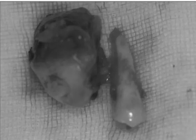
* 처치 및 경과 : 2003년 11월 5일 조직검사를 시행하고 결과상 양성치성종양으로 일차진단되었고 임상 및 방사선학적 소견으로도 양성종양으로 판단되어 2003년 12월 26일 국 소마취하에 상악 우측 제 2소구치, 제 1대구치 발치와 함 께 병소를 적출하였다. 병소는 인접조직과 용이하게 분 리되었으며 적출물의 크기는 18×19×21mm였다(Fig. 5, 6). 적출 후 지혈을 시행하고 바셀린 거즈로 드레싱하여 상피재생을 유도하였다. 2004년 1월 10일 조직검사결과 CGCOT로 진단되었으며 술 후 6개월이 지난 후에 적출부 의 전정성형술을 시행하였다.
현재 약 1년간의 정기적 관찰을 시행한 결과 재발은 나타 나고 있지 않다.
* 조직학적 소견: 호산성 육아세포들이 판상을 이루고 있으
Fig. 1. Intraoral photograph, Rt. upper 1,2 premolar and 1 molar buccal area swelling.
Fig. 3.Axial CT scan, Rt. upper maxillary buccal side lesion with thin bony wall and inner calicified material.
Fig. 4.Coronal CT scan.
Fig. 2. Panoramic view reveals alveolar bone resorption between Rt. upper second premolar and first molar.
며, 치성상피세포들이 섬모양으로 육아세포들 사이 산재 되어 있었다. 작은 석회화된 조직도 발견되었다(Fig. 7).
* 면역조직화학적 소견 : Cytokeratin이 치성상피세포에 침착되
고 Vimentin이 육아세포의 세포질에 침착됨이 관찰되었 다. S-100단백은 육아세포에 음성반응을 보였다(Fig. 8- 10).
Fig. 5.Conservative excisional margin. Fig. 6.Specimen, continued with second premolar.
Fig. 7. Tumor is composed of sheets of eosinophilic granular cells, and islands of odontogenic epithelium interspersed among the granular cells.
There are foci of small calcifications.
Fig. 8.CK (+, odontogenic epithelium, perimembra- nous staining pattern).
Fig. 9.S-100 (-, differentiates this case from granular cell tumor of soft tissue, which shows positive reaction for S-100 protein).
Fig. 10.Vimentin (+, granular cell, cytoplasmic staining pattern).
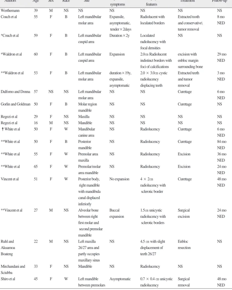
Table 1.Cenetral granular cell odontogenic tumors
Authors Age Sex Race Site Signs and Radiographic
Treatment Follow-up
symptoms features
Werthemann 39 M NS NS NS NS NS NS
Couch et al 55 F B Left mandibular Expansile, Radiolucent with Extracted tooth 8 mo
molar area asymptomatic, loculated borders and conservative\ NED
tender×2days tumor removal
*Couch et al 59 F B Left mandibular Duration×2y Loculated NS NS
cuspid area radiolucency with
focal densities
*Waldron et al 60 F B Left mandibular Expansion 2.0㎝ Radiolucent excision with 29 mo
cuspid area indistinct borders with enbloc margin NED
foci of calcifications surrounding bone
**Waldron et al 53 F B Left mandibular duration×19y, 2.0 × 3.0㎝ cystic Extracted teeth 3 mo
molar area expansile, radiolucency and tumor NED
asymptomatic displacing teeth removal
Dalforno and Donna 57 NS NS Left mandibular NS NS Curettage 6 mo
molar area NED
Gorlin and Goldman 50 F B Molar region NS NS Curettage NS
mandible
Regezi et al 29 F NS Maxilla NS NS NS NS
Regezi et al 16 M NS Mandible NS NS NS NS
�White et al 50 F W Mandibular NS Radiolucency Curettage 6 mo
canine area NED
**White et al 50 F B Posterior NS Radiolucency Curettage 84 mo
mandible NED
**White et al 55 F W Premolar area NS Radiolucency Excision 36 mo
maxilla NED
**White et al 65 F W Premolar/molar NS Radiolucency Excision 24 mo
area mandible NED
Vincent et al 51 F W Posterior body, No expansion 4 × 2㎝ Curettage 48 mo
right mandible radiolucency with NED
with mandibula sclerotic border
canal displaced inferiorly
**Vincent et al 27 M NS Alveolar bone Buccal 1.5㎝ unicystic Surgical 24 mo
between right expansion radiolucency with excision NED
first molar and sclerotic borders
second premolar mandible
Ruhl and 22 M NS Left maxilla NS 4.5 ㎝ with slight Enbloc NS
Akuamoa 26/27 area and displacement of resection
Boateng partly occupies teeth 26/27
maxillary sinus
Mirchandani and 33 F NS Mandible NS Radiolucency NS NS
Sciubba
Shiro et al 45 F W Left mandible Asymptomatic 0.7 × 0.4 ㎝ unicystic Surgical 48 mo
between premolars radiolucency removal NED
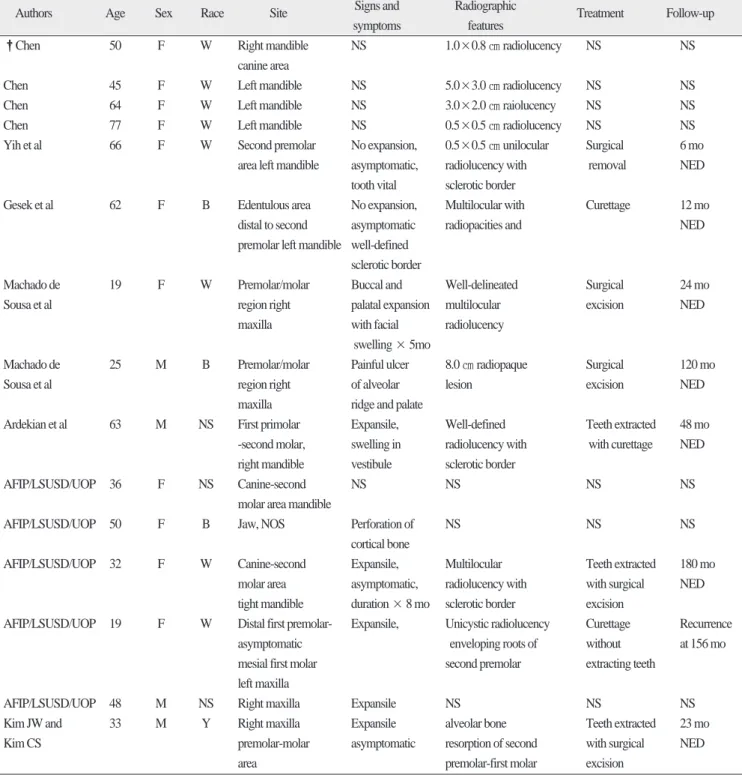
Table 2.continued
Authors Age Sex Race Site Signs and Radiographic
Treatment Follow-up
symptoms features
�Chen 50 F W Right mandible NS 1.0×0.8 ㎝ radiolucency NS NS
canine area
Chen 45 F W Left mandible NS 5.0×3.0 ㎝ radiolucency NS NS
Chen 64 F W Left mandible NS 3.0×2.0 ㎝ raiolucency NS NS
Chen 77 F W Left mandible NS 0.5×0.5 ㎝ radiolucency NS NS
Yih et al 66 F W Second premolar No expansion, 0.5×0.5 ㎝ unilocular Surgical 6 mo
area left mandible asymptomatic, radiolucency with removal NED tooth vital sclerotic border
Gesek et al 62 F B Edentulous area No expansion, Multilocular with Curettage 12 mo
distal to second asymptomatic radiopacities and NED
premolar left mandible well-defined sclerotic border
Machado de 19 F W Premolar/molar Buccal and Well-delineated Surgical 24 mo
Sousa et al region right palatal expansion multilocular excision NED
maxilla with facial radiolucency
swelling × 5mo
Machado de 25 M B Premolar/molar Painful ulcer 8.0 ㎝ radiopaque Surgical 120 mo
Sousa et al region right of alveolar lesion excision NED
maxilla ridge and palate
Ardekian et al 63 M NS First primolar Expansile, Well-defined Teeth extracted 48 mo
-second molar, swelling in radiolucency with with curettage NED right mandible vestibule sclerotic border
AFIP/LSUSD/UOP 36 F NS Canine-second NS NS NS NS
molar area mandible
AFIP/LSUSD/UOP 50 F B Jaw, NOS Perforation of NS NS NS
cortical bone
AFIP/LSUSD/UOP 32 F W Canine-second Expansile, Multilocular Teeth extracted 180 mo
molar area asymptomatic, radiolucency with with surgical NED tight mandible duration × 8 mo sclerotic border excision
AFIP/LSUSD/UOP 19 F W Distal first premolar- Expansile, Unicystic radiolucency Curettage Recurrence
asymptomatic enveloping roots of without at 156 mo
mesial first molar second premolar extracting teeth left maxilla
AFIP/LSUSD/UOP 48 M NS Right maxilla Expansile NS NS NS
Kim JW and 33 M Y Right maxilla Expansile alveolar bone Teeth extracted 23 mo
Kim CS premolar-molar asymptomatic resorption of second with surgical NED
area premolar-first molar excision
*same patient
**AFIP accessioned cases
�same patient NS, not stated
NED, no evidence of disease NOS, not otherwise specific
AFIP: Armed Forced Institute of Pathology, Washinton, DC LSU: Louisiana State University School of Dentistry, New Orleans UOP: University of Pacific School of Dentistry, San Francisco”
Ⅲ. 총괄 및 고찰
Werthemann1)에 의해 sponginocytic adamantinoma란 이름으로 처음 보고된 이후 현재의 CGCOT란 이름으로 정착되기 까지 그 임상병리학적 소견으로 인해 이 질환을 명명하는데 있어 많은 혼란이 있었다. 그 대표적인 명칭이 granular cell odonto- genic fibroma9)와 악골의“granular cell tumor”10)이다.
Odontogenic fibroma, ameloblastic fibroma와 CGCOT와의 감별 되는 점은 이 종양은 대부분이 mesenchymal origin이라는 점에 서 다르다. Odontogenic fibroma와 임상적으로 유사한 연령대에 서 발생하고, stellate reticulum lend가 없는 상피세포를 가진다는 점에서 CGCOT는 odontogenic fibroma의 한 변종이라 생각되어 졌다. 그리고 granular cell tumor와는 달리 백악질과 유사한 석회 화조직이 나타난다는 점에서 central granular cell odontogenic fibroma라 불려졌다2). 그러나 Gardner11)는 odontogenic fibroma의 특징은 cell rich fibroblastic stroma에 있다고 하며 CGCOT에는 이 러한 특징이 없는 것을 들며 이 두 가지는 구별되는 병변이라 고 주장하였다7).
White 등10)은 granular cell의 origin은 mesenchym이라 하였다. 이 들은 육아세포의 세포질에서 다수의 lysosomal like granule을 발 견하였다. Chen 등12)primary lysosome과 autophagic vacuole들을 발견하였고 granular cell이 Langerhans cell의 antimarker인 OKT6(CD1) antigen에 양성반응을 보이는 것을 근거로 하고 Langerhans cell origin일 것이라 보고하였다. 그러나 Chen은 Langerhans cell의 ulatrstructural marker인 Birbeck granule을 언급하 지 못했다2).
면역조직화학적 연구가 계속되면서 육아세포가 mesenchy- mal nature를 가지는 세포에 특이성을 보이는 vimentin에 양성반 응9)을 보이며, epithelial differentiation cell 의 marking에 사용되는 cytokeratin에 음성반응8)을 보임이 확인되어 CGCOT의 육아세 포는 mesenchymal origin임을 알게 되었다. 또한 Cytokeratin은 치 성상피에는 양성반응을 보이고, 보통의 육아세포 종양에서 양 성반응을 보이는 S-100 단백5,8,13)에는 음성반응을 나타내어 CGCOT는 odontogenic origin2)으로 받아들여지고 있다. 이와 같 은 면역조직화학적 검사는 CGCOT의 진단에 중요한 근거가 된다. Lysosomal granule들은 육아세포내부의 다양한 변화 형태 라고 생각되고 있으며, phagocytic activity는 종양세포가 histocyt- ic cell line에서 유래된 것임을 뒷받침한다7).
CGCOT는 그 임상적, 조직학적 소견상 odontogenic tumor이며 granular cell은 histocytic cell lineage를 가지는 mesenchymal origin 으로 생각된다.
CGCOT는 현재까지 재발은 극히 드물며 그 경과가 매우 좋 은 것으로 보고되고 있어 보존적인 절제술이 추천된다.
Ⅳ. 결 론
경북대학교 구강악안면외과에 상악 우측 제 2소구치, 제1 대 구치부위의 무통성 종창을 주소로 내원한 33세 남자 환자에서 관련치아의 발치를 포함한 병소의 절제술을 시행하였다. 조직 검사결과 치밀하게 응집된 판상의 거대 호산성 육아세포들 사 이에 섬상의 치성상피세포가 산재되어있고 백악질과 유사한 석회화된 조직들이 관찰되었다. 면역조직화학적 검사 결과 치 성상피는 CK에 양성반응을 보였고, 육아세포는 vimentin에 양 성, CK와 S-100단백에 음성반응을 보여 CGCOT로 진단되었다.
술 후 정기적 관찰을 시행한 결과 현재까지 재발 등의 합병 증은 없었으며 결손치의 보철적 수복계획에 있다.
참고문헌
1. Werthemann A: Uber spongiozytares Adamantinom. Oncologia 1950;3:193-207.
2. Brannon RB, Goode RK, Eversole LR, et al.: The central granular cell odontogenic tumor: Report of 5 new cases. Oral Surg Oral Med Oral Pathol Oral Rdiol Endod 2002;94:614-21.
3. Kramer IRH, Pindborg JJ, Shear M: Histological typing of odonto- genic tumors(ed2). Heidelberg, Germany, Springer-verlag 19924.
Machado de Sousa SO, Soares de Araujo N, Melhado RM, Cavalcanti de Araujo V. Central odontogenic granular cell tumor: immunohisto- chemical study of two cases. J Oral Maxillofac Surg 1998;56:787-91.
5. Gesek DJ JR, Adrian JC, Reid EN: Central odontogenic granular cell tumor : a case report including light microscopy, immunohistochem- istry, and literature review. J Oral Maxillofac Surg 1995;53:945-9.
6. Ardekian L, Manor R, Gaspar R, Laufer D: Central granular cell odontgenic tumor: case report and review of literature. J Oral Maxillofac Surg 1998;56:1343-5.
7. Meer S, Altini M, Coleman H, Daya N: Central granular cell odonto- genic tumor: Immunohistochemistry and ultrastructure. Am J Otolaryngol 2004;25;73-8.
8. Yih WY, Thompson C, Meshul CK, Bartley MH: Central odonto- genic granular cell tumor of the jaw: report of case and immunohisto- chemical and electron microscopic study. J Oral Maxillofac Surg 1995;53:453-9.
9. Vincent SD, Hammond HL, Ellis GL, Juhlin JP: Central granular cell odontogenic fibroma. Oral Surg Oral Med Oral Pathol 1987;63:715- 21.
10. White DK, Chen SY, Hartman KS, Miller AS, Gomez LF: Central granular cell odontogenic tumor of the jaw(the so-called granular cell ameloblastic fibroma). Oral Surg Oral Med Oral Pathol 1978;45:396- 405.
11. Gardner DG: Central odontogenic fibroma current concepts. J Oral Pathol Med 1989;25:556-61.
12. Chen SY: Central granular cell tumor of the jaw. An electron micro- scopic and immunohistochemical study. Oral Surg Oral Med Oral Pathol 1991;72:75-81.
13. Ruhl GH, Akuamoa-Boateng E: Grnular cells in odontogenic and non odontogenic fibroma. Virchows Arch A Pathol Anat Histopathol 1989;415:403-9.