Odontogenic fibroma (ODF) originates from odonto- genic ectomesenchyme. ODF can be further divided into central (intraosseous) odontogenic fibroma (CODF) and peripheral (extraosseous) odontogenic fibroma (PODF) according to the anatomical sites involved.1
The CODF is a rare odontogenic neoplasm. Regezi et al2were unable to identify any ODFs among 706 odonto- genic tumors retrieved from 54,534 oral specimens. Han- ders et al3 identified only 19 CODFs from over 80,000 accessioned oral specimens. In 40,000 consecutive oral biopsies from a Canadian population, there were only 25 CODFs (0.06%) and 36 PODFs (0.09%).4 Buchner et al5 have shown 23 PODFs (0.02%) and 16 CODFs (0.02%) among 91,178 oral lesions. Recently Lin et al6reported 15 cases of ODFs including 3 cases of central type. Although PODF is relatively rare, it is the most common peripheral odontogenic tumor (51%, 23/45).7 Moreover, PODF is more common than its central counterpart by a ratio of 1.4 : 1.7
Here, a case of CODF is presented because due to its ex- tensive bone destruction with partially ill-defined margin, differential diagnosis involved a wide range of pathosis.
Case report
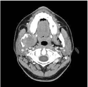
A 17-year-old male was referred to our hospital for eva- luation of a painless swelling on the right cheek, which had been identified 3 or 4 months earlier. His medical and surgical history was noncontributory. The extraoral exami- nation showed swelling on the right cheek causing some facial asymmetry (Fig. 1). The intraoral examination re- vealed a slight swelling on the right mandiblular ramus region, extending to the right retromolar area (Fig. 2). The mass was firm with normal color and no fluctuation was evident on palpation. There was no associated cervical lymphadenopathy. The panoramic radiographic examina- tion showed the presence of a multilocular radiolucency with partially ill-defined border between the right mandi- bular condyle and the distal root of the right mandibular third molar. Since the radiolucency was not associated with the crown of the impacted third molar and seemed to involve the mandibular foramen, the first radiologic im- pression involved neural origin tumor along with more ag- gressive lesions such as desmoplastic fibroma, juvenile ag- gressive fibromatosis, or fibrosarcoma (Fig. 3). CT exami- nation showed an expansile solid ovoid slightly enhancing mass involving the right mandibular condyle and ramus, measuring around 3.5×3.2 cm giving the impression of an ameloblastoma (Figs. 4-6). Microscopic examination revealed a non-encapsulated tumor consisting of bland
─ 85 ─
Central odontogenic fibroma: a case report
Kyung-Soo Nah
Department of Oral and Maxillofacial Radiology, School of Dentistry, Pusan National University, Busan, Korea ABSTRACT
Central odontogenic fibroma is a rare odontogenic neoplasm that originates from odontogenic ectomesenchyme.
Here, a case of central odontogenic fibroma in a 17-year-old male is reported. Since the present case showed a mul- tilocular radiolucency with partially ill-defined border between the right mandibular condyle and the distal root of the right mandibular third molar, differential diagnosis involved a wide range of pathosis from benign lesions like ameo- loblastic fibroma and odontogenic myxoma to more aggressive lesions such as desmoplastic fibroma, juvenile aggres- sive fibromatosis, or fibrosarcoma. (Imaging Sci Dent 2011; 41 : 85-8)
KEY WORDS : Fibroma; Odontogenic Tumors; Radiography; Diagnosis
*This work was supported by a 2-Year Research Grant of Pusan National University.
Received March 31, 2011; Revised May 13, 2011; Accepted May 20, 2011 Correspondence to : Prof. Kyung-Soo Nah
Department of Oral and Maxillofacial Radiology, School of Dentistry, Pusan Nation- al University, Busan 626-810, Korea
Tel) 82-55-360-5260, Fax) 82-55-360-5029, E-mail) [email protected]
Imaging Science in Dentistry 2011; 41 : 85-8 DOI: 10.5624/isd.2011.41.2.85
Copyright ⓒ 2011 by Korean Academy of Oral and Maxillofacial Radiology
This is an Open Access article distributed under the terms of the Creative Commons Attribution Non-Commercial License (http://creativecommons.org/licenses/by-nc/3.0) which permits unrestricted non-commercial use, distribution, and reproduction in any medium, provided the original work is properly cited.
Imaging Science in Dentistry∙pISSN 2233-7822 eISSN 2233-7830
fibroblast cells with wavy cytoplasm. A few strands and nests of odontogenic epithelium were observed (Figs. 7 and 8). Based on the clinical, radiographic, and histopathologic findings, the definitive diagnosis was CODF. The patient showed no clinical signs of recurrence 2 years after surgi- cal excision.
Discussion
According to Covani et al,8the CODF was believed to originate from mesenchymal odontogenic tissue such as
dental papilla, periodontal ligament, or dental follicle.
Considering the histogenesis of the lesion in the WHO classification,9it has been suggested that the epithelium- poor type of CODF is derived from the dental follicle, whereas the epithelium-rich type arises from the periodon- tal ligament.
─ 86 ─ Central odontogenic fibroma: a case report
Fig. 1.Extraoral photograph shows a swelling on the right cheek causing facial asymmetry.
Fig. 2.Intraoral photograph shows normal-colored mucosal right cheek swelling extending to the retromolar area.
Fig. 3.Cropped panoramic image shows a multilocular radiolu- cency with partially ill-defined border between the right mandibu- lar condyle and the distal root of the right mandibular third molar.
Fig. 4.Axial CT image shows an expansile solid ovoid mass involv- ing the right mandibular condyle and ramus measuring around 3.5
×3.2 cm.
The peak incidence of CODFs have been observed in the second decade of life and shows a female predilection.10 This lesion was originally thought to occur almost exclu- sively in the mandible, and was mostly found in the pos- terior mandible.11Clinically, CODF manifests as a painless swelling and has a slow growth that results in cortical ex- pansion.10,11 Clinical symptoms such as pain and pares- thesia are uncommon.12
Radiologically, most cases of CODFs are radiolucent, unilocular, and with well-defined borders similar to other
radiolucent lesions such as traumatic bone cyst, amelo- blastoma, odontogenic cysts, and central giant cell granu- lomas.13Araki et al14reported that the diagnosis of CODF by radiographic findings was extremely difficult, espe- cially when the lesion was associated with a crown of the unerupted tooth that might resemble a dentigerous cyst.
Since the present case showed a multilocular radiolucent lesion involving ramus and even condylar process with partially ill-defined borders, the differential diagnosis involved a wide range of pathosis from benign lesions such as ameoloblastic fibroma, odontogenic myxoma to more aggressive lesions such as desmoplastic fibroma, juvenile aggressive fibromatosis or fibrosarcoma. Most of the CODF cases reported3,6,8,12were small and associated with
─ 87 ─
Kyung-Soo Nah
Fig. 5.Axial CT image shows slight enhancement of the lesion.
Fig. 6.Coronal CT image shows massive bone resorption of the right mandibular condylar process and ramus.
Fig. 7.Photomicrograph reveals a non-encapsulated tumor consist- ing of bland fibroblast cells. A few strands and nests of odontoge- nic epithelium is observed (H&E stain, ×100).
Fig. 8.Photomicrograph reveals bland fibroblast cells with wavy cytoplasm (H&E stain, ×400).
roots of teeth. The CODF case reported by Daniels10was somewhat bigger in size, however it resembled a dentiger- ous cyst. Handers et al3 reported that there were only two mandibular lesions among the 19 CODF cases, and one of the mandibular lesions with a large multilocular radio- lucency involving most of the ramus was similar to this case except for the fact that the margin was scalloped and showed well-defined buccal cortical expansion suggesting benign nature of the lesion. Kaffe and Buchner13 stated that the majority of CODFs were unilocular radiolucent lesions with well-defined border, however they might also appear as multilocular lesions and might exhibit a mixed radiolucent-radiopaque appearance with poorly defined or diffused borders in rare cases. The great variability in radiologic appearance of the CODFs means that it should be considered in the differential diagnosis of all radiolu- cencies found in the jaws.
References
1. Neville BW, Damm DD, Allen CM, Bouquot JE. Oral and maxillofacial pathology. 2nd ed. Philadelphia: WB Saunders;
2002. p. 633-5.
2. Regezi JA, Kerr DA, Courtney RM. Odontogenic tumors;
analysis of 706 cases. J Oral Surg 1978; 36 : 771-8.
3. Handlers JP, Abrams AM, Melrose RJ, Danforth R. Central odontogenic fibroma: clinicopathologic features of 19 cases and review of the literature. J Oral Maxillofac Surg 1991; 49 : 46-54.
4. Daley TD, Wysocki GP, Pringle GA. Relative incidence of odontogenic tumors and oral and jaw cysts in a Canadian po-
pulation. Oral Surg Oral Med Oral Pathol 1994; 77 : 276-80.
5. Buchner A, Merrell PW, Carpenter WM. Relative frequency of central odontogenic tumors: a study of 1,088 cases from Northern California and comparison to studies from other parts of the world. J Oral Maxillofac Surg 2006; 64 : 1343-52.
6. Lin HP, Chen HM, Vu CH, Yang H, Kuo RC, Kuo YS, et al.
Odontogenic fibroma: a clinicopathological study of 15 cases.
J Formos Med Assoc 2011; 110 : 27-35.
7. Buchner A, Merrell PW, Carpenter WM. Relative frequency of peripheral odontogenic tumors: a study of 45 new cases and comparison with studies from the literature. J Oral Pathol Med 2006; 35 : 385-91.
8. Covani U, Crespi R, Perrini N, Barone A. Central odontogenic fibroma: a case report. Med Oral Patol Oral Cir Bucal 2005;
10(Suppl 2) : E154-7.
9. Philipsen HP, Reichart PA, Sciubba JJ, van der Waal I. Odon- togenic fibroma. In: Barnes L, Eveson JW, Reichart PA, Sid- ransky D. World Health Organization classification of tumours.
Pathology and genetics of tumours of head and neck tumours.
Lyon: IARC; 2005. p. 317.
10. Daniels JS. Central odontogenic fibroma of mandible: a case report and review of the literature. Oral Surg Oral Med Oral Pathol Oral Radiol Endod 2004; 98 : 295-300.
11. Gardner DG. Central odontogenic fibroma current concepts. J Oral Pathol Med 1996; 25 : 556-61.
12. Daskala I, Kalyvas D, Kolokoudias M, Vlachodimitropoulos D, Alexandridis C. Central odontogenic fibroma of the mandi- ble: a case report. J Oral Sci 2009; 51 : 457-61.
13. Kaffe I, Buchner A. Radiologic features of central odontogenic fibroma. Oral Surg Oral Med Oral Pathol 1994; 78 : 811-8.
14. Araki M, Nishimura S, Matsumoto N, Ohnishi M, Ohki H, Komiyama K. Central odontogenic fibroma with osteoid for- mation showing atypical radiographic appearance. Dentomaxil- lofac Radiol 2009; 38 : 426-30.
─ 88 ─ Central odontogenic fibroma: a case report