Imaging Science in Dentistry 2015; 45: 109-15 http://dx.doi.org/10.5624/isd.2015.45.2.109
Central odontogenic fibroma(COF) is an extremely rare benign ectomesenchymal neoplasm that accounts for 0.1%
of all odontogenic tumors.1-4 It has two major types: (1) the simple type and (2) the World Health Organization (WHO) type.3,5,6
This tumor appears in both the mandible and maxilla(55
% and 45%, respectively). It has been reported to occur in a wide age group with a strong female predilection.6-8
Radiographically, COF is usually radiolucent and some- times can have a mixed radiolucent-radiopaque appear- ance. Most COFs are well-defined unilocular lesions, but they can be multilocular or have poorly-defined borders in rare cases.1-3,9
Periosteal reaction occurs when cortical bone reacts to one of a range of possible stimulants.10 A Codman trian- gle develops when a portion of the periosteum is lifted off of the cortex by a tumor, pus, or hemorrhage at a leading edge. This aggressive form of periosteal reaction is com- monly seen in osteosarcomas and occasionally in infec-
tion and metastases.11 The current literature contains no report of periosteal reaction accompanying COF.
The aim of this report is to present a case of COF in the mandibular right molar region of a four-year-old boy that had a very rare unusual radiographic and cone-beam com- puted tomography(CBCT) appearance with periosteal reaction.
Case Report
A four-year-old boy was referred to the Oral and Maxil- lofacial Radiology Department at the School of Dentistry of the University of Mashhad. His chief complaint was a swelling in the right inferior border of the lower jaw, which had been present for one week and exhibited slow growth. He reported no pain or other symptoms. His par- ents had incidentally noticed the swelling. He had visited a general dentist, the lesion was initially diagnosed as a dentoalveolar abscess, and the patient was referred to the Oral and Maxillofacial Radiology Department for further investigation. His medical and familial history was unre- markable.
On the extraoral and intraoral examinations, no trismus, lymphadenopathy, paresthesia, or mucosal changes were
Central odontogenic fibroma (simple type) in a four-year-old boy: atypical cone-beam computed tomographic appearance with periosteal reaction
Najme Anbiaee1, Hamed Ebrahimnejad1,*, Alireza Sanaei1
1Department of Oral and Maxillofacial Radiology, Maxillofacial Diseases Research Center, School of Dentistry, Mashhad University of Medical Sciences, Mashhad, Iran
AbstRACt
Central odontogenic fibroma(COF) is a rare benign tumor that accounts for 0.1% of all odontogenic tumors. A case of COF(simple type) of the mandible in a four-year-old boy is described in this report. The patient showed asymptomatic swelling in the right inferior border of the lower jaw for one week. A panoramic radiograph showed a poorly-defined destructive unilocular radiolucent area. Cone-beam computed tomography showed expansion and perforation of the adjacent cortical bone plates. A periosteal reaction with the Codman triangle pattern was clearly visible in the buccal cortex. Since the tumor had destroyed a considerable amount of bone, surgical resection was performed. No recurrence was noted.(Imaging Sci Dent 2015; 45: 109-15)
Key woRds: Fibroma; Odontogenic Tumors; Cone-Beam Computed Tomography
Copyright ⓒ 2015 by Korean Academy of Oral and Maxillofacial Radiology
This is an Open Access article distributed under the terms of the Creative Commons Attribution Non-Commercial License(http://creativecommons.org/licenses/by-nc/3.0) which permits unrestricted non-commercial use, distribution, and reproduction in any medium, provided the original work is properly cited.
Imaging Science in Dentistry·pISSN 2233-7822 eISSN 2233-7830 Received January 31, 2015; Revised April 5, 2015; Accepted April 12, 2015
*Correspondence to : Dr. Hamed Ebrahimnejad
Oral and Maxillofacial Radiology Department, Faculty of Dentistry, Mashhad Uni- versity of Medical Sciences, PO Box: 91735-984, Mashhad, Iran
Tel) 98-51-38829501, Fax) 98-51-38829500, E-mail) [email protected]
*표 선 두께
=
1/0.3ptobserved. No dental caries were present in the right man- dibular quadrant. The expansion involved an area measur- ing approximately 3cm×2cm in the right inferior border of the lower jaw anterior to the mandibular angle. It had a non-ulcerated smooth surface. On palpation, the swelling was firm in consistency and non-tender.
A panoramic radiograph showed a poorly-defined de- structive expansive radiolucent area in the posterior body of the mandible near the angle region. The internal pat- tern was unilocular. The lesion had perforated the inferior cortical border of the mandible. Thinning of the cortical outlines of the follicles of the first and second permanent
Fig. 1. A panoramic radiograph shows a poorly-defined radiolucent lesion in the posterior mandibular region, perforating the inferior man- dibular cortex.
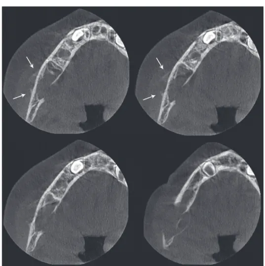
Fig. 2. Axial cone-beam computed tomography images show lingual expansion and perforation of the buccal and lingual cortical plates.
Note the periosteal reaction(Cod- man triangle) in the buccal cortex (arrows).
molars was observed(Fig. 1).
CBCT was performed for further evaluation. On the ax- ial view, lingual expansion and perforation of buccal and lingual cortical plates with some faint septae were seen. A periosteal reaction(Codman triangle) was clearly visible in the buccal cortex(Fig. 2).
The panoramic and cross-sectional reformatted CBCT images also confirmed the previous findings. They showed a poorly-defined multilocular radiolucent lesion with some wispy septae. Periosteal new bone formation was seen in the buccal aspect of the lesion. The buccal, lingual, and inferior cortical plates of the mandible were perforated.
No tooth displacement was observed, but the cortical out-
lines of the follicles of the first and second permanent mol- ars were eroded and perforated. The cortical border of the inferior alveolar nerve canal could not be seen throughout the lesion and seemed to be destructed(Fig. 3).
An incisional biopsy was performed through an extra- oral approach. Since the patient was uncooperative, the biopsy was performed under general anesthesia. Gross examination of the biopsy specimen showed several soft tissue pieces that jointly measured approximately 25mm
×15mm×8mm, whitish in color and elastic in consis- tency.
Histopathological examination of the incisional biop-
Fig. 4. Plump fibroblasts(arrow) within a collagenous background.
No epithelial remnants are found(H&E stain, 100×)
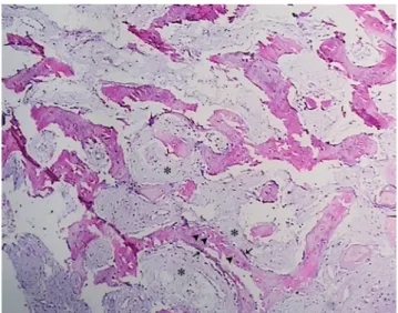
Fig. 5. Fibrous stroma with fibroblastic cells. Some myxoid chang- es(asterisks) in the stroma, normal bone trabecules containing osteocyte lacunae(arrowheads), and osteoblastic margins(arrows) are seen(H&E stain, 100×)
Fig. 3. Panoramic (A) and cross-sectional (B) reformatted cone-beam computed tomography images show a multilocular lesion with some faint septae. The lesion has perforated the lingual, buccal, and inferior mandibular cortical plates, the cortical outlines of the follicles of the molars, and the inferior alveolar nerve canal. Periosteal new bone formation is seen in the buccal cortical plate(arrows).
A B
sy specimen of the lesion revealed a non-encapsulated tumor consisting of a benign neoplastic proliferation of odontogenic ectomesenchymal tissue. The biopsy showed fibrous connective tissue stroma with plump fibroblastic cells alongside collagen bundles. No odontogenic epithe- lial cells were found in the sections. Some myxoid chang- es in the stroma and normal bone trabecules containing osteocyte lacunae and osteoblastic margins were seen (Figs. 4 and 5). A diagnosis of COF(simple type) was es- tablished.
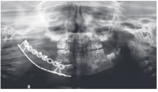
Since the tumor had destroyed a considerable amount of bone, surgical resection and bone grafting were perform- ed(Fig. 6). The surgical procedure consisted of segmental resection of the mandible with immediate bone grafting reconstruction via the intraoral route. The autogenous bone
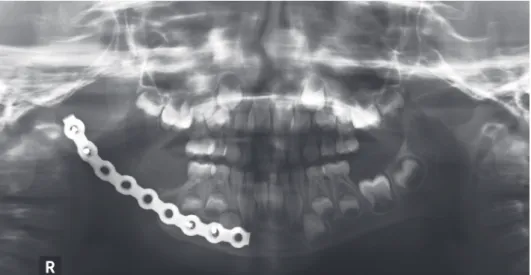
was harvested from the iliac crest. The histopathology of the postoperative tissue was consistent with the incisional biopsy diagnosis. Following the resection, the patient was maintained on systemic antibiotics but this failed to pre- vent infection. The graft site was infected after one month and the infected bone graft was removed(Fig. 7). Graft failure was most likely due to wound dehiscence and bone exposure. A six-month follow-up panoramic radio- graph showed that the patient’s condition was stable, with no recurrence(Fig. 8).
discussion
COF is classified as a rare benign tumor that originates from ectomesenchymal tissues such as the dental papillae,
Fig. 6. Postoperative panoramic radiograph.
Fig. 7. One-month follow-up radio- graph. The infected bone graft had been removed.
periodontal ligament, or dental follicle.3,12,13 It is most often found in females, and the ratio of incidence in the maxilla and mandible is approximately 1:1, with a slight tendency to favor the mandible. It involves the anterior part of the maxilla, whereas mandibular lesions affect the premolar and molar areas.6,14-17
COF can be subclassified according to histological fea tures. The simple type(the epithelium-poor type) is com- posed of collagen bundles interspersed with plump fibro- blasts. Small nests or islands of inactive odontogenic epithelium may or may not be present. Foci of dystrophic calcification may be seen. Some believe that this lesion belongs to the spectrum of odontogenic myxoma and should be classified as a myxofibroma. The WHO type (the epithelium-rich type) has a more complex pattern and contains long strands or isolated nests of odontogenic epithelial rests. It may have dysplastic dentin, osteoid, or cementum-like calcifications.3,5,6,14-16,18,19 Our case resem- bled the simple type.
COF is usually asymptomatic, involving slow expan- sion of the cortical bones.6,14,20,21 Our case occurred in the mandible of a four-year-old boy and manifested as an asymptomatic firm swelling.
Radiographically, COF is mostly unilocular with well- defined borders. Larger lesions show multilocular radio- lucency, with radiopaque areas sometimes seen in the interior of the tumor. In rare cases, COF can be poorly defined. Aggressive types can cause root resorption or displace teeth.1-3,9,14 On CT and CBCT scans, COF usu- ally presents as an expansile homogenous mass that can erode and perforate the adjacent cortical boundaries. Thin and straight septae can also be noted in some lesions.9,22-25 Less common CT findings include calcified materials
within the lesion, a diffuse sclerotic border, and peripheral osteosclerosis.26,27 The present case demonstrated a very unique malignancy-like radiographic appearance of COF with periosteal reaction.
Periosteal reaction occurs when cortical bone reacts to one of several possible underlying insults.10 In the jaws, periosteal reaction can occur in patients with inflammato- ry lesions, osteomyelitis, and malignant tumors, but peri- osteal reactions rarely occur in cases of benign lesions, such as eosinophilic granuloma and osteoid osteoma.
Periosteal reaction is more common in younger people and in patients with sarcomas compared to carcinomas.
Usually, a single-layer or multi-layered(lamellated or on- ion-skin) pattern appears in osteomyelitis and benign le- sions, whereas an spiculated, sunray, or Codman triangle appearance is mostly seen in malignant tumors.28,29
The imaging presentation of periosteal reaction is cha- racterized by the intensity, aggressiveness, and duration of the underlying etiologic factor. With slow-growing lesions, the periosteum has enough time to respond to the patho- logical process, resulting in a solid, continuous periosteal reaction. In rapidly growing bone lesions, the periosteum is unable to produce new bone as fast as the growing pa- thology. Consequently, a discontinuous pattern of perios- teal reaction is seen.11
If the periosteum is significantly elevated, it can break, forming an acute angle(Codman triangle). This is usual- ly seen in malignant bone tumors such as osteosarcoma, Ewing’s sarcoma, fibrosarcoma, juxtacortical chondrosar- coma, malignant fibrous histiocytoma, metastatic tumors, and in some other rapidly growing lesions such as aneu- rysmal bone cyst, giant cell tumor, or in reactive process- es(osteomyelitis and subperiosteal hematoma).10,11,30-32 To
Fig. 8. A six-month follow-up pa- noramic radiograph shows the pa- tient’s stable condition with no re- currence.
the best of our knowledge, periosteal reaction accompany- ing COF has not been previously reported. In the present case, the young age of the patient and rapid tumor growth led to a periosteal reaction. It seems that periosteal reac- tion is more closely related to the age of the patient and duration of the lesion than to the etiology of the tumor.
The radiographic differential diagnosis includes malig- nant tumors, such as osteosarcoma and Ewing’s sarcoma.
Osteosarcoma can exhibit a Codman triangle appearance and can be radiolucent with some initial ossification chan- ges, resembling the septae seen in our case. Ewing’s sar- coma occurs in young patients and can induce a periosteal reaction. Aggressive benign lesions, such as aneurysmal bone cyst and central giant cell granuloma, can also be included in the differential diagnosis list. In the jaws, an- eurysmal bone cyst can cause expansion, and it frequently occurs in the same region as our patient’s lesion. Further- more, both aneurysmal bone cyst and central giant cell granuloma can have a multilocular appearance with some wispy septae.30
The treatment of COF is enucleation and efficient cur- ettage. Recurrence is not common.6,14 Since the tumor in our case showed aggressive behavior, resection of the lesion and installation of a bone graft were performed.
In conclusion, the purpose of presenting this case was to describe an extremely rare manifestation of central odontogenic fibroma(simple type) in a four-year-old boy.
Periosteal reaction with a Codman triangle pattern is very rare in benign tumors, and the present case is the first case of COF reported to occur with a periosteal reaction. It can be concluded that aggressive benign lesions are capable of inducing periosteal reaction in children.
References
1. Daniels JS. Central odontogenic fibroma of mandible: a case report and review of the literature. Oral Surg Oral Med Oral Pathol Oral Radiol Endod 2004; 98: 295-300.
2. Kaffe I, Buchner A. Radiologic features of central odontogen- ic fibroma. Oral Surg Oral Med Oral Pathol 1994; 78: 811-8.
3. Covani U, Crespi R, Perrini N, Barone A. Central odontogenic fibroma: a case report. Med Oral Patol Oral Cir Bucal 2005;
10 Suppl 2: E154-7.
4. Cicconetti A, Bartoli A, Tallarico M, Maggiani F, Santaniello S.
Central odontogenic fibroma interesting the maxillary sinus. A case report and literature survey. Minerva Stomatol 2006; 55:
229-39.
5. Barnes L, Eveson JW, Reichart P, Sidransky D. World Health Organization classification of tumours. Pathology and genet- ics: head and neck tumours. Lyon: IARC Press; 2005.
6. Waldron CA. Odontogenic cysts and tumots. In: Neville BW,
Damm DD, Allen CM, Bouquot J. Oral and maxillofacial pa- thology. 3rd ed. St. Louis: Saunders Elsevier; 2009. p. 726-7.
7. Daskala I, Kalyvas D, Kolokoudias M, Vlachodimitropoulos D, Alexandridis C. Central odontogenic fibroma of the mandi- ble: a case report. J Oral Sci 2009; 51: 457-61.
8. Balaji P, Gupta A, Govindraju P, Vasan V, Jambunath U. Cen- tral odontogenic fibroma of maxilla: a rare case. Int J Sci Stud 2015; 2: 189-92.
9. Salgado H, Mesquita P. Central odontogenic fibroma of the maxilla - a case report. Rev Port Estomatol Med Dent Cir Maxilofac 2014; 55: 49-54.
10. Wenaden AE, Szyszko TA, Saifuddin A. Imaging of periosteal reactions associated with focal lesions of bone. Clin Radiol 2005; 60: 439-56.
11. Rana RS, Wu JS, Eisenberg RL. Periosteal reaction. AJR Am J Roentgenol 2009; 193: W259-72.
12. Bodner L. Central odontogenic fibroma. A case report. Int J Oral Maxillofac Surg 1993; 22: 166-7.
13. Venugopal S, Radhakrishna S, Raj A, Sawhney A. Central odontogenic fibroma. J Indian Soc Periodontol 2014; 18: 240- 14. Thankappan P, Chundru NS, Amudala R, Yanadi P, Ra-3.
hamthullah SA, Botu M. Central odontogenic fibroma of sim- ple type. Case Rep Dent 2014; 2014: 642905.
15. Gaikwad PT, Kulkarni MM, Saluja H, Nikam A, Yemle S, Sabnis SL. Central odontogenic fibroma: a case report and review of literature. Int J Oral Maxillofac Pathol 2013; 4: 29- 16. Veeravarmal V, Madhavan RN, Nassar MM, Amsaveni R. 32.
Central odontogenic fibroma of the maxilla. J Oral Maxillofac Pathol 2013; 17: 319.
17. Ramer M, Buonocore P, Krost B. Central odontogenic fibroma - report of a case and review of the literature. Periodontal Clin Investig 2002; 24: 27-30.
18. Gardner DG. Central odontogenic fibroma current concepts. J Oral Pathol Med 1996; 25: 556-61.
19. Brazao-Silva MT, Fernandes AV, Durighetto-Junior AF, Car- doso SV, Loyola AM. Central odontogenic fibroma: a case report with long-term follow-up. Head Face Med 2010; 6: 20.
20. Schussel JL, Gallottini MH, Braz-Silva PH. Odontogenic fi- broma WHO-type simulating periodontal disease: report of a case. J Indian Soc Periodontol 2014; 18: 85-7.
21. de Matos FR, de Moraes M, Neto AC, Miguel MC, da Silveira EJ. Central odontogenic fibroma. Ann Diagn Pathol 2011; 15:
481-4.
22. Talukder S, Agarwal R, Gupta P, Santosh BS, Misra D. Cen- tral odontogenic fibroma(WHO Type): a case report and review of literature. J Indian Acad Oral Med Radiol 2011; 23:
259-62.
23. Nah KS. Central odontogenic fibroma: a case report. Imaging Sci Dent 2011; 41: 85-8.
24. Hwang EH, Lee SR. Central odontogenic fibroma of the sim- ple type. Korean J Oral Maxillofac Radiol 2002; 32: 227-30.
25. Eversole LR. Odontogenic fibroma, including amyloid and ossifying variants. Head Neck Pathol 2011; 5: 335-43.
26. Ikeshima A, Utsunomiya T. Case report of intra-osseous fibro- ma: a study on odontogenic and desmoplastic fibromas with a review of the literature. J Oral Sci 2005; 47: 149-57.
27. Araki M, Nishimura S, Matsumoto N, Ohnishi M, Ohki H, Komiyama K. Central odontogenic fibroma with osteoid for- mation showing atypical radiographic appearance. Dentomax- illofac Radiol 2009; 38: 426-30.
28. Ida M, Tetsumura A, Kurabayashi T, Sasaki T. Periosteal new bone formation in the jaws. A computed tomographic study.
Dentomaxillofac Radiol 1997; 26: 169-76.
29. An SY, Shin HI, Choi KS, Park JW, Kim YG, Benavides E, et al. Unusual osteoid osteoma of the mandible: report of case
and review of the literature. Oral Surg Oral Med Oral Pathol Oral Radiol 2013; 116: e134-40.
30. White SC, Pharoah MJ. Oral radiology: principles and inter- pretation. 7th ed. St. Louis: Elsevier; 2014.
31. Schajowicz F. Juxtacortical chondrosarcoma. J Bone Joint Surg Br 1977; 59-B: 473-80.
32. Cory DA, Fritsch SA, Cohen MD, Mail JT, Holden RW, Scott JA, et al. Aneurysmal bone cysts: imaging findings and embo- lotherapy. AJR Am J Roentgenol 1989; 153: 369-73.