반상수막종은 두개강내 접형골평면(planum sphenoidale)에 서 자라는 경향이 있으며, 척추강내에서는 드물게 자란다(1).
그 중 척수를 완전히 에워싸는 반상수막종은 매우 드물고 정 확한 빈도는 알려져 있지 않다. 저자들은 최근 제 8-9번 흉추 디스크 위치부터 제 12 흉추-제 1 요추 디스크 위치에서 척 수를 완전히 에워싸는 반상수막종을 경험하여 CT와 MRI 소 견을 보고하고자 한다.
증례 보고
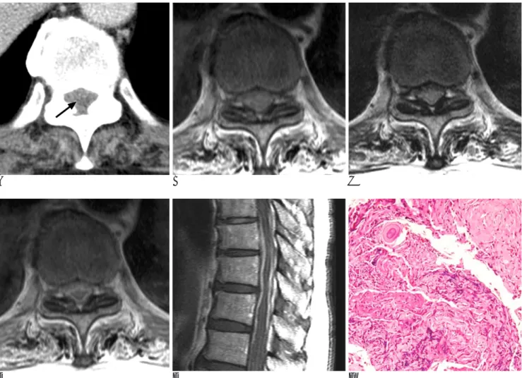
77세 남자 환자로 2개월 전부터 시작된 양측 하지의 근력약 화로 한방병원에서 치료 받다 8일전부터 심화된 하지마비 증 상으로 내원하였다. 신경학적 검사에서 운동장애는 양측 하지 모두 1등급을 보였고 감각이 전반적으로 감퇴되어 있었으나 대 소변장애는 없었다. 전산화 단층촬영에서 제 8-9번 흉추 디스 크 위치부터 제 12 흉추-제 1 요추 디스크 위치에 이르는 미 만성 병변이 경막내부에 보였고 음영은 척수보다 약간 높은 정 도였으며 척수를 완전히 둘러싸면서 압박하는 양상을 보였다 (Fig. 1A). 자기공명영상에서는 T1강조영상에서 척수의 백질 과 유사하였고(Fig. 1B), T2 강조영상에서는 척수의 백질보다 약간 낮은 신호강도를 보였는데(Fig. 1C) 이는 조직병리소견 (Fig. 1F)과 비교하여 볼때 섬유화의 동반에 의한 것으로 사 료된다. 황색인대의 비후와 함께 심한 척수 압박으로 인한 척 수병증이 의심되었다. 조영증강시 전제적으로 균일한 조영증강 양상을 보였고. 병변의 아래 경계는 정상 수막에서 조영증강되 는 부분과 정확히 구분이 어려웠다(Fig. 1D, E). 수술은 제 9
흉추 부터 제 12 흉추까지 척추후궁절제술(laminec-tomy)을 시행하였는데, 병변부위의 경막이 매우 두꺼워져 있었고 경막 을 열자 고무와 같은 질감(rubbery consistency)의 연한 갈색 의 미만성 종괴가 있었으며 중증도의 혈관분포정도 (vascularity)를 갖고 있었고 종괴는 거미막 (arachnoid menbrane)과 척수와 약간의 유착이 있었으나 신경근과는 관 계없이 위치하고 있었다. 종괴의 완전 적출은 불가능하였고 조 직병리소견상 이행형 수막종(transitional type meningioma)으 로 확진 되었다(Fig. 1F). 감압술 후 환자는 증상이 호전되어 퇴원하였다.
고 찰
척추 수막종은 원발성 척추종양의 약 25%를 점유하여 신경 성 종양 다음으로 두번째로 흔한 종양이다(2, 3). 척추 수막종 환자의 약 80%는 50-60대 여성이며 흔한 발병부위는 흉추, 경추, 요추 순이다. 성별에 따라 발병부위의 차이가 있어 흉추 와 경추의 발병비율이 여성에서 약 8:1, 남성에서 약 1:1로 알 려져 있다(2). 척추 수막종은 경막과 지주막이 근접해 있고 지 주막 융모의 밀도가 높은 신경근 출구부위(nerve root exit site)에서 주로 발생하며(2, 4), 조직학 분류상 수막성 수막종 (meningotheliomatous meningioma)이 가장 흔하다(2, 4, 5).
임상증상은 신경근병보다 진행성 척수병 증상이 특징적이며 국 소 통증(regional pain)과 하지 이상감각(lower extremity paresthesias)이 가장 흔하고, 감각운동장애(sensorimotor deficits)가 다음으로 흔한 증상이다(2).
반상수막종은 두개강내 접형골평면에서 주로 발생하고, 척 추강내에서의 발생빈도는 979예의 원발성 척추종양을 대상으 로 한 논문에서 1예가 기록되어있을 정도로 매우 드문 종양이 다(6). 특히 척추강내에서 척수주위를 둘러싸는 미만성의 고
─ 375 ─ 대한영상의학회지 2005;52:375-377
흉추 반상수막종: 증례 보고1
박현선・이상호2・최영근2・이상윤3・강호영・윤득희・조병준
척추 수막종은 원발성 척추종양의 약 25%를 점유하여 신경성 종양 다음으로 흔한 종양이나, 척추 반상수막종(en plaque meningioma)은 매우 드물어 발생 빈도가 알려져 있지 않다. 저자 들은 흉추에서 척수를 완전히 에워싸는 반상수막종을 경험하였기에 증례 보고 및 문헌 고찰하 는 바이다.
1우리들병원 영상의학과
2우리들병원 신경외과
3부천대성병원 영상의학과
이 논문은 2005년 3월 9일 접수하여 2005년 4월 29일에 채택되었음.
리양 종괴(collar-like mass)를 보이는 수막종은 매우 드물어, Stechison 등 이 흉 추 에 생 긴 석 회 화 된 반 원 형 태 의 (hemicircumferential) 반상수막종과 완전히 척수를 둘러싼 반 상수막종 등 2예(7), Niijima 등이 흉요추 이행부위에 생긴 골 화된 반상수막종 1예(1), Gamache 등이 경추에 생긴 석회화 된 반상수막종 1예(8), Messori 등이 경추에 생긴 석회화된 반상수막종 1예를 보고하였다(9). Stechison 등이 보고한 증 례 중 T12-L2에 위치한 반상수막종이 말단척수원뿔(distal conus medullaris)과 말총(cauda equina)을 완전히 에워싼 형 태를 보여 저자의 증례와 유사하나 이 예의 경우 myelogram 만 시행하고 CT 혹은 MRI를 시행하지 않아 석회화 없이 척 수를 완전히 에워싸는 반상수막종의 CT와 MRI 영상소견이 보 고된 논문은 없다.
척추 수막종에서 석회화를 보이는 경우는 1-5%로 보고 되 고 있다(4, 5, 7). 그러나 반상수막종에서 석회화를 보이는 빈 도는 알려진 바 없고 저자들의 증례에서는 석회화가 없었으나
이전에 보고된 반상수막종의 석회화 빈도를 볼 때(1, 7-9), 전체 척추 수막종의 석회화 빈도와는 다를 것으로 생각되며 보 다 많은 증례 연구가 필요하겠다.
조직학적으로 이전에 보고된 반상수막종은 대부분 수막성 수 막종이었으나 저자들의 증례는 Stechison 등의 2예중 하나와 같은 이행성 수막종이었다.
반상수막종은 인접한 구조물을 밀고 압박하는 피막화된 (encapsulated) 수막종과 다르게 광범위한 종양 기질 (extensive tumor matrix)을 갖고 인접한 구조물을 침윤하는 경향이 있어 전체 제거가 어렵고 재발가능성이 높으며 척수손 상의 위험도 높다고 한다(1, 7, 9). 저자들의 경우 환자의 나 이가 많고 종양이 척수를 완전히 에워싸고 있어 완전 적출은 어려워 척추후궁절제술과 함께 종양일부를 제거하여 감압수술 을 시행하였다.
결론적으로, 진행성 척수병의 임상소견이 있는 환자에서 MRI 검사소견상 척수를 완전히 에워싸는 경막병변이 T1과 T2강조
─ 376 ─
박현선 외: 흉추 반상수막종
A B C
D E F
Fig. 1. A 77-year-old man with back pain and paraplegia.
A. Transaxial CT scan obtained at the T10-11 disc level shows intradural extramedullary mass which encases the spinal cord com- pletely(arrow).
B, C. Axial T1- (B) and T2- (C) weighted MR images show slightly low to intermediate signal intensity mass.
D, E. Axial (D) and sagittal (E) T1-weighted postcontrast MR images show diffuse well enhancing circumferential mass at the T9-10 to the T12-L1 level.
F. Photomicrography (Hematoxylin and eosin, ×100) shows that the tumor is a transitional meningioma with psammoma bodies.
영상에서 척수와 동일한 신호강도를 보이고, 조영제를 사용한 T1강조영상에서 균일한 조영증강을 보이는 경우에 반상수막 종의 가능성을 고려해야 하겠다.
참 고 문 헌
1. Niijima K, Huang YP, Malis LI, Sachedev VP. Ossified spinal meningioma en plaque. Spine 1993;18:2340-2343
2. Souweidance MM, Benjamin V. Spinal cord meningiomas.
Neurosurg Clin North Am 1994;5:283-291
3. Solero CL, Fornari M, Giombini S, Lasio G, Oliveri G, Cimino C, et al. Spinal meningiomas: review of 174 operated cases.
Neurosurgery 1989;25:153-160
4. Levy WJ Jr, Bay J, Dohn D. Spinal cord meningioma. J Neurosurg 1982;57:804-812
5. Roux FX, Nataf F, Pinaudeau M, Borne O, Devaux B, Meder JF.
Intraspinal meningiomas: review of 54 cases with discussion of poor prognosis factors and modern therapeutic management. Surg Neurol 1996;46:458-463
6. Onofrio BM. Intradural extramedullary spinal cord tumors. Clin Neurosurg 1978;25:540-555
7. Stechison MR, Tasker RR, Wortzman G. Spinal meningioma en plaque. Report of Two cases. J Neurosurg 1987;67:452-455 8. Gamache FW Jr, Wang JC, Deck M, Heise C. Unusual appearance
of an en plaque meningioma of the cervical spinal canal: a case re- port and literature review. Spine 2001; 26: E87-E89
9. Messori A, Rychlicki F, Salvolini U. Spinal epidural en-plaque meningioma with an unusual pattern of calcification in a 14-year- old girl: case report and review of the literature. Neuroradiology 2002; 44: 256-260
─ 377 ─ 대한영상의학회지 2005;52:375-377
J Korean Radiol Soc 2005;52:375-377
Address reprint requests to : Hyeon Seon Park, M.D., Department of Diagnostic Radiology, Wooridul Spine Hospital 47-7 Chungdam-dong Gangnam-gu, Seoul 135-100, Korea.
Tel. 82-2-513-8195 Fax. 82-2-513-8175 E-mail: [email protected]
En Plaque Meningioma in Thoracic Spine: Case Report1
Hyeon Seon Park, M.D., Sang-Ho Lee, M.D.2, Young-Geun Choi, M.D.2, Sang Yeun Lee, M.D.3, Ho Yeong Kang, M.D., Deug Hee Yoon, M.D., Byung-June Jo, M.D.
1Department of Diagnostic Radiology, 2Neurosurgery, Wooridul Spine Hospital
3Department of Diagnostic Radiology, Puchon Daesung Hospital
Spinal en plaque meningioma is rarely found in the spinal canal, although lateral sphenoid wing menin- gioma displays a propensity for growth en plaque. We encountered a case of completely circumferential spinal en plaque meningioma, which is an even rarer condition. Herein, we report the CT & MRI findings along with a review of the related literature.
Index words :Meningioma
Magnetic resonance (MR) Computed tomography (CT)