176
J Korean Assoc Maxillofac Plast Reconstr Surg 2011;33(2):176-179
Case Report
원고 접수일 2010년 10월 14일, 게재 확정일 2011년 1월 28일 책임저자 노량석
(700-721) 대구시 중구 삼덕동 2가 188-1, 경북대학교 치의학전문대학원 구강악안면 외과학교실
Tel: 053-600-7562, Fax: 053-426-5365, E-mail: [email protected]
RECEIVED October 14, 2010, ACCEPTED January 28, 2011 Correspondence to Lyang Seok Noh
Deptment of Oral & Maxillofacial Surgery, School of Dentistry, Kyungpook National University
188-1, Samduck-dong 2-ga, Jung-gu, Daegu 700-721, Korea Tel: 82-53-600-7562, Fax: 82-53-426-5365, E-mail: [email protected]
CC This is an open access article distributed under the terms of the Creative Commons Attribution Non-Commercial License (http://creativecommons.org/licenses/
by-nc/3.0) which permits unrestricted non-commercial use, distribution, and reproduction in any medium, provided the original work is properly cited.
하악 소구치 부위에 발생한 석회화상피성치성종양이 혼재된 선양치성종양: 증례보고
노량석ㆍ조형우ㆍ최소영ㆍ김진수
경북대학교 치의학전문대학원 구강악안면외과학교실
Abstract
Combined Adenomatoid Odontogenic Tumor and Calcifying Epithelial Odontogenic Tumor in the Mandible: Case Report
Lyang Seok Noh, Hyung-Woo Jo, So-Young Choi, Chin-Soo Kim
Department of Oral & Maxillofacial Surgery, School of Dentistry, Kyungpook National University
Adenomatoid odontogenic tumors represent 3 to 7 percent of all odontogenic tumors. These tumors are more common in the maxilla than the mandible and usually include the anterior region. Clinically, the most common symptom is painless swelling and the tumor is associated with an unerupted tooth, typically a maxillary or mandibular cuspid. The adenomatoid odontogenic tumor appears radiographically as a unilocular radiolucency around the crown of an impacted tooth, resembling a dentigerous cyst. More often, it contains fine calcifications. Histopathologically, there is a thick wall cystic structure with a prominent intraluminal proliferation of the odontogenic epithelium. The most striking pattern is varying-sized solid nodules of spindle-shaped or cuboidal epithelial cells forming nests or rosette-like structures with minimal stromal connective tissues.
Conspicuous within the cellular areas are structures of tubular or duct-like appearance. The duct-like spaces are lined with a single row of cuboidal or low columnar epithelial cells, of which the ovoid nuclei are polarized away from the luminal surface. Small foci of calcification may also be scattered throughout the tumor. These have been interpreted as abortive enamel formations. In some adenomatoid odontogenic tumors, the material has been interpreted as dentoid or cementum.
Key words: Adenomatoid odontogenic tumor, Foci of calcification
서 론
선양치성종양(adenomatoid odontogenic tumor, AOT)은 서서히 성장하는 양성치성종양으로, 골내와 골외부위에서 발생한
다. 치성종양 중 3∼7%를 차지하며, 발생빈도가 비교적 드문
종양으로 하악보다는 상악에 호발하며, 특히 전치부에 잘 발생한
다[1]. 이 질환은 1905년에 Steensland에 의해 epithelioma ada-
mantinum으로 명명된 이후, adeno-ameloblastoma 등 다양한
Lyang Seok Noh: Combined Adenomatoid Odontogenic Tumor and Calcifying Epithelial Odontogenic Tumor in the Mandible: Case Report
177
Vol. 33 No. 2, March 2011
Fig. 1. Panoramic radiograph and
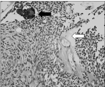
CBCT. Well defined, unilocular ra- diolucent lesion with internal radio- opaque foci on impacted #34 peri- coronal area.Fig. 2. CEOT-like area observed globular calcification (black ar-
row) and amyloid materials (white arrow) (H&E stain, ×10).용어로 사용되었다[2]. 방사선학적으로는 매복치의 치관주위로 단방성의 방사선 투과성으로 나타나며 미세한 석회화 물질을 포함 하는 경우가 많다. 조직 병리학적으로는 관 양상의 입방세포와 원주세포로 구성되어 있는 치성상피가 도강 내로 두드러지게 증식 하여 두터운 벽을 가지는 낭종성 구조로 되어 있다[1]. 몇몇 증례에 서는 조직 병리학적으로 명확한 세포 변연을 가지는 다면상 상피 세포, 결합체, 세포질내 균질체를등의 석회화 상피성 종양(calci- fying epithelial odontogenic tumor, CEOT) 특징이 혼재된 선양치성종양/석회화상피성치성종양 혼합 병소가 보고되었다[3].
본 증례보고에서는 13세 여아의 하악소구치부위에 석회화상피 성치성종양의 특징이 혼재된 선양치성종양을 문헌 고찰과 함께 보고하는 바이다.
증례보고
13세 여아가 좌측 하악부위의 낭성 병소로 경북대학교 치과병 원 구강악안면외과로 의뢰되었다. 내원 당시 하악 좌측 제1유구치 잔존되어 있었으며, 하악 좌측 제1소구치는 맹출지연되어 있었다.
파노라마 방사선 사진상에서 경계가 명확한 낭성 병소가 미맹출된 하악 좌측 제1소구치 치관을 중심으로 관찰되었고 매복 소구치의 여포간극(follicular space)이 증가되어 있었다. Cone beam computed tomography (CBCT)상에서 작은 크기의 석회화 물 질이 매복치관 상방에 산재되어 있었다(Fig. 1).
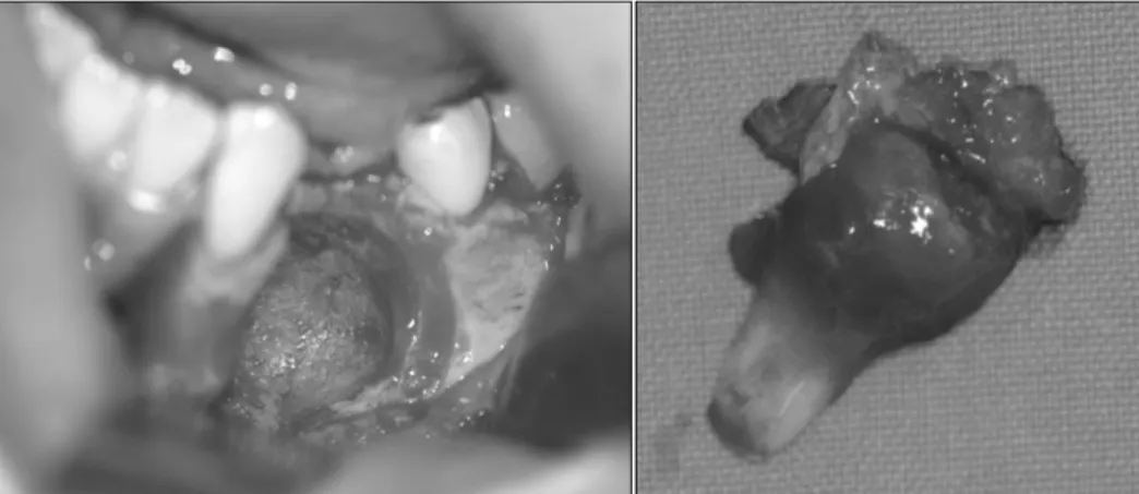
국소마취하에 하악 좌측 제 1유구치 발치와 함께 조직검사를 시행하였다. 조직 검사상 석회화상피성치성종양으로 진단되었다 (Fig. 2).병소부의 치료는 전신마취 하에 치료가 진행되었다. 치 은부위를 절개한후 병소부 제거와 함께 매복된 좌측 하악 제1소구 치 발치를 시행하였다(Fig. 3). 병소는 명확한 구형의 섬유성 캡슐 모양이었다. 술 후 1주일 경과하여 봉합사를 제거하였으며 조직 검사 결과는 선양치성종양으로 진단되었다(Fig. 4). 정기적 관찰 시행 중 발치부위 공간유지 위해 소아치과로 의뢰되어 공간 유지 장치를 제작, 장착하였으며, 3년간 예후관찰 중이며 재발소 견은 보이지 않았다(Fig. 5).
고 찰
선양치성종양은 이전에는 혼합성 치성 종양으로 분류되었으나 2005년 WHO 분류시 치성 상피 기원 양성종양으로 10대에 호발 하며 남성보다는 여성에서 발생빈도가 높다. 치료 방법으로는 적출술 및 소파술이 추천되며 재발율은 0.2%로 극히 드물다[4,5].
호발부위는 상악 전치부며, 병소 부위 및 형태에 따라 fol- licular, extrafollicular, peripheral type으로 분류할 수 있다.
Follicular, extrafollicular 형태는 골내 발생하는 병소로 전체
선양치성종양의 96%를 차지한다. 골내 발생 병소중 매복치와
관련된 follicular type은 71%정도의 높은 발생율을 나타내며,
경계가 명확하고, 매복된 치아의 치관부위에 단방성 방사선 투과
성을 나타낸다. 반면에 extrafollicular type은 매복치와 관련없으
며, 경계가 명확하고, 맹출된 영구치의 치근 사이, 치근부위에서
단방성의 방사선 투과성을 나타낸다. Peripheral type은 전체
178
노량석: 하악 소구치 부위에 발생한 석회화상피성치성종양이 혼재된 선양치성종양: 증례보고J Korean Assoc Maxillofac Plast Reconstr Surg Fig. 4. Light micrograph showing the typical appearance of AOT,
swirling spindle cells (black arrow) and duct-like formation (white arrow) (H&E stain, ×10).
Fig. 3. Resected tumor.
선양치성종양의 4.4%를 차지하며, 대부분 상악 전치부 치은부위 에 발생한다[1,6].
방사선학적 특징으로 매복치를 동반한 방사선 투과상을 나타내 며 경계는 명확하고 피질골성 또는 과골성 변연을 보인다. 방사선 투과성 부위는 치관의 하방으로 연장되고 투과성 내부에 석회화 물질이 산재되어 나타난다. 종양이 성장함에 따라 병소에 포함된 치아의 맹출을 방해하며 악골은 팽윤되나, 피질골의 천공은 드문 편이다. 병소내 석회화 물질이 관찰되지 않는 경우에는 함치성낭 과 감별이 힘든데, follicular type의 선양치성종양 중 77%가 초진시 함치성낭으로 진단된다. 함치성낭의 경우 방사선 사진 상에서 백악법랑경게부 상방 부위에 낭성 병소가 존재하는 반면 선양치성종양은 병소가 치관 하방으로 연장되거나 치아 전체를 둘러싼다. 이 밖에 병소내부에 석회화 양상을 보이는 법랑모세포 섬유치아종, 석회화 치성낭, 석회화상피성치성종양 등과 감별이 어렵다[1].
선양치성종양의 조직 병리학적 특징으로 관(duct-like)형태의
치성 상피와 다양한 유도 변화(inductive chage)를 가지는 결체 조직으로 구성되며, 부분적 낭성 부위와 종괴 부분으로 구성된다.
1) Encapsulation, 2) duct-like formation, 3) swirling spindle cells with rosette-like areas, 4) eosinophilic droplet materi- al, 5) trabecular or latticework epithelial proliferation의 특 질을 가진다[7]. 석회화상피성치성종양의 조직 병리학적 특징은 1) sheets of polyhedralcells, 2) nuclear pleomorphism, 3) amyloid material (intracellular and extracellular), 4) calcifi- cation of amyloid material, 5) well-defined cell border 6) intercellular bridges의 특징을 가진다[8,9].
종종 복합성 선양치성종양/석회화치성종양 경우가 조직학적으 로 발견된다. 이런 병소는 조직 병리학적으로 선양치성종양의 특징인 원주상피로 둘러싸인 도관상을 보이며 인접부위에 초자물 (amyloid)과 석회화 침착상을 보인다. 그리고 원주상의 세포가 2중으로 배열되어 장미 꽃송이 모양(rosette-like)양상을 나타낸 다. 동시에 석회화상피성치성종양과 유사한 경계가 명확한 다각 형(polyhedral) 상피성 종양세포와 종양실질 사이에 층판상의 석회괴가 나타난다. 그러나 이러한 특징들은 석회화치성종양에서 발견되는 고형성, 침투성 세포라기보다는 석화화 물질과 연관하 여 나타나며, 석회화치성종양의 특징인 전형적인 다형태성(pleo- morphism)과는 차이점을 가진다. 특이한 점은 선양치성종양의 특징인 장미 꽃송이 모양(rosette-like)이 나타나는 부위에서 석 회화상피성치성종양 유사세포가 동시에 나타난다는 것이다. 이러 한 점으로 미루어 선양치성종양과 석회화 상피성치성종양은 법랑 기관에서 유래되어 동일한 기원임을 추측할 수 있다.
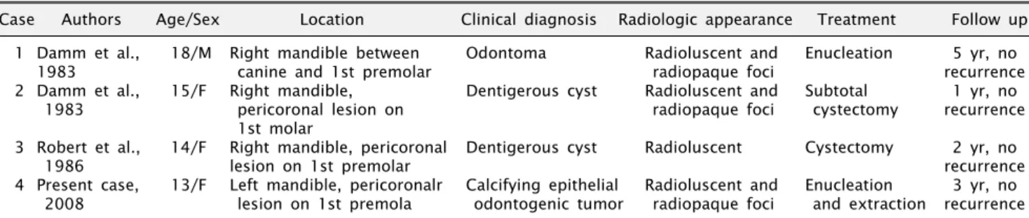
석회화상피성치성종양이 혼재된 선양치성종양은 1983년 Damm 등[3]에 의하여 처음으로 보고되었으며, 이전 보고들과 현재보고와의 비교는 다음과 같다(Table 1). 대부분의 경우 하악 에서 보고되었으며, 젊은 나이에 발생하였으며, 방사선학적으로 방사선 투과성과 방사선 불투과성 foci가 나타났으며, peri- coronal lesion을 나타냈다. 재발은 드문 편으로 나타났다.
문헌고찰에 따르면 Giansanti 등[10]은 선양치성종양은 주로
Lyang Seok Noh: Combined Adenomatoid Odontogenic Tumor and Calcifying Epithelial Odontogenic Tumor in the Mandible: Case Report
179
Vol. 33 No. 2, March 2011
Fig. 5. No recurrence has been
found after 3 year but a long-term follow-up is essential.Table 1. Reported cases of combined adenomatoid odontogenic tumor and calcifying epithelial odontogenic tumor
Case Authors Age/Sex Location Clinical diagnosis Radiologic appearance Treatment Follow up 1 Damm et al., 18/M Right mandible between Odontoma Radioluscent and Enucleation 5 yr, no
1983 canine and 1st premolar radiopaque foci recurrence
2 Damm et al., 15/F Right mandible, Dentigerous cyst Radioluscent and Subtotal 1 yr, no
1983 pericoronal lesion on radiopaque foci cystectomy recurrence
1st molar
3 Robert et al., 14/F Right mandible, pericoronal Dentigerous cyst Radioluscent Cystectomy 2 yr, no
1986 lesion on 1st premolar recurrence
4 Present case, 13/F Left mandible, pericoronalr Calcifying epithelial Radioluscent and Enucleation 3 yr, no 2008 lesion on 1st premola odontogenic tumor radiopaque foci and extraction recurrence
상악에서 발생하며, 젊은 여성에서 호발한다. 반면에 석회화상피 성치성종양은 주호 하악에서 발생하며, 남성과 여성에서 동일하 게 발생하며, 평균 발생 나이는 41세이다. 이러한 임상적 관점에 서 차이를 보이지만 두 종양은 발생학적으로 같은 기원으로 고려 된다.
본 증례에서는 좌측 하악부위 낭성 병소로 본원에 내원한 13세 남자 환자에서 임상 소견, 방사선학적 소견 및 조직학적 소견을 통해 석회화상피성치성종양의 특징이 혼재된 선양치성 종양으로 진단되어 인접구조물, 즉 하치조 신경 손상을 최소로 하면서 외과 적 적출술을 시행하였으며 3년 동안 추적 관찰 결과 재발이 없이 양호한 치유 상태를 보이고 있다.
References