Successful Conservative Surgical Treatment of Ameloblastic Fibroma in the Posterior Maxilla : A Case Report
Youngeun Lee
1, Hyojung Ahn
1, Sooeon Lee
1, Euncheol Kim
2, Sungchul Choi
11
Department of Pediatric Dentistry, School of dentistry, Kyung Hee University
2
Department of Maxillofacial Tissue Regeneration, School of Dentistry and Research Center for Tooth and Periodontal Tissue Regeneration, School of Dentistry, Kyung Hee University
Ameloblastic fibroma (AF) is a rare odontogenic ectomesenchymal tumor that is frequently seen in the first two decades of life, and occurs in the mandible. The most proper management of AF has been a recent topic of debate because of its recurrence and malignant transformation. This report describes AF in a 4-year-old male, which was a unilocular radiolucency on the maxillary right primary molar area with a scalloped border and cor- ticated margin. The tumor was treated conservatively with enucleation and curettage, and the decision was made to preserve the right primary second molar. A biopsy confirmed it as AF. During the 43 months of follow- up, the patient had no evidence of recurrence or malignant transformation. Moreover, the radiographic examina- tion revealed the generation of tooth germ to be a permanent second premolar. This report shows a case of AF in the posterior maxilla of a 4-year-old boy and discusses the conservative therapeutic approach to this tumor.
Therefore, the age of the patients should be an important consideration when choosing conservative or radical surgery in a young AF patient.
Key words : Ameloblastic fibroma, Mixed odontogenic tumor, Enucleation Abstract
Corresponding author : Sungchul Choi
Department of Pediatric Dentistry and Institute of Oral Biology, School of Dentistry, Kyung Hee University, 130-702, Seoul, Republic of Korea Tel: +82 2 958 9339 / Fax: +82 2 965 7247 / E-mail: [email protected]
Received May 23, 2013 / Revised September 12, 2013 / Accepted September 12, 2013
Ⅰ. Introduction
Ameloblastic fibroma (AF) is a tumor of mixed connec- tive and odontogenic tissue origin, most commonly found in younger age groups, between 15 and 25 years of age, with varied sex prediction from no preference to males being more affected than females
1,2). The posterior mandibular area is the most common anatomic site of AF
3).
There is controversy in the literature as to whether treatment should be conservative or aggressive. AF ex- hibits somewhat slow clinical growth, is well encapsulat- ed and shows an innocuous benign behavior, according
to most studies
3-5). A conservative treatment strategy, such as enucleation and curettage, is generally the treatment of choice, and the prognosis of these tumors is reported to be good
3,5). On the other hand, some re- searchers believe that AF is more aggressive than had been thought and a more radical therapy is needed on the basis of reviewing recurrent or malignantly trans- formed cases in the literature
6,7). According to this opin- ion, a more radical excision, block resection or segmental resection is recommended
8).
This report describes a rare case of AF, occurred in the posterior maxilla of a 4-year-old boy. The lesion was successfully treated conservatively.
※This research was supported by the Basic Science Research Program through the National Research Foundation of Korea, funded by the Ministry of Education, Science, and
Technology (2011-0013331).
Ⅱ. Case Report
A 4-year-old boy was referred to the Department of Pediatric Dentistry at the Kyung Hee Dental Hospital, Seoul, Republic of Korea, by his family dentist for evalu- ation of unerupted maxillary primary right molars. The medical, social and family histories were unremarkable.
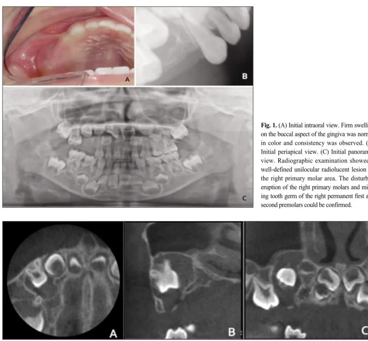
The intraoral examination revealed firm swelling on the buccal aspect of the gingiva in the primary right maxil- lary molar region (Fig. 1A). No discharge, redness, or fluctuation could be elicited from this area. The initial
intraoral and panoramic radiography showed a well-de- fined unilocular radiolucent lesion on the right primary molar area (Fig. 1B, C), and confirmed the presence of the unerupted teeth, and the missing tooth germ of the right permanent first and second premolars (Fig. 1C).
Cone beam computed tomography (CBCT) showed a well-defined monolocular radiolucent lesion around the primary first molar with a scalloped border and corticat- ed margin (Fig. 2). Sinus floor elevation, reactive mu- cosal thickening, and the partially indistinct cortication of the maxillary sinus floor could be observed (Fig. 2).
Fig. 1. (A) Initial intraoral view. Firm swelling on the buccal aspect of the gingiva was normal in color and consistency was observed. (B) Initial periapical view. (C) Initial panoramic view. Radiographic examination showed a well-defined unilocular radiolucent lesion on the right primary molar area. The disturbed eruption of the right primary molars and miss- ing tooth germ of the right permanent first and second premolars could be confirmed.
Fig. 2. CBCT view. (A) The transverse view showed a well-defined monolocular radiolucent lesion around the primary first molar with a scalloped border
and corticated margin. (B) Sagittal view. The lesion was extended to the palatal side and sinus floor. (C) Coronal view. Sinus floor elevation and reactive
mucosal thickening could be observed. The maxillary sinus floor showed partially indistinct cortication.
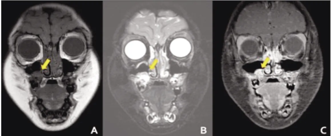
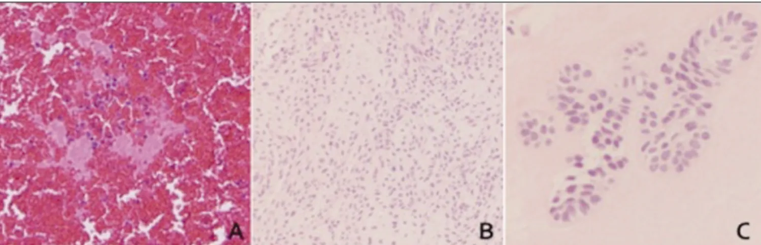
Magnetic Resonance Imaging (MRI) was taken to differ- entiate the lesion with the odontogenic cyst. The T1- weighted image (WI) of the MRI showed medium signal intensity, which confirmed the CBCT (Fig. 3A). In the T2WI, it had a higher signal intensity than the muscle (Fig. 3B). However, a gadolinium-enhanced T1WI showed an inhomogeneous high signal intensity, which had been enhanced by the contrast agent (Fig. 3C). The clinical and radiological diagnosis was AF. The cytologic smear by needle aspiration biopsy showed many lympho- cytes and a few histiocytes without malignant cells (Fig.
5A). Considering the patient’ s age and extent of surgery, the patient was treated by enucleation and curettage under general anesthesia, instead of resection.
The right primary first molar was also enucleated, whereas the decision was made to preserve the right pri- mary second molar. The right primary second molar was
exposed and a space maintainer was placed after it reached the occlusal plane (Fig. 4A, B). The pathological examination of the specimen revealed strands, cords, and islands of odontogenic epithelium in a primitive con- nective tissue stroma resembling dental papilla (Fig.
5B). Although a cell-free zone and/or a zone of hyalin- ization were occasionally found at the epithelial-mes- enchymal interface, no hard tooth structures were de- tected (Fig. 5C). Based on these results, the tumor was diagnosed as AF.
The patient was followed postoperatively for 43 months, but there was no sign of recurrence and malig- nant transformation. Postoperative panoramic radiogra- phy demonstrated completion of the bone healing with a new generation of tooth germ under the root of the right primary second molar area (Fig. 6).
Fig. 3. (A) In the T1-weighted images, a low signal appeared in the area consistent with the CT image. (B) In the T2- weighted images, a high signal appeared. (C) The same area showed an inhomogeneous enhanced signal in the gadolinium-T1-weighted image.
Fig. 4. (A) The lesion was completely enucleated and curetted. (B) A space maintainer was placed 1 month after surgery.
Ⅲ. Discussion
Ameloblastic fibroma is a rare tumor, accounting for only 2.5% of odontogenic tumors
2,5). The average age of presentation is in the teenage years
2,5). The majority of AF has been reported in the posterior mandible
3). The incidence rate of AF in posterior maxillary area is only half of that of posterior mandibular area
4). According to the data analysis from an extensive review of the pub-
lished studies on AF in the English literature (1949- 2005), 13.8% (17/123) of the cases was diagnosed as AF in children under five
3). Among them, maxillary AF was only 4.9% (6/123)
3). In this point of view, the rarity of this case, maxillary AF in a 4-year-old boy, can be fo- cused. Patients with maxillary AF usually present with intraoral findings including swelling of the alveolar process and noneruption of teeth
4), symptoms which also occur in our patient. This addresses the importance of routine oral examination in childhood and adolescence, which should be carried out thoroughly on both soft and hard tissues of the oral cavity.
Radiographically, AF is a unilocular lesion, occasional- ly multilocular when larger, with a smooth well-demar- cated border
2,5). Because lesions are frequently associated with unerupted teeth they may initially be interpreted as dentigerous cysts
9). In our patient’ s panoramic view and CBCT, well-defined unilocular radiolucency with a corticated margin and no expansion of the cortical plate in the maxillary posterior area were observed. Although CBCT can provide a good quality of image of the calcified tissue, the soft tissue is not distinguishable with this method. On the other hand, the MRI can discriminate a subtle difference among the soft tissues. This was why we chose the MRI to differentiate this lesion with the odontogenic cyst. Usually, the tumor in the head and neck region showed low signal intensity in the T1WI and a high signal in the T2WI
10). These findings agreed with our results of the MRI. However, it is important to note that the cyst has a low signal intensity in T1WI and high signal intensity in T2WI. To tell the difference, a gadolinium-enhanced MRI should be used. Hisatomi et Fig. 5. (A) A cytologic smear showed no malignant cells. (B, C) The stroma showed a fibromyxoid appearance with clusters of epithelial cells forming solid nests and glands [(A,B) magnification×100; (C) magnification×400; stain: H&E).
Fig. 6. Panoramic view (A) 18 months, (B) 36 months after the operation.
The radiographic image around the root of the right primary second molar
revealed the generation of the tooth germ (arrow).
al.
11)reported that an odontogenic tumor could be distin- guished from an odontogenic cyst, which shows peripher- al enhancement but no central enhancement. Our case showed inhomogeneous enhancement in the lesion in gadolinium-enhanced T1WI, enabling us to diagnose it as a tumor rather than a cyst.
Aspiration biopsy is a safe, quick and reliable proce- dure that can immediately differentiate inflammatory, reactive, cystic and neoplastic processes
12). A positive malignancy in this test can be considered as a final diag- nosis and treatment instituted accordingly. For the screening procedure, a needle aspiration biopsy was per- formed in this case. As a result, a negative fine needle aspiration (FNA) with inflammatory processes was ob- served. Scher et al.
13)stated that 13% of unsatisfactory FNA and 21% of negative FNA turned out to be a false- negative. Therefore, a biopsy is recommended in nega- tive and unsatisfactory FNA.
AF is a mixed tumor with both epithelial and mes- enchymal neoplastic proliferation. The epithelial compo- nent is described histologically as strands of cuboidal cells surrounding a reticulum similar to the source of embryonic enamel, which are ameloblasts. The mes- enchymal component looks similar to embryonic dental pulp but with less collagen. Ameloblastic fibroma has a slower growth rate than ameloblastoma and is less infil- trative. This results in a slow growing tumor that may expand bone and have smooth borders
3).
There is controversy over the management of AF. Both radical and conservative approaches are suggested by different authors. A wide excision of the tumor has been recommended unless the extent of surgery would result in significant cosmetic deformity
9,14). Others support enu- cleation or curettage as the initial treatment with a modified block resection reserved for recurrence
4). Furthermore, a majority of patients (91.5%) with AF were treated by conservative surgery
3). We opted for enucleation and curettage of the tumor with preservation of the maxillary right primary second molar because of its location, and cosmetic deformity and age. The radi- ographic image around the root of the right primary sec- ond molar revealed a putative permanent second premo- lar tooth germ, 3 years following the operation. This re- sult supports that a conservative surgical approach is meaningful in young patients. Chen at al.
3)emphasized the importance of the patient’ s age during treatment planning. Considering a significantly lower rate of malig- nant transformation, it was recommended to treat pa-
tients younger than 22 years by conservative strategy and by stepwise treatment principle with multiple recur- rence
3). However, recurrence rate, estimated as being be- tween 1.8 and 43.5%, long-term recall protocol is also important
14,15). It was reported that recurrence-free peri- od range from 1month to 96 months with a mean of 33.2 months
3). Also, conservative treatment with long-term (at least 10 years) annual follow-up evaluation indicat- ing no recurrence is recommended
5). This case was fol- lowed up during 43 months without recurrence. The pa- tient will continue to be evaluated annually.
Ⅳ. Summary
In this study, we report our experience with a case of AF in a 4-year-old boy treated with conservative surgi- cal enucleation and preservation of an impacted upper right second molar. After 43 months of follow-up, there were no signs of recurrence and complete spontaneous eruption of the preserved tooth was observed. Although its tendency to recur and undergo malignant transforma- tion makes dentists choose more aggressive surgical ex- cision, a conservative treatment strategy, such as enu- cleation and curettage, seems to be the most appropriate therapeutic option, especially for young patients.
References
1. Costa DO, Alves AT, Lourenco Sde Q et al. : Maxillary ameloblastic fibroma: a case report. Braz Dent J, 22:171-174, 2011.
2. Takeda Y : Ameloblastic fibroma and related lesions:
current pathologic concept. Oral Oncol, 35:535-40, 1999.
3. Chen Y, Wang JM, Li TJ : Ameloblastic fibroma: A review of published studies with special reference to its nature and biological behavior. Oral Oncol, 43:960-9, 2007.
4. Neville BW, Damm DD, Allen CM, Bouquot JE : Oral and Maxillofacial Pathology. 3rd ed., Saunders, St. Louis, 719-720, 2002.
5. Mosby EL, Russell D, Barker BF et al. : Ameloblastic fibroma in a 7-week-old infant : A case report and review of the literature. J oral Maxillofac Surg, 56:
368-372, 1998.
6. Reichart PA, Zobl H : Transformation of ameloblas-
tic fibroma to fibrosarcoma. Report of a case. Int J
Oral Surg, 7:403-7, 1978.
7. Takeda Y, Kaneko R, Suzuki A : Ameloblastic fibrosarcoma in the maxilla, malignant transforma- tion of ameloblastic fibroma. Virchows Arch A Pathol Anat Histopathol, 404:253-63, 1984.
8. Kousar A, Hosein MM, Ahmed Z, Minhas K : Rapid sarcomatous transformation of an ameloblastic fibro- ma of the mandible: case report and literature review. Oral Surg Oral Med Oral Pathol Oral Radiol Endod, 108:80-5, 2009.
9. Cohen DM, Bhattacharyya I : Ameloblastic fibroma, ameloblastic fibro-odontoma, and odontoma. Oral Maxillofac Surg Clin North Am, 16:375-84, 2004.
10. Langlais RP, van Rensburg LJ, Nortje CJ et al. : Magnetic resonance imaging in dentistry. Dent Clin North Am, 44:411-26, 2000.
11. Hisatomi M, Asaumi J, Kishi K et al. : MR imaging of epithelial cysts of the oral and maxillofacial region. Eur J Radiol, 48:178-82, 2003.
12. Silverman : Oral Cancer, Fifth Edition - American Cancer Society, Atlas of Clinical Oncology, BC Decker, 60-61, 2003.
13. Scher RL, Oostingh PE, Levine PA et al. : Role of fine-needle aspiration biopsy in the diagnosis of lesions of the oral cavity, oropharynx and nasophar- ynx. Cancer, 62:2602-6, 1988.
14. Zallen RD, Preskar MH, McClary SA : Ameloblastic fibroma. J Oral Maxillofac Surg, 40: 513-517, 1982.
15. Trodahl JN : Ameloblastic fibroma. A survey of cas- es from the Armed Forces Institute of Pathology.
Oral Surg Oral Med Oral Pathol, 33:547-58, 1972.
주요어: 법랑모세포섬유종, 진성 혼합 치성 종양, 적출술
상악 구치부에 발생한 법랑모세포섬유종의 성공적인 보존적 수술 : 증례 보고
이영은
1∙안효정
1∙이수언
1∙김은철
2∙최성철
11