Operative Treatment of Medial Epicondylitis: A Comparative Analysis of the Clinical Outcomes between the Suture Anchor Group and the Non-suture Anchor Group
Sang Jin Cheon , Woong Ki Jeon
Department of Orthopedic Surgery, Pusan National University Hospital, Busan, Korea
Background: The The purpose of this study was to make a comparative analysis of the clinical outcomes after the operative treatment of refractory medial epicondylitis between the suture anchor group and the non-suture anchor group.
Methods: We enrolled 20 patients (7 men and 13 women) with recalcitrant medial epicondylitis who were able to receive operative treatment in a minimum of an 18-month follow-up. The mean age was 48.6 years (range, 36–59 years). The patients were allocated into either the suture anchor group (7 patients) or the non-suture anchor group (13 patients). We evaluated clinical outcomes using the visual analog scale (VAS), the pain grading system of Nirschl and Pettrone, and postoperative grip strength.
Results: The VAS score decreased from 8.8 to 2.0 for the suture anchor group and from 8.6 to 1.3 for the non-suture anchor group (p=0.16). The postoperative grip strength was 95%, 93% of the non-treated arm in both groups (p=0.32). The postoperative satisfaction level was good in 5 patients and fair in 2 for the suture anchor group and excellent in 5 patients, good, in 4, and fair, in 4 for the non- suture anchor group (p=0.43). The clinical outcomes did not show a statistically significant difference between the two groups.
Conclusions: We found that patients with recalcitrant medial epicondylitis were treated reliably with satisfactory clinical outcomes whether or not suture anchors were used. We believe the use of suture anchors when more than 50% of the tendon origin is affected provides an effective and favorable treatment modality.
(Clin Shoulder Elbow 2015;18(4):221-228)
Key Words: Elbow; Medial epicondylitis; Suture anchor Clinics in Shoulder and Elbow Vol. 18, No. 4, December, 2015 http://dx.doi.org/10.5397/cise.2015.18.4.221
Received October 9, 2015. Revised October 29, 2015. Accepted November 3, 2015.
Correspondence to: Sang Jin Cheon
Department of Orthopedic Surgery, Pusan National University Hospital, 179 Gudeok-ro, Seo-gu, Busan 49241, Korea Tel: +82-51-240-7531, Fax: +82-51-247-8395, E-mail: [email protected]
Financial support: This work was supported by a 2-Year Research Grant of Pusan National University. Conflict of interests: None.
Introduction
Medial epicondylitis of the elbow is characterized by pain in the medial elbows during flexion of the wrists or during prona- tion of the forearm. It is not as common as lateral epicondylitis, but it is known to respond relatively well to conservative man- agement such as drug therapy, physiotherapy, and localized steroid injections.1) Sometimes medial epicondylitis is non- responsive to conservative management and it becomes severely debilitating for the patient. Operative treatments are recom- mended for such instances, and studies have shown that they lead to good clinical outcomes.2-4) However, the studies on the clinical outcomes after operative treatment of medial epicondy-
litis are few.
The extensor muscles such as the extensor carpi radialis brevis and the extensor carpi radialis longus attached to the lateral epi- condyle are affected during lateral epicondylitis. Thornton et al.5) reported that the use of suture anchors for the attachment of the extensor tendon origin to the lateral epicondyle yields a more stable, anatomical repair, which in turn improves grip strength and pain and expedites return to normal activities. Whilst only the extensor carpi radialis brevis is affected in lateral epicondyli- tis, the area of affected region is broader in medial epicondylitis which encompasses not only the pronator teres muscle and the flexor carpi radialis but also, in severe forms of the disease, the flexor digitorum superficialis and the flexor carpi ulnaris.6) The
operative treatment of medial epicondylitis is similar to that of the lateral epicondylitis comprising debridement of degenerative tissue, multiple drilling of the tendon origin, and the restoration which repairs the tendon origin to the medial epicondyle. The authors also believe that medial epicondylitis-induced damage on the flexor carpi radialis and on the pronator teres muscle should be repaired using suture anchors for enhanced stability and recovery.
Studies that describe the use of suture anchors according to the extent of lesions are insufficient. Thus, in our study we sought to address this issue and to investigate the difference in clinical outcome between those who received suture anchor and those who did not for the treatment of medial epicondylitis of the elbow and to determine the usefulness of suture anchors during medial epicondylar restoration.
Methods
Subjects of Study
Between October 2003 and July 2009, we enrolled a total of 20 patients with recalcitrant medial epicondylitis of the elbow who were able to receive operative treatment and partake in at least an 18-month follow-up. The average duration between the initial pain and when the patients received the operation was 22 months (range, 12–38 months). The patient sample com- posed of 7 men and 13 women with an average age of 48.6 years (range, 36–59 years). The patients were symptomatic on the right arm in 9 patients, on the left arm in 9, and bilaterally in 2 (however, these patients were operated on unilaterally). In terms of the treatment group, 7 patients were allocated into the suture anchor group and 13 patients into the non-suture anchor group. The occupation of patients varied widely, which included baseball players, golfers, tennis players, housewives, drivers, and waiters/waitresses. When we assessed the medical history of the patients, we found that 2 patients had had previous elbow trauma (10%) and 18 had a history of repetitive overuse of the elbows (90%). Also, in all the patients, we carried out a preop- erative physical examination that included the following assess- ment parameters: pain in the medial elbow, tenderness around the medial epicondyle, and pain in the medial epicondyle dur- ing isometric exercise.
The subjects in our study had received at least one year of conservative management such as physiotherapy, administra- tion of non-steroidal anti-inflammatory drug (NSAID), and local steroid injections. Yet their symptoms did not improve and pain persisted, which hindered their daily activities and occupational work, then we performed surgery. The surgery of every patient was carried out by the same surgeon. We took biopsy samples intra-operatively for all patients. And an intra-operative decision concerning the allocation of treatment, whether or not to use a suture anchor, was made according to how substantive the
lesion was in respect to the tendon origin. A preoperative assess- ment of pain and radiographic assessment were made. Postop- eratively, grip strength was assessed at one-year follow-up and pain and the level of treatment satisfaction were assessed at an average 35-month follow-up (range, 18–56 months).
Operative Treatment 1) Non-suture anchor group
The medial epicondyle was palpated with the elbow in 45o of flexion. From this palpation point, a 6 to 7 cm longitudinal line was marked over the predefined point of tenderness for the inci- sion. A incision was made into the soft tissue and the branches of the medial cutaneous nerve and the muscle fascia were softly retracted posteriorly. Another incision of around 2 cm was made along the muscle that originates from the common flexor tendon origin. The common flexor muscle was then spread apart to reveal the elbow joint. Through debridement, we removed the angiofibroblastic lesion from the pronator teres muscle of the medial epicondyle and from the flexor carpi radialis origin. Then we made multiple drillings at the anterior medial epicondyle. If an intraoperative assessment showed that the area of the lesion took up less than 50% of the tendon origin, the flexor-pronator unit was re-attached to the remaining epicondyle without using a suture anchor. Lastly, the surgical area was cleansed and the bleeding was stopped. The subcutaneous tissue and skin were sutured, taking care that the ulnar nerve was not damaged. A compression dressing was applied and the patient’s arm was im- mobilized using a long arm cast with the elbow in 30o to 40o of flexion.
2) Suture anchor group
The protocol for debridement of the lesion on the common flexor tendon origin of the medial epicondyle is the same as for the non-suture anchor group. But when an intra-operative as- sessment of the degenerative lesion is made and the lesion is broad (i.e., taking up more than 50% of the tendon origin), then a 2.4 mm metal suture anchor was used to restore the common flexor tendon at the medial epicondyle. The subsequent proce- dure was the same as that for the non-suture anchor group (Fig. 1).
Postoperative Rehabilitation
As the postoperative rehabilitation the patients underwent a long arm cast immobilization with the elbows in 30o to 40o of flexion for 48 to 72 hours, so we essentially restricted all active elbow motion during this period. Then, passive and active range of motion was begun carefully. The skin sutures were removed on the second week of operation, from when light daily activi- ties were allowed. From the 6th and 8th postoperative week, we began implementing muscle strengthening exercises. Depending on each patient’s progress, the return to sports and to pre-injury level of daily activities was commenced gradually from the 3rd and 4th postoperative month.
Postoperative Assessment 1) Pain
The postoperative assessment of pain was made using the visual analog scale (VAS) and using the grading system devised by Nirschl and Pettrone.7) The VAS, a scale from a score of 0 indicating no pain to a score of 10 indicating unbearable pain, was assessed preoperatively and at an average 35 months after the operation. To gain better insight of the patients’ level of pain, we evaluated pain also during exercise. At the final follow-up, we assessed the level of pain during forced palmar flexion of the wrist. The level of pain was designated as grade A (5 points) if patient experienced no pain; as grade B (4 points) if the patient experienced slight discomfort; as grade C (3 points) if the patient experienced discomfort yet could flex the wrist; as grade D (2 points) if the patient experience discomfort that prevented wrist flexion; and as grade E (1 point) if the patient complained of persistent, severe pain. The patients with pain grades A and B were classified as having a good outcome, whereas those with grades C, D, and E were considered as having a poor outcome.
Lastly, we measured the level of satisfaction of treatment using Nirschl and Pettrone’s satisfaction scale comprising of 4 grades:
excellent, good, fair, and failed. An ‘excellent’ outcome is a state of no pain and of return to normal level of activity, a ‘good’ out- come is a return to normal level of activity but with intermittent mild pain, a ‘fair’ outcome is a return to daily activities without pain but only when lifting heavy items or when pain is thought to have resolved by 75%, and a ‘failed’ outcome is when preop- erative symptoms have not improved.
2) Grip strength
We followed the measurement protocol for grip strength de- vised by Rosenberg et al.8) Using a hand dynamometer (JAMAR Hydraulic Hand Dynamometer; Lafayette Instrument, Lafayette, IN, USA), the grip strength was measured preoperatively, on the 6th postoperative week, and at the 1-year follow-up. Grip strength was measured with the patient sitting in neutral posture, with the shoulders in adduction, the elbow flexed 90o, and the lower arm and wrist in neutral position. A total of 3 repeat mea- surements were made with 2 to 3 minutes of intervening resting periods, and the repeats were averaged to give a mean. We thought that if patients, after 12 months of rehabilitation, were able to achieve active, strong motions using their treated arm to a level that is comparable to that of their contralateral untreated arm then the grip strength could be used as an assurance to pa- tients of a tangible recovery and to promote patient satisfaction.
We calculated the grip strength of the treated arm as a percent- age of the grip strength of the non-treated arm at the 1-year follow-up.
3) Radiography
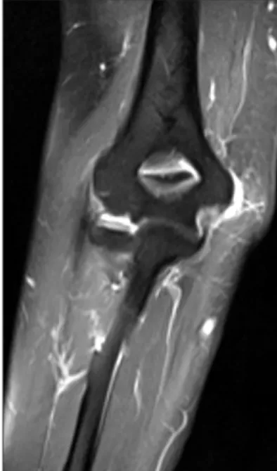
All patients were examined preoperatively using radiography, and in 14 patients magnetic resonance imaging (MRI) was car- ried out. The medial epicondylar region matching to that pre- defined through physical examination was located with the help of a radiologist. The medial epicondylitis is depicted as a region of high signal intensity (Fig. 2). Postoperatively, we examined the plain radiographs for the correct placement of the suture anchor and for the dissipation of calcification around the medial epi- condyle.
A B
C D
Fig. 1. (A, B) Intraoperative findings shows broad (>50%) degenerative tissue on com- mon flexor tendon origin. (C, D) After debridement of angiofibroblastic lesions, 2.4 mm metal suture anchor used for repairing tendon origins.
Statistical Analysis
All statistical analyses were performed using the SPSS statisti- cal analysis program ver. 12.0 (SPSS Inc., Chicago, IL, USA). Sta- tistical significance was defined using the Wilcoxon signed rank test and the 95% confidence interval (p<0.05).
Results
All patients had a degenerative lesion either at the anterior compartment or in the deep muscles of the common flexor ten- don. The average age of the patients in the suture anchor group was 49.1 years. The preoperative VAS score of 8.8 decreased to a postoperative value of 2.0. The pain during forced palmar flex- ion of the wrist was grade A in 4 patients (57.1%) and grade B in 3 patients (42.9%), giving an average of 4.6. We found that the postoperative satisfaction level of treatment was ‘good’ in 5 pa- tients (71.4%) and ‘fair’ in 2 patients (28.6%) using the Nirschl/
Pettrone score. The average grip strength of the treated arm, taken as the percentage of the contralateral, non-treated arm, was 95% (range, 92%–98%) at the final follow-up, indicating that it was not significantly different from that of the contralateral arm (Table 1, 2).
The average age of the patients in the non-suture anchor group was 48.4 years. As in the suture group, the preoperative VAS score of 8.6 improved to 1.3 postoperatively. The pain dur- ing forced palmar flexion was grade A in 7 patients (53.8%) and grade B in 6 patients (46.2%), showing an average of 4.5. The postoperative treatment satisfaction level was ‘excellent’ in 5 pa- tients (38.5%), ‘good’ in 4 (30.8%), and ‘fair’ in 4 (30.8%). The
average grip strength of the treated arm was 93% (range, 91%–
97%) of the counterlateral, non-treated arm at the final follow- up, indicating again that a significant difference is not seen with the contralateral arm (Table 1, 2).
We made a comparative analysis of the treatment outcomes between the suture anchor group and the non-suture anchor group. We found that the average preoperative and postopera- tive VAS scores did not show a statistically significant difference between the two groups (p=0.16). Neither did the postopera- tive grip strengths (p=0.32) nor the pain at forced palmar flexion (p=0.43) show statistically significant differences between the two groups (Table 2).
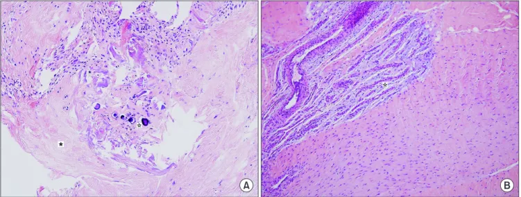
The preoperative physical examination had showed that all 20 patients (100%) were positive for medial epicondylitis. A pathological evaluation of the resected tissue showed that 15 patients had Nirschl’s stage 4 lesions (75.0%) and 5 patients had Nirschl’s stage 3 lesions (25.0%). When we compared the pre- and postoperative radiographic results, we found that in the 8 patients who had showed the signs of micro-calcificaiton and osteopenia (40.0%) these signs disappeared on the postopera- tive radiograms (Fig. 3, 4).
Discussion
The medial epicondylitis of the elbow is commonly called the Fig. 2. The magnetic resonance imaging of a 54-year-old women with medial
epicondylitis shows high signal intensity on medial epicondylar area of the elbow.
Table 1. Postoperative Satisfaction between Suture Anchor Group and Non- suture Anchor Group
Outcome With anchor (n=7) Without anchor (n=13)
Excellent 0 (0) 5 (38.5)
Good 5 (71.4) 4 (30.8)
Fair 2 (28.6) 4 (30.8)
Failure 0 (0) 0 (0)
Total 7 (100) 13 (100)
Values are presented as number (%).
Table 2. Changes between Preoperative and Last Follow-up Clinical Out- comes
Characteristic With anchor
(n=7) Without anchor (n=13) p-value
Age (yr), mean 49.1 48.4 -
Preoperative VAS, mean 8.8 8.6 0.16
Postoperative VAS , mean 2.0 1.3
Pain during forced palmar
flexion, mean 4.6 4.5 0.43
Postoperative grip strength
ratio to opposite side (%) 95 93 0.35
p<0.05, Wilcoxon rank-sum test.
VAS: visual analog scale.
‘golfer’s elbow’. It shows a prevalence of 0.4%, without showing a gender disparity in prevalence, and it presents mostly in 30 to 40 years old. Risk factors for medial epicondylitis include repeti- tive arm use, excessive exercise, obesity, and smoking. Although the pathophysiology of medial epicondylitis has not been clearly defined, it is thought that accumulation of repetitive focal trau- ma at the medial ligament origin or the flexor-pronator origin on the medial epicondyle leads to pain, swelling, tenderness, and motion restriction that are typical in medial epicondylitis.
Repetitive external rotation and valgus stress on the elbow cause excessive extension and microtrauma of the common flexor muscles, which originate at the medial epicondyle.
Pathologically, the normal collagen structure of the tendon is de- stroyed because of the recruitment of acute and chronic inflam- matory cells, the fibroblastic response, the immature vascular re- sponse, and the incomplete healing process. At the initial phase of medial epicondylitis, the inflammation takes the form that is characteristic of synovitis; then, as the disease progresses, it takes a more degenerative form showing features such as micro-lac- erations, tendinous degeneration, calcification, and incomplete neurovascular responses.1) Nirschl and Ashman9) classified the le- sions secondary to medial epicondylitis into 4 phases: phase 1 is inflammatory and reversible; phase 2 is characterized by angio- fibroblastic degeneration; phase 3 is characterized by structural function loss; and phase 4, include fibrosis and calcification in addition to the features seen in phases 2 and 3.
Ollivierre et al.10) found of the 50 patients with medial epi- condylitis of the elbow who underwent operative treatment 28 patients had lesions in the flexor carpi radialis-pronator teres muscle interval. They also reported that patients showed signs of angiofibroblastic tendinosis and fibrillary degeneration of the collagen. Further, in 14 patients they found radiographic and pathologic signs of calcification. Intriguingly, they reported that the extent of preoperative pain at rest was correlated to the ex- tent of calcification.
In our study, the pathological results showed that 15 patients had Nirschl’s grade 4 lesion and showed signs of chronic inflam- mation and calcification (Fig. 5). Despite being positive for me- dial epicondylitis through physical examination, 7 of the 15 pa- tients who had grade 4 lesions did not show radiographic signs of calcification and showed satisfactory postoperative outcomes.
These findings suggest that even with the absence of radiograph- ic signs, the disease may be progressed pathologically; results from physical examination should be evaluated with those of radiographic and pathologic tests to make a well-informed diag-
A B
Fig. 3. A 55-year-old woman with medial epicondylitis. (A) White circle in- dicates small calcification on medial epicondylar area of the elbow. (B) After surgical treatment without anchor, small calcification has disappeared.
Fig. 4. A 54-year-old women with medial epicondylitis shows mild osteopenia lesion on periarticular area of the elbow. G2 suture anchor was inserted on medial epicondylar area of elbow. The postoperative range of mo- tion was full and postoperative visual analog scale decreased from 9 to 1.
nosis and decision regarding treatment plan.
Typically, at the initial stages of medial epicyondylitis the pa- tient feels pain during flexion of the wrists or during pronation of the forearm. But as clinical course of the disease progresses, the patient may feel pain or motion restriction even at rest or during extension and may present with concomitant ulnar nerve paraly- sis.2,3,6)
The anatomical attachment of the flexor-pronator unit to the medial epicondyle is made tendinously. From the radialis to- wards the ulanris, the pronator teres, the flexor carpi radialis, the palmaris longus, the flexor digitorum superficialis, and the flexor carpi ulnaris are attached to the medial epicondyle of the elbow.
The flexor digitorum superficialis is attached to the deep medial epicondyle, whereas the rest (pronator teres, flexor carpi radia- lis, palmaris longus, and flexor carpi ulnaris) are attached to the shallower medial epicondyle. As a common flexor tendon, the described flexor-pronator unit of the lower arm has its origin at the medial epicondyle at least partially if not completely. For this reason, this structure can be easily damaged during sports-relat- ed exercises that require excessive external rotation or impose valgus stress on the elbows such as throwing a ball or swimming.
Further, the ulnar nerve, which passes between the medial in- termuscular septum and the medial head of the triceps muscle, runs near the medial epicondyle and traverses to the lower arm through the ulnar nerve groove. Elbows of patients with medial epicondylitis and valgus deformity may often sustain co-injuries such as ulnar nerve injury, nerve compression, neuritis, and en- trapment neuropathy as a result of an inflammatory reaction.2)
For the diagnosis of medial epicondylitis, a detailed self-re- ported medical history of the patient must be taken and a physi- cal examination of pain and tenderness of the medial elbow and of pain of the medial epicondyle during isometric exercise
should be made. Plain radiography, ultrasonography, or MRI can be used for the differential diagnosis of medial epicondylitis.
The disease should be differentiated from other elbow diseases that cause discomfiture of the medial elbow similar to that seen in medial epicondylitis.3) Discomfiture may arise through vari- ous causes including instability of the medial collateral ligament, ulnar nerve subluxation and inflammation, shoulder lesions, el- bow pain induced by friction from the triceps medial head, and tendinitis.
For the treatment of medial epicondylitis of the elbows, con- servative management is implemented prior to operative treat- ment. Conservative management includes drug therapy using NSAIDs, heat therapy, electrotherapy, physiotherapy using mas- saging, cast immobilization, and local steroid injections.6,11) Most patients respond well to conservative treatments, and Descatha et al.12) found that in 81% of patients, symptoms resolved within 3 years of management. However, if despite such conservative management pain persists for more than 6 to 12 months or symptoms worsen, then it becomes an indication for operative treatment.
The order of procedure during an operative treatment is as follows: a healing response is promoted through debridement of the degenerative tissue; focal vascularity is promoted; and the tendon origin is reattached to the medial epicondyle. Concomi- tant injuries such as the injuries of the ulnar nerve or the medial collateral ligament are treated at simultaneously. Especially, it is thought that medial epicondylitis are associated with degenera- tive tissue is larger and deeper than those associated with the lateral epicondylitis, which tends only to include the extensor carpi radialis brevis. Thus, we anticipate that the anatomical restorative effects of using suture anchors for the repair of the tendon origin to the epicondyle could be more pronounced in
A B
Fig. 5. (A) Calcification pattern at medial epicondylar area: The dark puple circles and hollow asterisk indicate calcification and black asterisk indicates collagen.
(B) Chronic inflammation pattern at medial epicondylar area: Hollow asterisk indicates angiofibroblastic lesion that shows spindle shape’s fibroblast (A, B: H&E,
×20).
medial epiconylitis than in the lateral epicondylitis.
In the 10 elbows with lateral epicondylitis that were nonre- sponsive to conservative management, Deng et al.13) reported a satisfactory clinical outcome at the 12th months postoperative follow-up after debridement of the extensor muscle lesion and repair using a suture anchor. Similarly, Thornton et al.5) found that when 22 patients with chronic lateral epicondylitis received anatomical restoration using suture anchors a satisfactory clinical outcome was seen in 18 patients. Ollivierre et al.10) reported that pain was resolved either partially or completely in every patients who received treatment for the medial epicondylitis. Other studies also describe results of satisfactory outcome after surgi- cal treatment of the lateral or medial epicondylitis.1-5) Vinod and Ross14) found that when the 60 recalcitrant medial epicondylitis of the elbow were treated operatively, which required resection of the lesion and re-attachment of the common flexor tendon to the epicondyle using suture anchors, rehabilitation and return to daily activity could be expedited and satisfactory results could be achieved at the final follow-up.
Like, in our study, we evaluated the use of suture anchors for the treatment recalcitrant medial epicondylitis. We made a comparative analysis of the clinical outcomes between those in the suture anchor group and those in the non-suture anchor group. We found that in 14 patients (70%) a ‘satisfactory’ or bet- ter outcome of treatment was seen; nevertheless, in all patients we found a greater than ‘fair’ outcome. However, there were no ‘excellent’ outcomes in the suture anchor group. This could be because for patients to be eligible for the suture anchor treat- ment their degeneration tissue had to be relatively larger in size than that found in the non-suture anchor group. Therefore, this gave an inherent difference in the two treatment group that could bias the treatment outcomes. For example, those in the suture anchor group may show a relatively slower recovery than the non-suture anchor group because their lesion was larger to begin with.
The use of suture anchors is relatively easy, and they are known to be effective tools for anatomical reduction. This is exemplified by the fact that despite having large lesions those in the suture anchor group achieved full range of motion of the elbow within 3 to 5 days of surgery and showed an expedited return to daily activities. Even though our results show that the suture anchor group and the non-suture anchor group do not show a statistically significant difference in clinical outcome, with a longer follow-up the suture anchor group may exhibit a clini- cally significant enhancement in outcome over the non-suture anchor group.
In our study, we evaluated the following parameters of clinical outcome: two measurements of pain, treatment satisfaction, and grip strength. We found there were no statistically significant dif- ferences in these parameters between the suture anchor group and the non-suture anchor group. The use of suture anchors for
the medial epicondylitis has been dictated by individual clinical status of the patient or has relied on the discretion of the surgeon after the intra-operative inspection of the pathologic tissue. Now a clear threshold or guideline should be outlined for a consistent approach. Anatomically, the common flexor tendon origin at the medial epicondyle has a more rhomboid shape, giving it a larger surface area for attachment, than that of the lateral epicon- dyle.15) A greater surface area of attachment means that there may be an exaggerated loss in stability when there is a large degenerative tissue in medial epicondylitis. Thus, an anatomical restoration at the tendon and recovery may be difficult. With this argument, we used suture anchors to repair common flexor tendons to the medial epicondyle when the lesions occupied a space larger than 50% of the tendon origin. More studies like ours on the effect of selective treatment based on surface area of the degenerative tissue on the lateral and the medial epicondyle on treatment outcome are needed.
There are a few limitations to this study. First is the small sample size that reduces the power of this study. Second, a bias was introduced during the allocation of treatment to patients. A non-random approach was taken to treat patients with severe le- sions using a suture anchor and those without, not with a suture anchor. This meant that the sample populations in the two treat- ment groups could be epidemiologically different. A prospective study with a greater sample size and an extended follow-up would unequivocally determine the effectiveness and the clini- cal outcomes of suture anchors during the operative treatment of medial epicondylitis.
Conclusion
We found that when patients with recalcitrant medial epicon- dylitis of the elbows were treated operatively satisfactory clinical outcomes were seen. We found that the clinical factors were improved in both the suture anchor group and in the non-suture anchor group even though lesion size was a factor in determin- ing the mode of treatment the patient received. We conclude that the use of suture anchors for medial epicondylitis with le- sions that are deep and affect more than 50% of the tendon origin can be considered as an effective treatment modality.
References
1. Ciccotti MC, Schwartz MA, Ciccotti MG. Diagnosis and treat- ment of medial epicondylitis of the elbow. Clin Sports Med.
2004;23(4):693-705.
2. Gabel GT, Morrey BF. Operative treatment of medical epi- condylitis. Influence of concomitant ulnar neuropathy at the elbow. J Bone Joint Surg Am. 1995;77(7):1065-9.
3. Kurvers H, Verhaar J. The results of operative treatment of me- dial epicondylitis. J Bone Joint Surg Am. 1995;77(9):1374-9.
4. Vangsness CT Jr, Jobe FW. Surgical treatment of medial epicondylitis. Results in 35 elbows. J Bone Joint Surg Br.
1991;73(3):409-11.
5. Thornton SJ, Rogers JR, Prickett WD, Dunn WR, Allen AA, Hannafin JA. Treatment of recalcitrant lateral epicondylitis with suture anchor repair. Am J Sports Med 2005;33(10):1558-64.
6. Jobe FW, Ciccotti MG. Lateral and medial epicondylitis of the elbow. J Am Acad Orthop Surg. 1994;2(1):1-8.
7. Nirschl RP, Pettrone FA. Tennis elbow. The surgical treatment of lateral epicondylitis. J Bone Joint Surg Am. 1979;61(6):832-9.
8. Rosenberg N, Soudry M, Stahl S. Comparison of two methods for the evaluation of treatment in medial epicondylitis: pain estimation vs grip strength measurements. Arch Orthop Trau- ma Surg. 2004;124(6):363-5.
9. Nirschl RP, Ashman ES. Tennis elbow tendinosis (epicondylitis).
Instr Course Lect. 2004;53:587-98.
10. Ollivierre CO, Nirschl RP, Pettrone FA. Resection and repair for medial tennis elbow. A prospective analysis. Am J Sports Med.
1995;23(2):214-21.
11. Tschantz P, Meine J. Medial epicondylitis. Etiology, diagno- sis, therapeutic modalities. Z Unfallchir Versicherungsmed.
1993;86(3):145-8.
12. Descatha A, Leclerc A, Chastang JF, Roquelaure Y; Study Group on Repetitive Work. Medial epicondylitis in occupa- tional settings: prevalence, incidence and associated risk fac- tors. J Occup Environ Med. 2003;45(9):993-1001.
13. Deng Y, Tang K, Li H, et al. Short-term effectiveness of suture anchor after debridement of extensor tendon insertion for recalcitrant lateral epicondylitis. Zhongguo Xiu Fu Chong Jian Wai Ke Za Zhi. 2013;27(1):1-6.
14. Vinod AV, Ross G. An effective approach to diagnosis and sur- gical repair of refractory medial epicondylitis. J Shoulder Elbow Surg. 2015;24(8):1172-7.
15. Alcid JG, Ahmad CS, Lee TQ. Elbow anatomy and structural biomechanics. Clin Sports Med. 2004;23(4):503-17.