Spontaneous Rupture of the Extensor Carpi Radialis Brevis and Radial Collateral Ligament of the Elbow in a Recreational Golfer: Surgical Experience of Repair of a Chronic Retracted Tendon and Ligament
Jin-Young Park, Jin-Young Bang1
Center for Shoulder, Elbow and Sports Medicine, Neon Orthopaedic Clinic, Seoul, 1Department of Orthopaedic Surgery, Haeundae Paik Hospital, Inje University College of Medicine, Busan, Korea
Lateral epicondylitis with rupture of the radial collateral ligament of the elbow has not been reported in the literature. We report on a case of a recreational golfer who had not received steroid injection and had no trauma history. The patient was treated with open surgi- cal repair. At 2 years follow-up, satisfactory clinical and radiological outcomes were observed with return to pre-injury level. The authors report this case and review the literature.
(Clin Shoulder Elbow 2016;19(1):39-42)
Key Words: Open extensor carpi radialis brevis repair; Lateral epicondylitis; Radial collateral ligament rupture
Clinics in Shoulder and Elbow
CiSE
Copyright © 2016 Korean Shoulder and Elbow Society. All Rights Reserved.
This is an Open Access article distributed under the terms of the Creative Commons Attribution Non-Commercial License (http://creativecommons.org/licenses/by-nc/4.0) which permits unrestricted non-commercial use, distribution, and reproduction in any medium, provided the original work is properly cited.
pISSN 2383-8337 eISSN 2288-8721
CASE REPORT
Clinics in Shoulder and Elbow Vol. 19, No. 1, March, 2016 http://dx.doi.org/10.5397/cise.2016.19.1.39
Received October 4, 2015. Revised February 1, 2016. Accepted February 18, 2016.
Correspondence to: Jin-Young Bang
Department of Orthopaedic Surgery, Inje University Haeundae Paik Hospital, 875 Haeun-daero, Haeundae-gu, Busan 48108, Korea Tel: +82-51-797-0990, Fax: +82-51-797-2203, E-mail: [email protected]
Financial support: None. Conflict of interests: None.
Lateral epicondylitis, or tennis elbow, is a common muscu- loskeletal disorder. Lateral epicondylitis was first reported in 1873.1) Subsequently, Nirschl and Pettrone2) performed patho- logical examination of extensor carpi radialis brevis (ECRB) at- tachment in the lateral epicondyle of the humerus and reported this change not only as a pathological condition of acute in- flammation but also as a tendon condition of angiofibrosis. The anatomy of the lateral epicondyle is composed of the ECRB, the extensor digitorum communis, and the lateral collateral liga- ment.3) Degeneration of the common extensor tendon can prog- ress and the rupture can spread to a closed structure. In general, the lateral ulna collateral ligament was known to stabilize in posterolateral rotatory instability, while, according to one report, in terms of elbow stability, the radial collateral ligament (RCL) is more important than the lateral ulnar collateral ligament because RCL is essentially isometric and can provide stability throughout the range of elbow flexion.4) In this article, we describe a spon- taneous rupture of the ECRB with RCL and its open repair in a recreational golfer.
Case Report
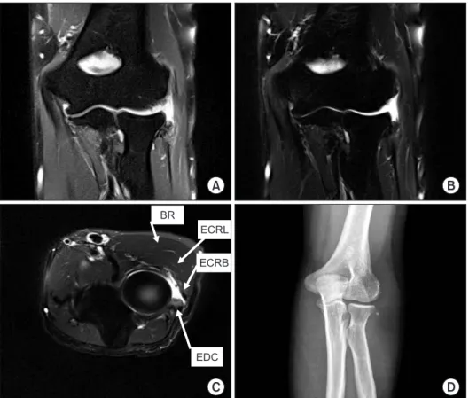
A healthy, right-handed, 45-year-old male visited our shoul- der-elbow center for left elbow pain which occurred 3 months ago without trauma. The patient was a week day golfer for several years and a single handicapper. There was tenderness of the lateral epicondyle and a positive index on Cozen’ test (with flexed at 90o and with the forearm in pronation) and Mill’s test (with the patient’s hand closed, the wrist in dorsiflexion and the elbow extended). Pain and apprehension was induced in the table top relocation test and varus stress test for assessment of instability but baseline radiographs of elbow showed unremark- able findings. No steroid injection history of the elbow was detected. His preoperative clinical scores were as follows: Mayo elbow performance score (MEPS): 40, visual analogue scale score for pain: 30%. A left-sided magnetic resonance imaging (MRI) showed a complete tear of the ECRB tendon from the lat- eral epicondyle (20 mm retraction and 20 mm width). In addi- tion, a complete tear of the RCL at the humeral attachment site and dystrophic calcification around the lateral epicondyle were
40
www.cisejournal.orgClinics in Shoulder and Elbow Vol. 19, No. 1, March, 2016
observed on a left-sided MRI (Fig. 1). Conservative treatments including nonsteroidal antiinflammatory drugs (NSAIDs), extra- corporeal shock wave therapy, orthoses, and physical therapy were administered for 6 months; however those strategies failed to relieve lateral epicondylitis and instability symptoms. There- fore, surgical treatment was decided.
Description of the Procedure
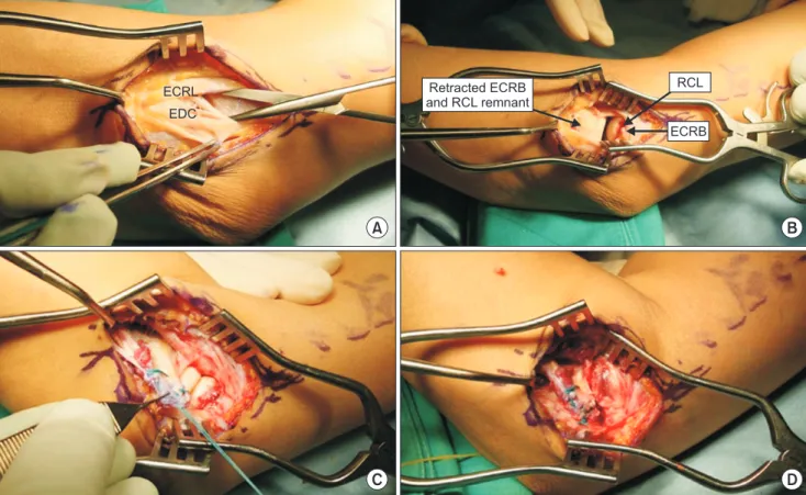
The patient was placed in a supine position and checked for varus instability using fluoroscopic imaging. A curvilinear incision was made 2 cm proximal and 3 cm distal to the lateral epicon- dyle. After the deep fascia was incised, the origin of the com- mon extensor tendon was approached by elevating the inferior border of the extensor carpi radialis longus muscle. Amorphous tissue was visualized, and complete tears were observed at the origins of the ECRB muscle and the RCL (Fig. 2A). The proximal margins of the tears were retracted to the distal portion revealing fibrotic scar tissue (Fig. 2B). Degenerative tissue of the proximal torn tendon was debrided. After four holes were drilled into the tendon origin site, the torn tendon and ligament was reattached to the origin site with one suture anchor (STATAK soft tissue anchor 2.5 mm; Zimmer, Warsaw, IN, USA) using the Krackow method with the elbow in flexion and the wrist in extension (Fig.
2C, D). Adequate soft tissue was available for joint coverage.
Tension of the repaired tendon was confirmed at the 30o wrist flexion position. Additional sutures were added between the
remnant ligament of the RCL and the repaired extensor tendon.
Then, a layer by layer closure was performed.
Rehabilitation and Clinical Outcome
After the operation, the elbow, wrist, and hand were im- mobilized with 90o elbow flexion, 0o wrist extension, and 0o finger extension in a long arm splint for 2 weeks. Subsequently, the splint was replaced with a functional brace at 90o of elbow flexion. Full range of motion was started at 6 weeks postopera- tively. The patient was pain free and playing golf and skiing at 6 months after surgery. ECRB tendon was intact at postoperative 1 year MRI (Fig. 3). He returned to golf as a single handicapper 2 years postoperatively. The range of motion was full, and the Cozen’ and Mill’s test was negative, and there was no instability pain. The modified MEPS improved from 40 preoperatively to 94 postoperatively. The visual analogue scale score for satisfac- tion was 90%.
Discussion
Damage to the ECRB is the most common etiology for lateral elbow pain. Although the term golfer’s elbow is used to describe medial epicondylitis, the more common problem is actually lat- eral epicondylitis.5) This condition is caused by repetitive forceful extension of the forearm accompanied by a twisting motion, particularly if associated with excessive gripping of a golf club.6,7)
Fig. 1. (A, B) Common extensor tendon tear was observed in a T2 coronary image. (C) ECRB tendon tear was observed in a T2 axial image. (D) Dystrophic calcifi cation was ob- served in the elbow antero-posterior X-ray.
ECRL: extensor carpi radialis longus muscle, ECRB: extensor carpi radialis brevis muscle, EDC: extensor digitorum communis muscle, BR: brachioradialis muscle.
A B
C D
BR
ECRL ECRB
EDC
Spontaneous Rupture of ECRB with Radial Collateral Ligament Jin-Young Park and Jin-Young Bang
www.cisejournal.org
41
Repetitive overuse of a tendon can exceed the normal tolerance of the tendon. When the tolerable rate of stretch of the tendon fibers is exceeded, the internal stress becomes greater than the ultimate tensile strength of the tendon, resulting in a tear.8) Dzugan et al.9) reported on acute radial ulno-humeral ligament
injuries with chronic lateral epicondylitis. These cases suffered traumatic events. Kalainov and Cohen10) reported three cases of atraumatic lateral epicondylitis and subsequently reported clinical findings consistent with posterolateral rotatory instabil- ity of the elbow. These cases had undergone steroid injections
ECRL EDC
ECRB Retracted ECRB RCL
and RCL remnant
A B
C D
Fig. 2. (A) Detached ECRB tendon from the origin site between ECRL and EDC. (B) Retracted ECRB and torn RCL was observed aft er retraction of ECRL and EDC. (C) retracted ECRB and RCL was repaired through Krachow suture. (D) Th e image aft er ECRB repair using suture anchor.
ECRB: extensor carpi radialis brevis, ECRL: extensor carpi radialis longus, EDC: extensor digit communis, RCL: radial collateral ligament.
Fig. 3. Extensor carpi radialis brevis tendon was reattached by suture anchor at postoper- ative 6 months magnetic resonance imaging.
42
www.cisejournal.orgClinics in Shoulder and Elbow Vol. 19, No. 1, March, 2016
for lateral epicondylitis. No reports are available on ECRB and RCL rupture in a patient with lateral epicondylitis without a memorable traumatic event or steroid injection. Degeneration of common extensor tendon and repetitive stressful motion can induce rupture of the common extensor as well as rupture of the surrounding tissue. Treatment options for lateral epicondylitis include NSAIDs, laser therapy, physical therapy, acupuncture, and autologous blood injection, but, unfortunately, there is little objective evidence for their effectiveness. Surgery is often recommended when conservative strategies fail to relieve the symptoms (refractory case). In this case, the patient had pain due to instability and the instability is believed to have a role in reducing the effect of the conservative therapy. We postulate that sport activity using excessive tensile force can induce rup- ture of the RCL in the degenerative changes at the origin of the extensor tendon.
In conclusion, A patient with a lateral epicondylitis lesion who sustains an elbow injury through consistent exercise may develop an additional lesion involving the RCL. A physical ex- amination for RCL instability is required for patients with chronic symptoms of lateral epicondylitis. When there is lateral epicon- dylitis associated with damage of the RCL, repair of the RCL for relieving instability symptom is recommended.
References
1. Runge F. Zur genese und behandlung des schreibekrampfes.
Berl Klin Wochenschr. 1873;10:245-8.
2. Nirschl RP, Pettrone FA. Tennis elbow. The surgical treatment of lateral epicondylitis. J Bone Joint Surg Am. 1979;61(6):832-9.
3. Cohen MS, Hastings H 2nd. Rotatory instability of the elbow.
The anatomy and role of the lateral stabilizers. J Bone Joint Surg Am. 1997;79(2):225-33.
4. Moritomo H, Murase T, Arimitsu S, Oka K, Yoshikawa H, Su- gamoto K. The in vivo isometric point of the lateral ligament of the elbow. J Bone Joint Surg Am. 2007;89(9):2011-7.
5. Batt ME. A survey of golf injuries in amateur golfers. Br J Sports Med. 1992;26(1):63-5.
6. Stockard AR. Elbow injuries in golf. J Am Osteopath Assoc.
2001;101(9):509-16.
7. Ahmad Z, Siddiqui N, Malik SS, Abdus-Samee M, Tytherleigh- Strong G, Rushton N. Lateral epicondylitis: a review of pathol- ogy and management. Bone Joint J. 2013;95(9):1158-64.
8. Kraushaar BS, Nirschl RP. Tendinosis of the elbow (tennis el- bow). Clinical features and findings of histological, immunohis- tochemical, and electron microscopy studies. J Bone Joint Surg Am. 1999;81(2):259-78.
9. Dzugan SS, Savoie FH 3rd, Field LD, O’Brien MJ, You Z. Acute radial ulno-humeral ligament injury in patients with chronic lateral epicondylitis: an observational report. J Shoulder Elbow Surg. 2012;21(12):1651-5.
10. Kalainov DM, Cohen MS. Posterolateral rotatory instability of the elbow in association with lateral epicondylitis. A report of three cases. J Bone Joint Surg Am. 2005;87(5):1120-5.