Ossification of the Coracoacromial Ligament in Subacromial Impingement Syndrome: A Case Report
Kyupill Moon , Youn Soo Hwang, Kyung Taek Kim, Jin Wan Kim, Jeong Hoon Chae
Department of Orthopaedic Surgery, Dong-Eui Medical Center, Busan, Korea
Here, a case of a 59-year-old man with rotator cuff tear and impingement syndrome caused by an ossified coracoacromial ligament is presented. Ossification of the coracoacromial ligaments can occur because of degenerative changes due to trauma or repeated stress, which can lead to impingement syndrome. Therefore, when coracoacromial ligament ossification is present, rotator cuff damage due to impingement syndrome should be considered. Here, we conducted arthroscopic subacromial decompression, removal of the ossified coracoacromial ligament, and supraspinatus and subscapularis tendon repairs. We achieved satisfactory surgical outcomes without re- lapse; therefore, we report this case with a literature review.
(Clin Shoulder Elbow 2017;20(3):167-171)
Key Words: Shoulder; Coracoacromial ligament; Ossification; Impingement syndrome Clinics in Shoulder and Elbow Vol. 20, No. 3, September, 2017
https://doi.org/10.5397/cise.2017.20.3.167
Received March 3, 2017. Revised May 19, 2017. Accepted May 19, 2017.
Correspondence to: Kyupill Moon
Department of Orthopaedic Surgery, Dong-Eui Medical Center, 62 Yangjeong-ro, Busanjin-gu, Busan 47227, Korea Tel: +82-51-850-8937, Fax: +82-51-850-8943, E-mail: [email protected]
IRB approval (No. DEMC-2017-03).
Financial support: None. Conflict of interests: None.
The coracoacromial ligament (CAL) forms the coracoacromial arch, which together with the acromion and coronoid processes act as resistance structures against superior translation of the humeral head. The CAL fibers, which are spread out under- neath the acromion, are very rigid and inelastic. These fibers are known to cause subacromial impingement syndrome (SAIS).1)
In rare cases, calcification and ossification occur in the CAL, which may be caused by chronic degenerative changes to the CAL because of trauma, repeated stress or abnormal calcium and phosphorus metabolism.2,3) These serve as diagnostic clues to shoulder impingement syndrome and/or rotator cuff damage.1)
We experienced a case of a 59-year-old man with rotator cuff tear and impingement syndrome caused by an ossified CAL. We conducted arthroscopic subacromial decompression, removal of the ossified CAL, and supraspinatus and subscapularis tendon repairs. We achieved satisfactory surgical outcomes without re- lapse; therefore, we report this case with a literature review.
Case Report
A 59-year-old male patient was admitted with chief com- plaints of sharp pain and night pain in the right shoulder, which he experienced after playing golf a week earlier. Although the patient did not have any specific medical history, he often played golf, which required frequent use of his shoulders. The patient had complained of intermittent right shoulder pain that started 6 months earlier and had not improved, despite conser- vative treatment at a private hospital.
Upon physical examination, sharp pressure in the anterior CAL of the right shoulder was palpable, while the range of mo- tion (ROM) in the shoulder was reduced to a forward flexion of 120° and abduction of 90°. The patient tested positive on the painful arc, empty can, and Neer and Hawkins impingement tests, which are typical signs of impingement syndrome (Constant score, 42 points). The preoperative blood tests revealed no spe- cific findings.
On the plain radiograph, a pillar-shaped ossification of the CAL was observed between the coronoid process and acromion
of the subacromial space in the right shoulder (Fig. 1). Magnetic resonance imaging (MRI) revealed ossification of the CAL in the subacromial space, as well as a partial-thickness tear in the bursal-sided supraspinatus tendon and superior subscapularis
tendon (Fig. 2).
The shoulder pain that had begun 6 months earlier and did not respond to conservative treatments, such as drugs and physi- cal therapy. For the rotator cuff tear and impingement syndrome
A B
C D
*
* * *
Fig. 2. (A, B) Magnetic resonance image(axial, sagittal views) showing the subacro- mial bony spur at the coracoacromial liga- ment attachment site and coracoacromial ossification in the subacromial space (ar- rows). (C, D) Bursa surface partial tear of the supraspinatus tendon at the far anterior portion was confirmed (asterisks).
Fig. 1. Preoperative plain radiographs (shoulder anteroposterior, supraspinatus outlet, and axial views) showing ossification along the coracoacromial ligament (arrows).
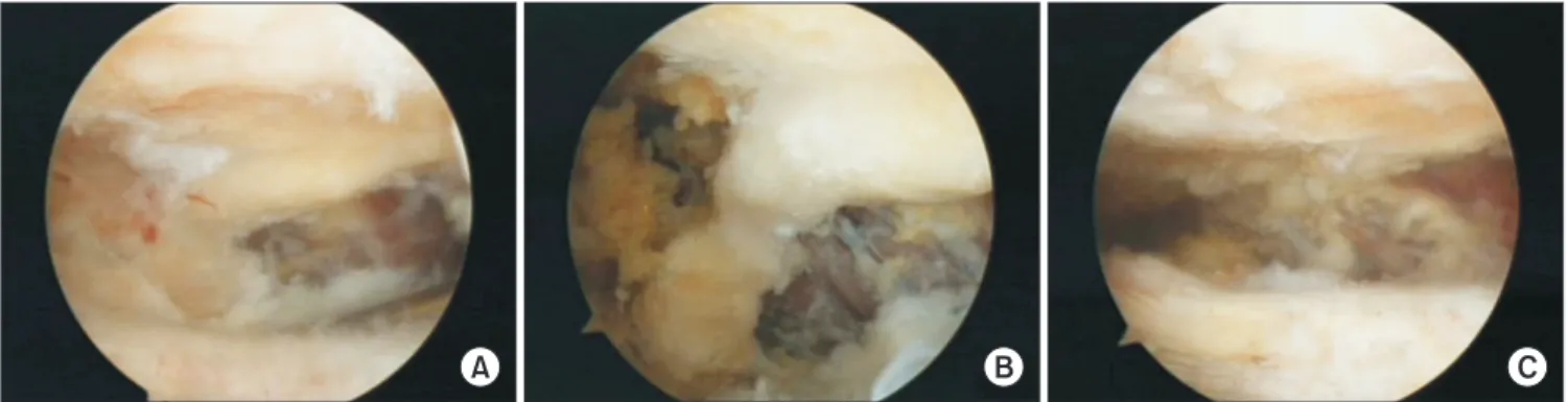
caused by ossification of the CAL with sudden exacerbation of symptoms after playing golf a week earlier, we conducted arthroscopic subacromial decompression, removal of the os- sified CAL, and rotator cuff repair using suture anchors under general anesthesia. The surgery was performed with the patient in a lateral decubitus position using conventional anterior and posterior approaches. An articular sided partial-thickness tear on the subscapularis tendon was observed on joint endoscopy via the posterior approach, which was repaired accordingly. The subscapularis partial-thickness tear is thought to have occurred incidentally, without relation to CAL ossification. Moreover, bur- sal synovial congestion and hypertrophy near the subacromial were observed. Additionally, subacromial osteophytes and CAL ossification, which has a form similar to the pillars in the calcified cave, were discovered. A burr tool was used to remove the CAL ossification thorough subacromial decompression (Fig. 3). Unlike on MRI, a full-thickness, small size (1.5 cm) tear in the far ante- rior portion of the supraspinatus tendon was observed; however, no patterns of acute tears, such as bleeding in the tear area, were observed. Therefore, rotator cuff repair was performed us- ing two suture anchors (Fig. 4).
The right shoulder pain dissipated immediately after sur- gery. The patient began performing passive joint motions after 4 weeks, followed by active joint motions after 6 weeks of immobilization using an abduction brace in accordance with
the rehabilitation program following rotator cuff repair. Three months postoperatively, the patient began performing muscle- strengthening exercises. At 1-year follow-up, the patient had no right shoulder pain, and his ROM had recovered to a normal range. Neer and Hawkins signs were all negative, indicating that there were no signs of impingement (Constant score, 65 points), while reformation of the CAL ossification was not found on the plain radiograph (Fig. 5).
Discussion
SAIS is a disease in which changes in the subacromial tissue, rotator cuff tendon, subacromial bursa, and biceps long head tendon occur owing to reduced subacromial space. This disease is the most common shoulder disease diagnosed in primary care institutions, accounting for 44% to 65% of all shoulder pains.4,5) SAIS may have internal causes, such as rotator cuff hypertrophy, rotator cuff calcium deposit, and hypertrophy of structures that pass underneath the coracoacromial arch, including the sub- acromial bursa, as well as external causes, such as subacromial osteophytes, acromial fracture, os acromiale, osteophytes in the acromioclavicular joint, and exostosis of the humeral greater tubercle. In particular, the anteroinferior acromion that connects to the CAL has received attention as an important factor in the pathogenesis of SAIS in the rotator cuff. Burns and Whipple6)
A B C
Fig. 3. Arthroscopic excision of the coracoacromial ligament ossification. (A) Coracoacromial ligament ossification is shown in the subacromial space. (B, C) Coracoacromial ligament ossification was excised using a shaver and burr.
A B
Fig. 4. Arthroscopic repair of the torn rota- tor cuff (supraspinatus tendon). (A) Full- thickness tear of the supraspinatus tendon at the far anterior portion was confirmed.
(B) Full-thickness tear of the supraspinatus tendon was repaired using suture anchors.
conducted a cadaveric study of the relationship between the CAL and the supraspinatus tendon and found that CAL caused greater impingement than the acromion or supraspinatus ten- don.
Sarkar et al.7) reported that the fibers near the acromion form a fibrovascular layer before attaching to the bone, and that chondrocytes can be found at this transitional area, between the ligament and bone. Fibro-cartilaginous formation at the tip of the spur can represent a site of enchondral ossification. Bone spurs probably develop because of tensile forces transmitted through the ligament, which are associated with thickening of the fibro- cartilaginous layer and disruption of the anchoring fibers of CAL at the anterior portion of the acromion.7)
A case report by Morimoto et al.3) indicated that calcification and ossification of the CAL can cause shoulder impingement syndrome, while Chen and Bohrer2) reported that these events occur together with calcification of the coracoclavicular liga- ment. Moreover, Reichmister et al.1) stated that, if ossification of the CAL is found on the plain radiograph of a patient with SAIS, then rotator cuff damage should be strongly suspected.
Morimoto et al.3) reported satisfactory outcomes after per- forming ossified CAL removal and rotator cuff tear repair in 37 athletes. Further, Reichmister et al.1) reported favorable out- comes when performing ossified CAL removal and rotator cuff tear repair on four patients suspected of having impingement syndrome and full-thickness rotator cuff tear based on arthro- gram or MRI findings among eight patients with CAL ossification detected on plain radiographs.
Fealy et al.8) stated that the CAL is made up of an anterolat- eral band and a posteromedial band, and that the anterolateral band is closely associated with SAIS, while emphasizing the im- portance of adequate removal of the visualized the anterolateral corner of the acromion during surgery for symptom alleviation.
During surgery, it is important to keep in mind the important role the CAL plays as a static restraint to superior humeral head migration. This is especially important for patients who have an anterosuperior portion massive rotator cuff tear, in whom sacri- ficing or removal of the CAL may cause a significant increase of glenohumeral translation and instability.
There is no definite explanation for the cause and pathogen- esis of rotator cuff disease, suggesting that the causes may be multifactorial. The relatively simple and easy to understand clas- sification of pathogenic mechanisms is divided into extrinsic fac- tors caused by mechanical impingement and intrinsic factors by degenerative changes in the rotator cuff. These two factors are also closely related, and once a disease occurs, one factor is not entirely involved in the progression of the disease. This mechani- cal impingement triggers regression of the rotator cuff (endog- enous factor), which again exacerbates the mechanical impinge- ment (exogenous factor). As a result of this vicious cycle, loss of function of the rotator cuffs is added, resulting in rupture.9) For successful treatment of the rotator cuff diseases, it is essential to understand the function and structure of the rotator cuff, as well as to clarify the pathogenesis and natural history of its disorder.
As shown in the literature review, if ossification of the CAL is found on plain radiographs and symptoms of impingement syn- drome are present, then an accompanying rotator cuff tear must be considered. If present, this condition may be treated success- fully via arthroscopic removal of the ossified CAL and repair of the torn rotator cuff.
References
1. Reichmister JP, Reeder JD, McCarthy E. Ossification of the coracoacromial ligament: association with rotator cuff pathol- ogy of the shoulder. Md Med J. 1996;45:849-52.
Fig. 5. One year postoperative radiographs (shoulder anteroposterior, supraspinatus outlet views) showing the complete removal of the coracoacromial ligament ossification without recurrence.
2. Chen YM, Bohrer SP. Coracoclavicular and coracoacromial lig- ament calcification and ossification. Skeletal Radiol. 1990;19:
263-6.
3. Morimoto K, Mori E, Nakagawa Y. Calcification of the cora- coacromial ligament. A case report of the shoulder impinge- ment syndrome. Am J Sports Med. 1988;16:80-1.
4. Koester MC, George MS, Kuhn JE. Shoulder impingement syn- drome. Am J Med. 2005;118:452-5.
5. Michener LA, McClure PW, Karduna AR. Anatomical and biomechanical mechanisms of subacromial impingement syn- drome. Clin Biomech (Bristol, Avon). 2003;18:369-79.
6. Burns WC, Whipple TL. The functional relationship of the coracoacromial ligament, supraspinatus tendon and biceps
tendon. Orthop Trans. 1989;13:237.
7. Sarkar K, Taine W, Uhthoff HK. The ultrastructure of the cora- coacromial ligament in patients with chronic impingement syndrome. Clin Orthop Relat Res. 1990;(254):49-54.
8. Fealy S, April EW, Khazzam M, Armengol-Barallat J, Bigliani LU.
The coracoacromial ligament: morphology and study of acro- mial enthesopathy. J Shoulder Elbow Surg. 2005;14:542-8.
9. Gramstad GD, Yamaguchi K. Anatomy, pathogenesis, natural history, and nonsurgical treatment of rotator cuff disorders, In:
Galatz LM, ed. Orthropaedic knowledge update: shoulder and elbow 3. Rosemont, IL: American Academy of Orthopae- dic Surgeons; 2008. 149-59.