Intramedullary Screw Fixation for Clavicle Shaft Fractures:
Comparison of the Anterograde versus the Retrograde Technique
Yong Girl Rhee , Nam Su Cho, Sung Whan Cho, Jong Hoon Song
Department of Orthopedic Surgery, College of Medicine, Kyung Hee University, Seoul, Korea
Background: The purpose of this study was to investigate the difference between two nailing approaches of intramedullary screw fixa- tion, the retrograde nailing versus the anterograde nailing, on the radiological and clinical outcomes in patients with clavicle shaft fractures.
Methods: From April 2002 to August 2014, we enrolled a total of 22 patients with clavicle shaft fractures to participate in this study.
Twelve patients received retrograde intramedullary nailing and 10 received anterograde nailing. The average duration of follow-up was 12 months. In all the patients, we took follow-up radiographs of the anteroposterior and the axial views to assess the postoperative ra- diological outcomes. We measured the visual analogue scale (VAS) score, American Shoulder and Elbow Surgeons (ASES) score, and the range of motion (ROM).
Results: Clinically, we did not find a statistically significant difference in the retrograde group and the anterograde group in terms of the duration to bone union, the VAS score the ASES score and the ROMs. Radiologically, we found that the difference in the clavicle short- ening of the affected arm and the unaffected arm did not show a statistically significant difference at the immediate postoperative as- sessment. we found that the difference in the clavicle shortening of the affected arm between the immediate postoperative and the final follow-up value did not show a statistically significant difference.
Conclusions: We found that both the retrograde nailing and the anterograde nailing gave a favorable outcome for clavicle shaft frac- tures. Although we saw evidence of clavicle shortening after intramedullary screw fixation, this was not a factor that influenced clinical outcome.
(Clin Shoulder Elbow 2016;19(1):8-14)
Key Words: Fracture; Clavicle; Anterograde; Retrograde Clinics in Shoulder and Elbow Vol. 19, No. 1, March, 2016 http://dx.doi.org/10.5397/cise.2016.19.1.8
Received April 1, 2015. Revised November 16, 2015. Accepted November 30, 2015.
Correspondence to: Yong Girl Rhee
Shoulder and Elbow Clinic, Department of Orthopedic Surgery, College of Medicine, Kyung Hee University, 23 Kyungheedae-ro, Dongdaemun- gu, Seoul 02447, Korea
Tel: +82-2-958-8370, Fax: +82-2-964-3865, E-mail: [email protected] Financial support: None. Conflict of interests: None.
Introduction
Clavicle shaft fractures are common and compose 2.6% of all fractures.1) They compose 88% of all fractures of the shoulder girdle and 80% of all fractures of the clavicle.2) Despite such a high frequency in prevalence, controversy still remains as to the relative benefits of surgical and conservative treatment for clavi- cle shaft fractures. Most authors propose that fractures should be treated using the figure of eight bandage. But recently, because of the use of bandages is troublesome, the patient’s desire for expedited return to activity, and the favorable outcomes associ- ated with surgical treatment, many patients now lean toward
receiving surgical treatment over conservative treatment. There are various approaches and techniques to surgically treat clavicle shaft fractures: intramedullary fixation using K-wires,3) Rush pins, Knowles pins, Steinmann pins,4) Hagie and Rockwood pins, elas- tic stable intramedullary nails,5) intramedullary screw fixation,6) external fixation,7) fixation with plating,8) and minimally invasive plate osteosynthesis (MIPO) fixation.9) Yet none of these have been singled out as the gold standard of treatment. The ten- dency to treat clavicle shaft fractures using plating has increased after the advent of MIPO fixation10) and intramedullary fixation is another widely used alternative for clavicle shaft fractures.11) Of the types of fixation, the intramedullary screw fixation is useful
in that it builds upon the weaknesses of simple intramedullary fixation of instability by fixing the far cortex; the technique pro- vides rotational stability, though not perfectly, through interfrag- ment compression. Through a retrograde intramedullary nailing of the screw, a technique introduced by Boehme et al.,12) many authors have shown good clinical outcomes.13-16) We devised an anterograde intramedullary nailing approach to treat clavicle shaft fractures and used that along with the previously described retrograde approach.
The purpose of this study was to investigate and compare the outcomes of retrograde intramedullary screw fixation and of anterograde intramedullary screw fixation for clavicle shaft frac- tures. We made a comparative analysis of the two approaches using the radiological and clinical outcomes. We hypothesized that the anterograde approach is superior to the retrograde ap- proach.
Methods
The institutional review board (IRB) an exemption for this study due to its retrospective design (KMC IRB 1513-04). From April 2002 and August 2014, we considered the following pa- tients with clavicle shaft fractures for enrollment in our study:
patients who received surgery for the fracture within 2 weeks of sustaining the injury and patients who were able to partake in at least one year of follow-up. Those with multiple fractures were excluded. A total of 22 patients fulfilled the inclusion criteria and were enrolled into our study. We retrospectively analyzed the patients. We determined that as indications for surgery patients must have a clavicle shortening of more than 1.2 cm or have skin tenting. The contraindications for surgery were as follows:
1) severe comminuted clavicle fractures; 2) proximal or distal 1/3 clavicle fractures; 3) open fractures; and 4) clavicle fractures with concomitant neurological symptoms. The study participants were 16 men and 6 women with an average age of 36.8 years (range, 19–76 years). The average duration of follow-up was 12 months (range, 4–23 months). We performed retrograde intramedullary nailing until 2006, but we concerned that this approach was associated with distancing between the posterior cortical bone of lateral fragment and the anteromedial cortical bone of medial fragment, devised an anterograde intramedul- lary nailing approach to address this and utilized this approach instead from 2014.
We classified the clavicle fractures according to the classifica- tion system established by Robinson et al.8): type 2a1 fractures are cortical alignment fractures without displacement; type2a2 fractures are cortical alignment fractured with angulation of the distal fracture fragment; type2b1 fractures are cortical displaced fractures with simple, wedge comminution; and type 2b2 frac- tures are cortical displaced fractures that are mutlifragmentary and segmental. In our study 20 patients had a type 2b1 fracture
and 2 patients had type 2b2 fractures. For the treatment of their clavicle shaft fractures, 12 patients received retrograde intramed- ullary nailing and were allocated into the retrograde group, and 10 received anterograde intramedullary nailing and were allo- cated in the anterograde group.
Assessment of Outcomes
We took radiographs of the preoperative antero-posterior and axial views of the shoulder in every patient to measure the length of the affected and the unaffected clavicle. The same ra- diographs were taken postoperatively at the following periods:
2 weeks, 4 weeks, 6 weeks, 3 months, 6 months, and 1 year.
We used the radiographs to assess clavicle length of both arms, the extent of bone union, strength of screw fixation, and bone nonunion. We determined bone union as the point either when range of motion (ROM) was restored without concomitant clini- cal pain or with radiologically assessed resolution of fracture line or when the callus fracture is remodeled into trabecular bone.
The clavicle shortening was measured as the difference between the clavicle length at the immediate postoperative follow-up and at the final follow-up. For the clinical assessment, we analyzed pain at the final follow-up using the visual analogue scale (VAS) score for subjective pain and the American Shoulder and Elbow Surgeons (ASES) score. The ROM for anterior flexion, external rotation, internal rotation, and abduction were assessed. We using a portable, hand-held Nottingham Mecmesin Myometer (Mecmesin Co., Nottingham, UK) measured muscle strength relative to the unaffected contralateral side. At the final follow- up, we also rated the level of subjective satisfaction and divided the resulting evaluation as either satisfied or unsatisfied.
Statistical Analysis
We performed the statistical analyses of the results using the IBM SPSS software package ver. 20.0 (IBM Co., Armonk, NY, USA). The results were analyzed using the Mann-Whitney test.
We considered statistical significance at a p-value of less than 0.05.
Surgical Techniques
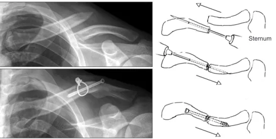
The surgery was performed with the patients under general anesthesia and in beach chair position. For the anterograde ap- proach (Fig. 1), we made a skin incision of around 2.5 cm fol- lowing the Langer line above the clavicular fracture. Then, after the soft tissue was dissected, we exposed the fracture site. We positioned the K-wire pointing towards the intramedullary of the proximal fracture fragment, then the K-wire was penetrated through the anterospoterior cortical bone of the proximal frac- ture fragment until it could be palpated subcutaneously (Fig. 2).
At the site of K-wire protrusion, we made a skin incision and pulled out the K-wire medially until it could no longer be seen at the fracture site. With the K-wire rested within the antero-pos-
terior cortical bone of the proximal fracture fragment, we used a bone forcep to perform reduction of the fracture. If the reduc- tion was incomplete in clavicle shaft fractures with large fracture fragments, we performed wiring of the fracture site (Fig. 3). After an anatomical reduction was confirmed, the K-wire was pulled out passing the fracture site until it rested at the antero-posterior cortical bone of the distal fracture fragment. Then, using a K- wire of the same length, we measured the length of the intra- medullary K-wire. After, we inserted the intramedullary K-wire until it completely pierced through the proximal posterosuperior cortical bone and was exposed to the cutaneous compartment.
Following the K-wire, we made a 3.5 mm drilling and inserted a cannulated screw after tapping (Fig. 4). Taking the tapper as the standard, we confirmed the intramedullary space. For women or pediatric patients with relatively small bodyweight, we used a
4.0 mm cannulated screw, whereas for patients of normal adult bodyweight, we used a 5.0 mm cannulated screw. The fracture site was exposed in the same way as the anterograde approach for the retrograde approach. The K-wire was positioned in the direction of the intramedulla of the distal fracture fragment and inserted until the posteroinferior cortical bone was penetrated.
We performed an anatomical reduction of the fracture site and inserted the K-wire into the proximal fracture fragment until it rested across the anterosuperior cortices. We made the mea- surement as described before and, following the K-wire, we inserted the cannulated screw (Fig. 5). An arm sling was applied for postoperative 4 weeks, and immediate postoperatively pas- sive joint motions were performed.
Sternum
Fig. 1. Anterograde technique of intra- medullary screw fixation.
Fig. 2. The K-wire was aimed obliquely toward the anterio-inferior cortex of
the medial fragment. Fig. 3. Wiring was used to correct an incomplete reduction.
Results
Clinical Outcomes
The retrograde group comprised 12 patients and the antero- grade group, 10 patients. The average age of the patients in the retrograde group was 33.8 years and in the anterograde group, 40.6 years. The average duration of follow-up was 13.2 months in the retrograde group and 14.1 months in the anterograde group (p>0.05). Eight patients in the retrograde group received wiring to hold the fracture fragments, and 6 received wiring in the anterograde group. The clavicle shaft fractures of the patients were classified according to the Robinson classification system as follows: in the retrograde group, none had type 2A1 or type 2A2 fractures, 11 patients had a type 2b1 fracture, and 1 patient had a type 2b2 fracture; in the anterograde group, none had type 2a1 or type 2a2 fractures, 9 patients had a type 2b1 fracture, and 1 patient had a type 2b2 fracture. The significant difference
in distribution in classification were not seen between the retro- grade and the anterograde group (p=0.453).
At the final follow-up, we found that none of the parameters of clinical outcome showed a statistically significant difference between the two treatment approaches. The VAS score was 1.5 ± 1.9 in the retrograde group and 0.5 ± 0.6 in the antero- grade group (p=0.283). The ASES score was 86.6 ± 11.5 in the retrograde group and 90.2 ± 9.1 in the anterograde group (p=0.579). For the ROMs of the joint, the anterior flexion was 163o ± 8.0o in the retrograde group and 165o ± 5.3o in the an- terograde group (p=0.697), external rotation was 64o ± 6.9o in the retrograde group and 61.5o ± 4.5o in the anterograde group (p=0.571), the internal rotation was T7.4 ± 1.8 in the retro- grade group and T6.9 ± 2.2 in the anterograde group (p=0.353), andabduction was 124o ± 16.2o in the retrograde group and 115o ± 9.9o in the anterograde group (p=0.07) (Table 1). The two groups did not show a statistically significant difference in muscle strength neither during forward elevation nor abduction.
The difference in the muscle strength during forward elevation
Fig. 4. A cannulated screw (5.0 mm) was inserted through the K-wire.
Sternum
Fig. 5. Retrograde technique of intramedul- lary screw fixation.
Table 1. Clinical Outcomes of the Retrograde Group and the Anterograde Group
Variable Retrograde
(n=12) Anterograde
(n=10) p-value*
VAS (points) 1.5 ± 1.9 0.5 ± 0.6 0.283
ASES (points) 86.6 ± 11.5 90.2 ± 9.1 0.579
Range of motion (degree)
Active further flexion 163 ± 8.0 165 ± 5.3 0.697 External rotation at the side 64 ± 6.9 61.5 ± 4.5 0.571 Internal rotation to the back T7.4 ± 1.8 T6.9 ± 2.2 0.353
Abduction 124 ± 16.2 115 ± 9.9 0.07
Values are presented as mean ± standard deviation.
VAS: visual analogue scale, ASES: American Shoulder and Elbow Surgeons.
*Statistically significance was set to p<0.05.
of the affected and unaffected side was 0.8 ± 1.5 kg in the retrograde group and 0.7 ± 1.3 kg in the anterograde group (p=0.241). At abduction, this difference was 1.8 ± 2.3 kg in the retrograde group and 1.4 ± 1.9 kg in the anterograde group (p=0.478).
Twenty out of 22 patients rated their level of subjective satis- faction of the surgical outcome as satisfactory. Two patients rated their outcome as unsatisfactory. The reason for the unsatisfactory outcome was found to be the case because of a bone nonunion in one patient from the retrograde group and because of a deep infection in one patient in the anterograde group, for whom we carried out an incision and drainage to address the complica- tion.
Radiological Outcomes
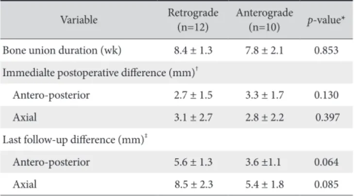
We found that 21 of 22 patients (95.5%) achieved a success- ful bone union after the surgical treatment. The single patient in whom bone nonunion was found was part of the retrograde group. The time to bone union was on average 8.4 ± 1.3 weeks in the retrograde group and 7.8 ± 2.1 weeks in the anterograde group, but a statistically significant difference was not seen between the groups (p=0.853). The postoperative difference in the affected and unaffected clavicle length in the antero- posterior view was 2.7 ± 1.5 mm in the retrograde group and 3.3 ± 1.7 mm in the anterograde group (p=0.130) and in the axial view was 3.1 ± 2.7 mm in the retrograde group and 2.8 ± 2.2 mm in the anterograde group (p=0.397), again not showing a statistically significant difference. At the final follow-up, nei- ther did we find a statistically significant difference between the preoperative and the immediate postoperative difference in the clavicle length of the affected arm: in the antero-posterior view, this was 5.6±1.3 mm in the retrograde group and 3.6 ± 1.1 mm in the anterograde group (p=0.064) and, in the axial view, 8.5 ± 2.3 mm in the retrograde group and 5.4 ± 1.8 mm in the anterograde group (p=0.085) (Table 2).
Complications
A total of 10 patients presented with postoperative compli- cations; the retrograde and the anterograde group each had 5 patients who presented with complications (Table 3). The follow- ing complications were seen in the retrograde group: 2 patients had delayed bone union, but with surveillance bone union was achieved and pain resolved; 1 patient had bone nonunion, so for whom the screw was removed; 1 patient had angulated fracture and decreased anterior flexion; and 1 patient had flar- ing and edema at the area of surgery, but with surveillance the symptoms spontaneously resolved. The following complications were seen in the anterograde group: 1 patient had deep infec- tion at the area of surgery, so incision and drainage were per- formed; 2 patients showed loosening of screw fixation, but with surveillance bone union was nevertheless achieved; and 2 pa- tients had skin tenting but pain was not complained, and screws were removed after bone union.
Discussion
In this study we found that intramedullary screw fixation for clavicle shaft fractures using either the retrograde or the antero- grade approach showed good postoperative clinical outcomes.
Conservative treatment is generally recommended for most clav- icle shaft fractures. But a recent study by Robinson et al.17) found that in 581 patients treated through conservative treatment, 21%
of them presented with bone non-union. Another study by Zlo- wodzki et al.18) found that in 159 patients who received conser- vative treatment around 15.1% showed non-union. Wick et al.19) found that a high prevalence of diastasis was associated with patients who received conservative treatment for clavicle shaft fractures, which may have led to the decreased clinical outcome with a nonunion rate of 10% to 15%. Therefore, it is becom- ing more evident that conservative measures alone may not be sufficient for the treatment of clavicular fractures. An alternative to conservative treatment is surgical treatment, of which there are several types. For the surgical treatment of the clavicle shaft fractures, the plating and screw fixation are the most commonly used tools.20) Many papers have shown that plating and screw Table 2. Bone Union and Clavicle Shortening in the Retrograde Group and
the Anterograde Group
Variable Retrograde
(n=12) Anterograde
(n=10) p-value*
Bone union duration (wk) 8.4 ± 1.3 7.8 ± 2.1 0.853 Immedialte postoperative difference (mm)†
Antero-posterior 2.7 ± 1.5 3.3 ± 1.7 0.130
Axial 3.1 ± 2.7 2.8 ± 2.2 0.397
Last follow-up difference (mm)‡
Antero-posterior 5.6 ± 1.3 3.6 ±1.1 0.064
Axial 8.5 ± 2.3 5.4 ± 1.8 0.085
*Statistically significance was set to p<0.05. †Unaffected side-affected side.
‡Difference between the values of the immediate postoperative and the last follow-up clavicle length of the affected side.
Table 3. Complications
Variable Retrograde (n=12) Anterograde (n=10)
Non-union (n) 1 0
Delayed union (n) 2 0
Angulation (n) 1 0
Superficial infection (n) 1 0
Deep infection (n) 0 1
Screw problem (loosening) (n) 0 2
Skin tenting (n) 0 2
fixation is associated with a higher rate of bone union and en- hanced clinical outcome than conservative treatment.8,18,21,22)
However, plating and screw fixation require a large skin inci- sion23) and a relatively long duration of surgery. Especially, after removal of the plate, the screw holes may induce a stress riser leading to unnecessary refractures. Further, a large incision that is required at the time of fixation and later at the removal of the plate may increase the risk of infection.24) Conversely, authors such as Ferran et al.25) and Liu et al.26) have reported that the complications and clinical outcomes of intramedullary fixation using either plating or screw fixation did not show a clinically significant difference.
The intramedullary screw fixation compared to plating re- quires a smaller skin incision and less periosteal peeling. Further, it has relative advantages in that the screw itself can sustain some of the weight itself alleviating burden on the fracture site and exert compression on the fracture site. Khalil6) found that a better fixation, stability, and clinical outcome can be achieved during intramedullary fixation using a screw over plating. In this study, we found that the average ASES score was 88.4 showing a satisfactory outcome with intramedullary screw fixation. We did not find a statistically significant difference in terms of the clinical scores (VAS score, ASES score, and the ROMs) between patients of the retrograde group and the anterograde group.
Abo El Nor27) reported a postoperative clavicle shortening of an average 4 mm after intramedullary screw fixation. In our study, the average postoperative clavicle shortening was 5.8 mm in the antero-posterior view and 9.3 mm in the axial view at the final follow-up. We found that the extent of clavicle shorten- ing was greater in the retrograde group than in the anterograde group, but no difference was found statistically. Hill et al.28) found that a clavicle shortening of more than 20 mm and Laza- rides and Zafiropoulos,29) a clavicle shortening of more than 15 mm were associated with a poor prognosis. Conversely, Nowak et al.30) found that clavicle shortening was not an accurate prog- nostic marker for clinical outcome and that there were other better markers of prognosis. In this study, we found that although clavicle shortening occurred more in the retrograde group than in the anterograde group, the clinical outcomes between two were not statistically different showing that the extent of clavicle shortening were not associated with clinical outcome or were not severe enough to influence it.
The posterior thick trapezius muscle must be dissected so that the screw can be inserted deeply in the retrograde approach.
During this approach, because the screw length cannot be as- sessed accurately, it is difficult to choose an appropriate screw.
Further, because the posterosuperior-anteromedial cortical in- terval where the intramedullary screw is laid has the tendency to lengthen, the retrograde has another disadvantage in that sometimes it is difficult to remove the screw after bone union. To address this problem, we used a screw that as 2.6 mm longer for
the retrograde rather than the one used for the anterograde ap- proach. The anterograde approach has the advantage in that it enters from above the clavicle and that screw fixation is relatively easy. A few studies on the bicortical fixation during intramedul- lary fixation of clavicle shaft fractures have found that when the cortices are penetrated with screws, a resulting stress riser increases the chances of a fracture especially during elevation or the abduction of the arm. But in our study, bicortical fixation did neither limit the forward elevation or abduction of the arms nor induce any fractures. Other authors have reported that in the instance a curvature of the clavicle that necessitates the use of a straight screw to stabilize cortices of both ends of the fracture site, the existing fracture may be inadvertently extended or a new fracture may be induced during this process. To avoid this, in our study we pre-inserted guide pins to position the K-wire and to help ultimately to guide directionality and positioning of the screw. Because excessive interfragmentary compression in comminuted fractures can lead to clavicle shortening, wiring especially for segmental comminuted fractures can be used for prophylactic prevention of clavicle shortening.
Several limitations to this study exist: the size of the sample number was small, being only 22; the study was a retrospective study; time-span during which the two treatment approaches were performed does not overlap at all; and an inter-observer difference in measuring the clavicle length may exist. We at- tempted to address some of these limitations by selecting a sin- gle surgeon to carry out all the treatments, both the anterograde and the retrograde approach and two physicians who received training from the same fellowship program to measure the clav- icle length. Yet prospective large scale studies that compare the outcomes prospectively are required.
Conclusion
We found that the retrograde and the intramedullary screw fixation show comparatively favorable clinical outcomes for clavicle shaft fractures. Our results show that both approaches of intramedullary screw fixation are ideal options for the surgical treatment of these fractures. Interestingly, we found that post- operative clavicle shortening was not an influencing factor of clinical outcome. Although intramedullary screw fixation is not a method that gives the most stabilization, it can still be regarded as one of the beneficial treatment options for clavicular fractures for its capacity to stabilize the far cortex.
References
1. Postacchini F, Gumina S, De Santis P, Albo F. Epidemiology of clavicle fractures. J Shoulder Elbow Surg. 2002;11(5):452-6.
2. Nowak J, Mallmin H, Larsson S. The aetiology and epidemiol- ogy of clavicular fractures. A prospective study during a two-
year period in Uppsala, Sweden. Injury. 2000;31(5):353-8.
3. Ripstein CB. Kirschner wire fixation in fractures of the clavicle.
Can Med Assoc J. 1948;59(3):255-7.
4. Payne DE, Wray WH, Ruch DS, Zura RD, Moorman CT. Out- come of intramedullary fixation of clavicular fractures. Am J Orthop (Belle Mead NJ). 2011;40(6):E99-104.
5. Hartmann F, Hessmann MH, Gercek E, Rommens PM. Elastic intramedullary nailing of midclavicular fractures. Acta Chir Belg. 2008;108(4):428-32.
6. Khalil A. Intramedullary screw fixation for midshaft fractures of the clavicle. Int Orthop. 2009;33(5):1421-4.
7. Schuind F, Pay-Pay E, Andrianne Y, Donkerwolcke M, Rasquin C, Burny F. External fixation of the clavicle for fracture or non- union in adults. J Bone Joint Surg Am. 1988;70(5):692-5.
8. Robinson CM, Goudie EB, Murray IR, et al. Open reduction and plate fixation versus nonoperative treatment for displaced midshaft clavicular fractures: a multicenter, randomized, con- trolled trial. J Bone Joint Surg Am. 2013;95(17):1576-84.
9. Sohn HS, Shin SJ, Kim BY. Minimally invasive plate osteosyn- thesis using anterior-inferior plating of clavicular midshaft frac- tures. Arch Orthop Trauma Surg. 2012;132(2):239-44.
10. Sohn HS, Kim WJ, Shon MS. Comparison between open plat- ing versus minimally invasive plate osteosynthesis for acute dis- placed clavicular shaft fractures. Injury. 2015;46(8):1577-84.
11. Hill CE. Is intramedullary nailing more effective than non-op- erative treatment in adults with displaced middle-third clavicle fractures? J Orthop Traumatol. 2014;15(3):155-64.
12. Boehme D, Curtis RJ Jr, DeHaan JT, Kay SP, Young DC, Rock- wood CA Jr. The treatment of nonunion fractures of the midshaft of the clavicle with an intramedullary Hagie pin and autogenous bone graft. Instr Course Lect. 1993;42:283-90.
13. Marlow WJ, Ralte P, Morapudi SP, Bassi R, Fischer J, Waseem M. Intramedullary fixation of diaphyseal clavicle fractures using the rockwood clavicle pin: review of 86 cases. Open Orthop J.
2012;6:482-7.
14. Mudd CD, Quigley KJ, Gross LB. Excessive complications of open intramedullary nailing of midshaft clavicle fractures with the Rockwood Clavicle Pin. Clin Orthop Relat Res. 2011;
469(12):3364-70.
15. Neer CS 2nd. Nonunion of the clavicle. J Am Med Assoc.
1960;172:1006-11.
16. Rowe CR. An atlas of anatomy and treatment of midclavicular fractures. Clin Orthop Relat Res. 1968;58:29-42.
17. Robinson CM, Court-Brown CM, McQueen MM, Wakefield AE. Estimating the risk of nonunion following nonopera- tive treatment of a clavicular fracture. J Bone Joint Surg Am.
2004;86-A(7):1359-65.
18. Zlowodzki M, Zelle BA, Cole PA, Jeray K, McKee MD;
Evidence-Based Orthopaedic Trauma Working Group. Treat- ment of acute midshaft clavicle fractures: systematic review of 2144 fractures: on behalf of the Evidence-Based Orthopaedic Trauma Working Group. J Orthop Trauma. 2005;19(7):504-7.
19. Wick M, Müller EJ, Kollig E, Muhr G. Midshaft fractures of the clavicle with a shortening of more than 2 cm predispose to nonunion. Arch Orthop Trauma Surg. 2001;121(4):207-11.
20. Bernstein J. Nonoperative treatment compared with plate fixation of displaced midshaft clavicular fractures. J Bone Joint Surg Am. 2007;89(8):1866; author reply 1866-7.
21. McKee MD, Seiler JG, Jupiter JB. The application of the limited contact dynamic compression plate in the upper extremity: an analysis of 114 consecutive cases. Injury. 1995;26(10):661-6.
22. Poigenfürst J, Rappold G, Fischer W. Plating of fresh clavicular fractures: results of 122 operations. Injury. 1992;23(4):237-41.
23. S Thyagarajan D, Day M, Dent C, Williams R, Evans R. Treat- ment of mid-shaft clavicle fractures: a comparative study. Int J Shoulder Surg. 2009;3(2):23-7.
24. Böstman O, Manninen M, Pihlajamäki H. Complications of plate fixation in fresh displaced midclavicular fractures. J Trauma.
1997;43(5):778-83.
25. Ferran NA, Hodgson P, Vannet N, Williams R, Evans RO.
Locked intramedullary fixation vs plating for displaced and shortened mid-shaft clavicle fractures: a randomized clinical trial. J Shoulder Elbow Surg. 2010;19(6):783-9.
26. Liu HH, Chang CH, Chia WT, Chen CH, Tarng YW, Wong CY. Comparison of plates versus intramedullary nails for fixa- tion of displaced midshaft clavicular fractures. J Trauma.
2010;69(6):E82-7.
27. Abo El Nor T. Displaced mid-shaft clavicular fractures: surgi- cal treatment with intramedullary screw fixation. Arch Orthop Trauma Surg. 2013;133(10):1395-9.
28. Hill JM, McGuire MH, Crosby LA. Closed treatment of dis- placed middle-third fractures of the clavicle gives poor results.
J Bone Joint Surg Br. 1997;79(4):537-9.
29. Lazarides S, Zafiropoulos G. Conservative treatment of frac- tures at the middle third of the clavicle: the relevance of short- ening and clinical outcome. J Shoulder Elbow Surg. 2006;
15(2):191-4.
30. Nowak J, Holgersson M, Larsson S. Can we predict long-term sequelae after fractures of the clavicle based on initial find- ings? A prospective study with nine to ten years of follow-up. J Shoulder Elbow Surg. 2004;13(5):479-86.