Risk Factors for Recurrence of Anterior Shoulder Instability after Arthroscopic Surgery with Suture Anchors
Chang-Hyuk Choi , Seok-Jun Kim, Seung-Bum Chae, Jae-Keun Lee, Dong-Young Kim
Shoulder and Elbow Clinic, Daegu Catholic University Medical Center; Department of Orthopedic Surgery, Catholic University of Daegu School of Medicine, Daegu, Korea
Background: We investigated the risk factors for the recurrence of anterior shoulder instability after arthroscopic surgery with suture an- chors and the clinical outcomes after reoperation.
Methods: A total of 281 patients (February 2001 to December 2012) were enrolled into our study, and postoperative subluxation and dislocation were considered as recurrence of the condition. We analyzed radiologic results and functional outcome including the Ameri- can Shoulder and Elbow Surgeons Evaluation Form, the Korean Shoulder Society Score, and the Rowe scores.
Results: Of the 281 patients, instability recurred in 51 patients (18.1%). Sixteen out of 51 patients (31.4%) received a reoperation.
In terms of the functional outcome, we found that the intact group, comprising patients without recurrence, had a significantly better functional outcome than those in the recurrent group. Age and the size of glenoid defect at the time of initial surgery significantly dif- fered between intact and recurrent group (p<0.05). We found that the number of dislocations, the time from the initial presentation of symptoms to surgery, and the number of anchor points significantly differed between initial operation and revision group (p<0.05). The functional outcome after revision surgery was comparable to intact group after initial operation.
Conclusions: Eighteen percent of recurrence occurred after arthroscopic instability surgery, and 5.6% received reoperation surgery. Risk factors for recurrence were young age and the initial size of glenoid defect. In cases of revision surgery, good clinical outcomes could be achieved using additional suture anchor.
(Clin Shoulder Elbow 2016;19(2):78-83)
Key Words: Shoulder; Joint instability; Arthroscopic surgery; Suture anchors
Clinics in Shoulder and Elbow Clinics in Shoulder and Elbow Vol. 19, No. 2, June, 2016
http://dx.doi.org/10.5397/cise.2016.19.2.78
Received August 16, 2015. Revised March 31, 2016. Accepted April 23, 2016.
Correspondence to: Chang-Hyuk Choi
Department of Orthopedic Surgery, Catholic University of Daegu School of Medicine, 33 Duryugongwon-ro 17-gil, Nam-gu, Daegu 42472, Korea
Tel: +82-53-650-4276, Fax: +82-53-650-4272, E-mail: [email protected] Financial support: None. Conflict of interests: None.
Introduction
The surgical aims of arthroscopic reconstruction for anterior shoulder instability are to reconstruct the anterior labrum, to restore the shoulder glenoid cavity, and to address the abnor- mal loosening of the glenoid joint. The arthroscopic approach has been associated with a higher proportion of recurrence of shoulder instability than the open approach. But recently with developments in arthroscopic and surgical techniques, the post- operative clinical outcomes between the two approaches have now been reported to be comparable.1) Recurrence of anterior shoulder instability can be subdivided with respect to the sever-
ity of shoulder instability (apprehension, subluxation, or disloca- tion). To address the recurrence, the indication for reoperation can be made on the basis of either the prevalence or the extent of symptoms. Several factors are known to influence the postop- erative recurrence of anterior shoulder instability: factors relat- ing to the patient (age, gender, number of dislocations, sports engaged by the patient, etc.); pathologic factors (glenoid defects, humeral head defects, capsular redundancy, etc.); and surgical factors (number of suture anchors per suture, etc.).2-6) Because awareness of the risk factors and diagnostic parameters that a patient displays can influence the surgeon’s choice of surgery, it is of paramount interest to gain a better understanding of their
effects. In this study, we investigated the risk factors for the recur- rence of shoulder instability afer arthroscopic surgery with suture anchors and the clinical outcomes of reoperation.
Methods
We enrolled a total of 281 patients who had received ar- throscopic surgery for anterior shoulder instability by the same specialist between February 2001 and December 2012. The initial arthroscopic surgery had been indicated for those who had complaints of persistent shoulder instability. Those who had postoperative recurrence were also included in the recruitment.
The following exclusion criteria was applied during the recruit- ment process: posterior shoulder dislocation; multidirectional instability; severe bony Bankart lesion; huge Hill-Sach lesion (greater than 25%): shoulder hyperlaxity; and acute fracture-in- duced dislocation. We defined recurrence as postoperative sub- luxation (the diagnosis of subluxation is based on the patient’s subjective finding that their shoulder is ‘about to’ dislocate or on results of shoulder dislocation tests), as postoperative dislocation, or as complaints of shoulder instability. We tried reoperation on patients with more than two experiences of redislocation;
persistent instability during daily activities; severe limitations on sports activity; and reserved reoperation in cases of alleviation of symptoms with muscle strengthening exercises, or modification of activity.
Interscalene nerve block was used to anesthetize the patient in a beach chair position. The following arthroscopic portals were used to ascertain the appropriate arthroscopic windows:
the posterior portal, the anterolateral portal, and the anterior portal. Any preexisting sutures were removed, and revision surgery was performed provided that the patient’s labrum re- mained uncompromised. First, the glenoid was prepared to create a bleeding bed for optimal healing. Depending on the glenoid condition of each patient, we performed either simple suture with 3–4 anchors or, if bone defect was greater than 20%, a suture bridge technique. We inserted two bioabsorbable anchors (3.0 mm, PDLLA; Arthrex, Naples, FL, USA) at the 1 cm medial margin of the glenoid rim between the middle and the inferior glenohumeral ligaments. Using suture hooks, we in- serted the sutures through the glenoid capsule in a manner that allows the medial glenoid neck to be sufficiently covered and made mattress sutures. Then, at the upper and lower articular facets of the glenoid rim, we made two holes for 3.5 mm push- loc anchors (Arthrex) to complete the suture bridge repair. Un- less otherwise indicated, we used the Bio-suture tak (Arthrex) as suture anchors. With the advent of soft anchor Y-knot (CONMED, Utica, NY, USA), we used this particular anchor as the fourth suture anchor to minimize bone defect during simple sutures.
After suture repair, the patients were not placed in an abduction external rotation position since they were already in beach chair
position throughout the procedure, but they were directly asked to perform external rotation and internal rotation as they were to assess for engagment. After anterior labral suturing, additional remplissage was performed on patients with engagement. We confirmed labral integrity using probes and the stability of the glenoid capsule by examining the extent of laxity through re- expansion of the capsule after aspiration of the synovial fluid.
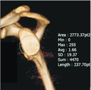
The average age of the patients at the time of initial surgery was 26 years (range, 17–58 years), and the average follow-up period was 92 months (range, 1 year–12 years 2 months). Our sample of patients included 248 male patients (88.3%) and 33 female patients (11.7%). For the functional assessment of the shoulders, we used the American Shoulder and Elbow Surgeons Evaluation Form (ASES), the Korean Shoulder Society Score (KSES), and the Rowe score. The extent of joint defect of the shoulders was measured through 3-dimensional computed to- mography and Sugaya’s method (Fig. 1).6) We analyzed the type and size of the Hill-Sach lesion in each patient and the propor- tion of postoperative recurrence. Then, we prospectively ana- lyzed the clinical outcomes of those who received reoperation for the recurrence. All statistical analyses were made with the chi-square test, two sample t-test, or binary logistic regression.
Statistical significance was set as a p-value of less than 0.05.
Results
We found that of the 281 patients a total of 51 patients presented with recurrent anterior shoulder instability (18.1%):
incomplete dislocation was seen in 26 patients (9.3%) and com- plete dislocation, in 25 patients (8.9%). The duration between operation and re-admission to hospital for redislocation was an average 3 months (range, 2–110 days). We reoperated on 16
Fig. 1. The method of glenoid defect size’s measurement.
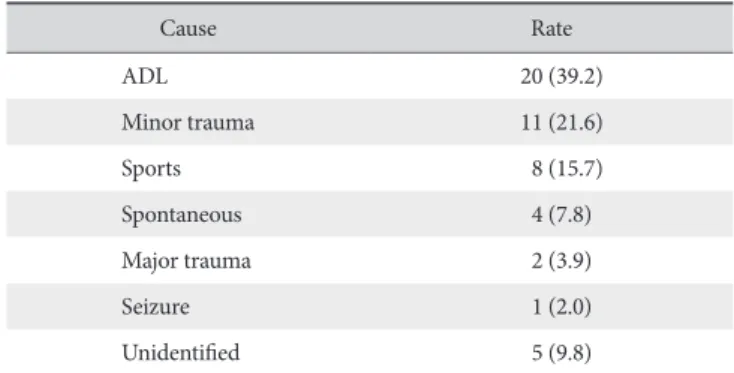
patients (31.4%) who despite conservative management such as modification of activity and muscle strengthening training had persistent subluxation or dislocation and whose symptoms did not ameliorate. In the recurrent group, the causes and the pro- portion of recurrence were as follows: activities of daily living in 39.2% (20/51); minor trauma in 21.6% (11/51); sports-related exercise in 15.7% (8/51); spontaneous redislocation in 7.8%
(4/51); major trauma in 3.9% (2/51); seizures in 2.0% (1/51);
and idiopathic in 9.8% (5/51) (Table 1).
Through a multivariate analysis, we found that there was a significant difference in the extent of the glenoid defect at the initial surgery phase between the two patient groups (p<0.05).
Although the number of anchors used did not differ between the two groups, it was close to being so (p=0.07). Gender, age, number of dislocations, and the time between first appearance of symptoms to receiving surgery did not show a statistically significant difference between the two groups (Table 2). To de- termine the factors attributable to recurrence, we carried out a multivariate analysis and found that age and the size of glenoid defect at the time of initial surgery were two significant factors (p<0.05) (Table 3). Further, the functional outcomes differed with statistical significance between the postoperative values of the intact group and the corresponding post-reoperative values of the recurrent group: in the intact group, the average ASES was 92 points; the average KSES, 94 points; and the average Rowe score, 94 points; and in the recurrent group, these correspond- ing post-reoperative values were 75 points, 76 points, and 66 Table 1. Causes of Redislocation
Cause Rate
ADL 20 (39.2)
Minor trauma 11 (21.6)
Sports 8 (15.7)
Spontaneous 4 (7.8)
Major trauma 2 (3.9)
Seizure 1 (2.0)
Unidentified 5 (9.8)
Values presented as number (%).
Table 2. Univariate Analysis between Intact Group and Recurrent Group Variable
Group
p-value Intact
(n=230) Recurrent
(n=51)
Sex (n) 0.634
Female 28 5
Male 202 46
Average age (yr) 33 33 0.804
Average No. of dislocations 7.5 7.7 0.856
Average time until 1st postoperative dislocation (mo) 63 57 0.677
Average No. of anchors 3.06 2.88 0.180
Average initial glenoid defect (%) 9.8 12.4 0.023*
*Statistically significant at p<0.05.
Table 3. Multivariate Analysis for Recurrence
Variable OR 95% CI for OR p-value
Sex
Female 1 - -
Male 0.819 0.067–10.081 0.876
Age 0.855 0.756–0.966 0.012*
No. of dislocations 0.943 0.811–1.097 0.449
Time until 1st postoperative dislocation (mo) 1.004 0.994–1.014 0.463
No. of anchors 1.223 0.511–2.925 0.652
Initial glenoid defect 1.215 1.038–1.423 0.016*
OR: odds ratio, CI: confidence interval.
*Statistically significant at p<0.05.
points, respectively (p<0.05).
Patients who suffered from redislocation and were indicated for reoperation incurred an average of 2.62 dislocations from the time of initial surgery to the time of reoperation, which was an average 11.4 months (range, 1–72 months). Through a com- parative analysis of the patients on the basis of whether or not they received reoperation or not (265 non-recipients of reopera- tion vs. 16 recipients of reoperation), we found that the number of suture anchors used differed with statistical significance (2.88 vs. 3.4) but age (33 vs. 33.5 years) and glenoid defect (12.4%
vs. 12.4%) did not. The number of suture anchor in reoperation group increased compared to initial operation group (3.47 vs.
3.02, p=0.019). The use of suture anchors in the 16 patients who receieved reoperation can be summarized as such: single- row suture anchor technique with three anchors was used in eight patients (average number of dislocations, 6.71 times;
average glenoid defect, 15.25%); single-row suture anchor technique with four anchors in four patients (average number of dislocations, 3 times; average glenoid defect, 15.75%); suture bridge in three patients (average number of dislocations, 2.7 times; average glenoid defect, 20.6%); and a remplissage in one patient (number of dislocations, 2 times; average glenoid defect, 10.31%). Only one patient in the recurrent group, on whom a reoperation had been performed using the single-row suture anchor technique with three anchors, had another redisloca- tion, which was caused by a traffic accident three months of receiving the reoperation. For this patient, we used four anchors during the re-revision, and we did not observe further redisloca- tions over the 12-month follow-up period. We did not observe redislocation in the other 15 patients who received a reopera- tion during the postoperative follow-up period, which was on average 23 months (range, 2–74 months). In these patients, we found that their functional scores were significantly enhanced from their preoperative values after reoperation: the ASES score improved from 75 to 88 points; the KSES score improved from 76 to 90 points; and the Rowe score, from 66 to 86 points (p<0.05).
Discussion
Risk factors for shoulder instability after arthroscopic surgery can be grouped as patient-related, pathologic, or surgical. Ex- amples of patient-related risk factors include age, as suggested by Ahmed et al.7) Simiarly, Flinkkilä et al.2) deemed an age of less than 20 years as the most important risk for shoulder instability.
However, we could not come to the same conclusion through the findings of our study because the average age between the two groups did not differ significantly (33 vs. 33 years). Examples of pathologic risk factors have been suggested by various au- thors: erosion of the joint and Hill-Sachs lesions were reported by Flinkkilä et al.;2) bone defect at the shoulder joint and Hill-
Sach lesions, by Ahmed et al.;7) and reinjury in the first postoper- ative year and major Hill-Sachs lesions, by Shibata et al.8) We as- sessed for engagement after arthroscopic reconstruction among our sample of patients and found that there was none. For this reason, we concluded that Hill-Sachs lesion would not have a significant influence on postoperative recurrence. Lastly, exam- ples of surgical risk factors include, as Boileau et al.3) reported, use of less than four sutures anchors at different anchor points and a sub-optimimal location of anchors around the glenoid le- sion. Likewise, Shibata et al.8) reported that the largest risk factor for recurrent shoulder instability was the use less than four suture anchors. In our study, we used an average of 3.5 suture anchors during the revision arthroscopy, and in agreement with previous findings we did not observe redislocations save for one incident.
Whether or not to increase the number of suture anchors could be determined arthroscopically by inspecting the integrity of the labrum.3) Using questionnaires, physical examination, and radio- logical assessments that are based on preoperative and patho- logic risk factors, Boileau et al.3) assessed age, level and type of activity, shoulder hyperlaxity, Hill-Sachs lesion, glenoid defect, and etc; the values of these parameters were integrated into the instability severity index score (ISIS). If a patient had a ISIS of over three points, Boileau et al.3) performed a Latarjet surgery;
however, in our study, no patients showed an ISIS of more than three points.
Itoi et al.4) reported that fibrous reconstruction can restore stability of the shoulders given that the glenoid defect does not exceed 21%. Yet when Mologne et al.5) carried out reconstruc- tion on shoulders with 25% of glenoid defect, they found that, except for 14% of patients who showed postoperative recurrent instability, the fracture was restorable without subsequent recur- rence. In addition, Sugaya et al.6) reported that arthrosopic repair in shoulders with 25% of glenoid defect was associated with a recurrence rate of 8%. These findings show that, given that the glenoid defect is less than 25%, we can anticipate a favorable arthroscopic reconstruction outcome. Accordingly, in our study, where the proportion of glenoid defect (calculated through Sugaya’s method) was 12.4% in the recurrent group and 9.8% in the intact group, we found that not only was the glenoid defect less than 25% in all study participants but also that as the sever- ity of glenoid defect increased as the proportion of recurrence increased.
In their report describing the type of suture technique used for reoperations for recurrent shoulder instability, Creighton et al.9) reported using an average 4.6 suture anchors in revision arthroscopic shoulder instability repair with concomitant cap- sular plication and rotator interval closure in 18 patients. They reported a satisfactory outcome in 13 patients (72%) and recur- rent instability in 3 patients (16%). In another study, Boileau et al.10) reported that all 22 patients returned to work and only one patient presented with recurrent anterior instability (5%)
after arthroscopic revision with concomitant labral reattachment with suture anchors; additionally, some patients had received inferior capsular application or rotator cuff interval closure. To accelerate recovery during revision arthroscopy for anterior shoulder instability, studies have introduced double-row suture anchor techniques and suture bridge techniques that promote glenolabral attachment.11-13) In their study that investigated the use of double anchor footprint fixation (DAFF) technique for ar- throscopic Bankhart repair, Yoneda14) have suggested that DAFF technique should be indicated for patients with glenoid defect of greater than 20% whereas DAFF technique in combination with bone grafting, for patients with glenoid defect of less than 20%. In this study, we found that suture bridge repair in three patients provided good clinical outcomes. Taking these results al- together, we suggest that the number of suture anchors and the extent of glenoid defect are factors that influence the prevalence of recurrence of anterior shoulder instability despite arthroscopic shoulder stabilization. We propose that alternative, prophylactic measures should be employed to prevent redislocations during arthroscopy and that at least four suture anchors or the DAFF technique be used during revision arthroscopy.
In our study, among patients who were reoperated on for recurrent anterior shoulder instability, an average of 2.62 recur- rences occurred between the time of initial surgery and reopera- tion, which was performed within a year of the initial surgery.
During the reoperation, we used the athroscopic approach that would provide the most stability for each patient, given that the patient had a non-compromised labrum: a single-row suture anchor technique for 12 patients with an average glenoid defect of 15.42%; a suture bridge technique for three patients with an average glenoid defect of 20.6% and a weak labrum or labral defect; a single-row suture anchor technique and a concomitant Remplissage technique for one patient with glenoid defect of 10.3%. In all the patients of the recurrent group, arthroscopic revision resulted in a satisfactory clinical outcome, attributable to the prompt response to reoperation and to uncompromised labral quality. Despite having a recurrent unstable shoulder, we found that the extent of glenoid defect did not worsen in most patients and that, in those with less than 25% of glenoid defect, arthroscopic revision alone resulted in a good clinical outcome.
When we compared the surgery outcomes of specifically the patients who received reoperation and those without recurrent dislocation, we found that the age did not differ significantly (33.5 vs. 33.57 years), but the number of suture anchors used did (3.47 vs. 3.02 anchors; p=0.019). We did not observe any post- operative complications, such as redislocations, in the patients who had received a reoperation. Further their post-reoperative functional outcomes (ASES, 88 points; KSES, 90 points; Rowe score, 86 points) did not differ significantly from the postopera- tive functional outcomes of those without recurrence.
There are several limitations to this study. Because the study
design was not retrospective in nature, at the time of surgery, the patients were not randomized with respect to the allocation of surgery; thus, the choice of treatment and the allocation into treatment group were determined on the basis of arthroscopic findings. Our study is also limited because our conclusions are based on findings from non-contemporary surgery outcomes, a phenomenon made unavoidable for two characteristics of our study: a small sample of patients and enrollment of patients over a long period of time.
Conclusion
Eighteen percents of patients who received arthroscopic surgery for anterior dislocation of the shoulders presented with postoperative redislocation. We performed reoperation in 5.6%
of the patients who had recurrence. Risk factors for recurrence were young age and the initial size of glenoid defect. The func- tional outcome was higher in patients without recurrence than in those with recurrence. But we found that in those who received reoperation, given that the labrum was in a good condition, the use of additional suture anchors that enhanced the stability of the suture, gave an improvement of symptoms that was compa- rable to those of first-time recipients of the surgery.
References
1. Abrams JS, Savoie FH 3rd, Tauro JC, Bradley JP. Recent ad- vances in the evaluation and treatment of shoulder instability:
anterior, posterior, and multidirectional. Arthroscopy. 2002;
18(9 Suppl 2):1-13.
2. Flinkkilä T, Hyvönen P, Ohtonen P, Leppilahti J. Arthroscopic bankart repair: results and risk factors of recurrence of instabil- ity. Knee Surg Sports Traumatol Arthrosc. 2010;18(12):1752-8.
3. Boileau P, Villalba M, Héry JY, Balg F, Ahrens P, Neyton L. Risk factors for recurrence of shoulder instability after arthroscopic Bankart repair. J Bone Joint Surg Am. 2006;88(8):1755-63.
4. Itoi E, Lee SB, Berglund LJ, Berge LL, An KN. The effect of a glenoid defect on anteroinferior stability of the shoulder af- ter Bankart repair: a cadaveric study. J Bone Joint Surg Am.
2000;82(1):35-46.
5. Mologne TS, Provencher MT, Menzel KA, Vachon TA, Dew- ing CB. Arthroscopic stabilization in patients with an inverted pear glenoid: results in patients with bone loss of the anterior glenoid. Am J Sports Med. 2007;35(8):1276-83.
6. Sugaya H, Moriishi J, Kanisawa I, Tsuchiya A. Arthroscopic os- seous Bankart repair for chronic recurrent traumatic anterior glenohumeral instability. Surgical technique. J Bone Joint Surg Am. 2006;88(Suppl 1 Pt 2):159-69.
7. Ahmed I, Ashton F, Robinson CM. Arthroscopic bankart repair and capsular shift for recurrent anterior shoulder instability:
functional outcomes and identification of risk factors for recur-
rence. J Bone Joint Surg Am. 2012;94(14):1308-15.
8. Shibata H, Gotoh M, Mitsui Y, et al. Risk factors for shoulder re-dislocation after arthroscopic Bankart repair. J Orthop Surg Res. 2014;9:53.
9. Creighton RA, Romeo AA, Brown FM Jr, Hayden JK, Verma NN. Revision arthroscopic shoulder instability repair. Arthros- copy. 2007;23(7):703-9.
10. Boileau P, Richou J, Lisai A, Chuinard C, Bicknell RT. The role of arthroscopy in revision of failed open anterior stabilization of the shoulder. Arthroscopy. 2009;25(10):1075-84.
11. Rhee KJ, Kim KC, Shin HD, Kim YM. Revision using modified transglenoid reconstruction in recurred glenohumeral instabil-
ity combined with anchor-induced arthropathy. Knee Surg Sports Traumatol Arthrosc. 2007;15(12):1494-8.
12. Millett PJ, Braun S. The “bony Bankart bridge” procedure: a new arthroscopic technique for reduction and internal fixation of a bony Bankart lesion. Arthroscopy. 2009;25(1):102-5.
13. Zhang J, Jiang C. A new “double-pulley” dual-row technique for arthroscopic fixation of bony Bankart lesion. Knee Surg Sports Traumatol Arthrosc. 2011;19(9):1558-62.
14. Yoneda M. New concept of arthroscopic bankart repair: dou- ble anchor footprint fixation (DAFF) technique. J Bone Joint Surg Am. 2005;24(11):1305-16.