Iliac Bone Graft for Recurrent Posterior Shoulder Instability with Glenoid Bone Defect
Sang-Hun Ko , Yun-Jae Cho
Department of Orthopedic Surgery, Ulsan University Hospital, Ulsan, Korea
Recurrent posterior shoulder instability is a debilitating condition that is relatively uncommon, but its diagnosis in young adults is increas- ing in frequency. Several predisposing factors for this condition have been identified, such as the presence of an abnormal joint surface orientation, an osteochondral fracture of the humeral head or glenoid cavity, and a postero-inferior capsuloligamentary deficit, but their relative importance remains poorly understood. Whilst, conservative treatment is effective in cases of hyperlaxity or in the absence of bone abnormality, failure of conservative treatment means that open or arthroscopic surgery is required. In general, soft-tissue recon- structions are carried out in cases of capsulolabral lesions in which bone anatomy is normal, whereas bone grafts have been required in cases where posterior bony Bankart lesions, glenoid defects, or posterior glenoid dysplasia are present. However, a consensus on the exact management of posterior shoulder instability is yet to be reached, and published studies are few with weak evidence. In our study, we report the reconstruction of the glenoid using iliac bone graft in a patient suffering recurrent posterior shoulder instability with severe glenoid bone defect.
(Clin Shoulder Elbow 2014;17(4):190-193)
Key Words: Joint instability; Posterior; Bone graft; Ilium
Posterior shoulder instability comprises of approximately 4%
of all recurrent shoulder dislocations. Therefore, it is a relatively rare condition that it is known to be associated with several oth- er conditions such as external trauma, inherited abnormalities, abnormal joint surface orientation like that from excess muscle relaxation, osteochondral fracture of the humeral head or the glenoid cavity, and postero-inferior capsuloligamentary deficit.1,2) Burkhead and Rockwood3) report that conservative treatment is possible in at least 80% of the cases where excess muscle re- laxation and bone defect are absent. However, when conserva- tive treatment fails, surgery is required such as a humeral head osteotomy, and an open or arthroscopic transglenoid suture capsulorrhaphy. However, a detailed protocol for the treatment of posterior shoulder instability is yet to be agreed on. Here, the authors report a case of severe posterior glenoid bone defect with shoulder instability, in which the glenoid underwent recon- struction by iliac bone graft.
Case Report
A 21-year-old male visited our Hospital due to discomfort from recurrent dislocation of the posterior shoulder on the right side. According to patient history, the patient experienced the initial posterior dislocation 4 years ago from an external trauma, and subsequently experienced continued discomfort and shoul- der instability during exercise, as well as recurrent posterior dislocation. According to physical examination, the functional ability of the shoulder joint was decreased due to these recur- rent dislocations. The patient’s range of motion decreased sub- stantially, where forward elevation was at 40 degrees, and exter- nal rotation was at 15 degrees. Also, the patient scored positive in all of the following tests for shoulder stability; the Jerk test, the posterior drawer test, and the posterior apprehension test. Ac- cording to X-ray, the right posterior shoulder was dislocated and posterior glenoid bone defect was observed. This was confirmed Clinics in Shoulder and Elbow
CiSE
Copyright © 2014 Korean Shoulder and Elbow Society. All Rights Reserved.
This is an Open Access article distributed under the terms of the Creative Commons Attribution Non-Commercial License (http://creativecommons.org/licenses/by-nc/3.0) which permits unrestricted non-commercial use, distribution, and reproduction in any medium, provided the original work is properly cited.
pISSN 2383-8337 eISSN 2288-8721
CASE REPORT
Clinics in Shoulder and Elbow Vol. 17, No. 4, December, 2014 http://dx.doi.org/10.5397/cise.2014.17.4.190
Received May 27, 2014. Revised July 20, 2014. Accepted July 29, 2014.
Correspondence to: Sang-Hun Ko
Department of Orthopedic Surgery, Ulsan University Hospital, 877 Bangeojinsunhwan-doro, Dong-gu, Ulsan 682-714, Korea Tel: +82-52-250-7120, Fax: +82-52-235-2823, E-mail: [email protected]
Financial support: None. Conflict of interests: None.
Iliac Bone Graft for Recurrent Posterior Shoulder Instability with Glenoid Bone Defect Sang-Hun Ko and Yun-Jae Cho
www.cisejournal.org
191
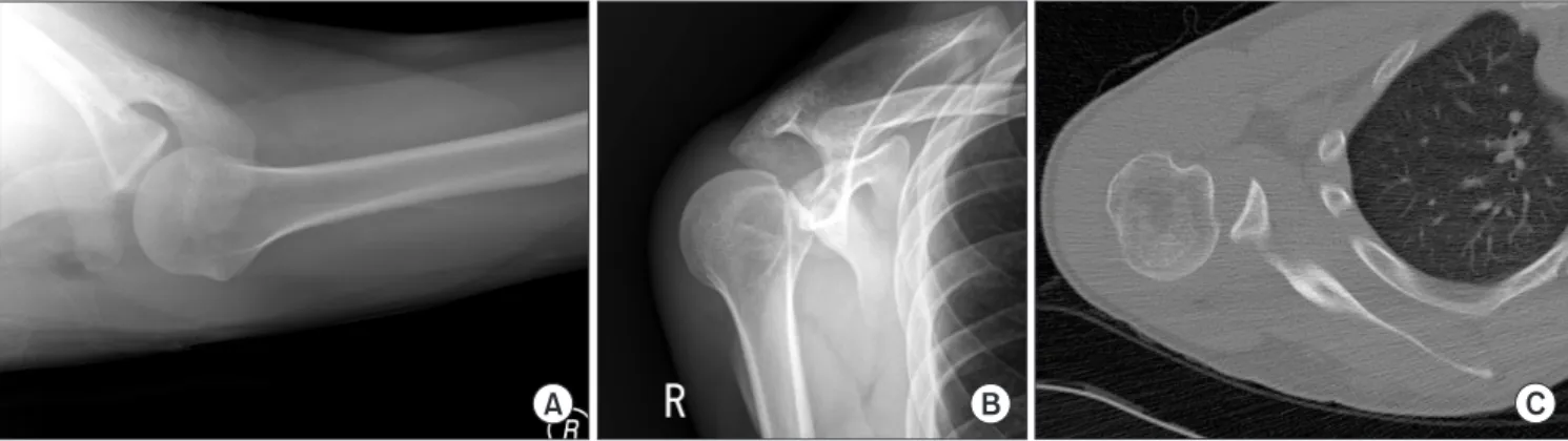
by dual-energy computed tomography (DECT), in which dislo- cation of the right posterior shoulder was observed and posterior glenoid bone defect was observed in at least 50% towards the left side. Besides, no abnormalities were observed in the rotator cuff or the biceps tendon (Fig. 1). The surgery method used was as follows; first, the patient was given general anesthesia and placed into the correct position. The patient was placed in a lat- eral position towards the left, and the body slanted backwards at an angle of approximately 30 degrees so that the shoulder was perpendicular to the floor. Secondly, key anatomical structures such as the coracoid process, the clavicle, and the scapular spine were marked. Afterwards, an incision of around 15 cm was made along the axillary line, above and across the shoulder joint, and another incision was made through the soft tissue. At this point, the arm was externally rotated to an angle of 90 degrees and the posterior deltoid lifted up, which was then approached via the deltoid splitting approach and pulled up in order to ex- pose the infraspinatus muscle and teres minor muscle. Each of these muscles was then pulled to the opposite sides, and the posterior capsuloligament approached through here. Then, the
posterior capsuloligament was cut into a T-shape, through which the posterior glenoid was reached. The membrane of the gle- noid bone was peeled off and flattened so as to secure an area onto which the autograft was attached. The transplant used was an iliac bone, measuring 2 cm in length and 3 cm in thickness, obtained from the eastern side of the iliac crest. The bone graft was appropriately situated onto the area of deficit and fixated using two cannulated screws of approximately 3.5 mm in size.
The shape of the bone graft was then polished using a grinder to fit into the glenoid space. Furthermore, stability was confirmed by approximating the range of motion of the shoulder joint.
Here, we found that the postero-inferior capsuloligament was loose, so plication was carried out using an ethibond. After the surgery, the patient was given an abduction brace with a cush- ion on the abdominal area to block internal rotation, and with this blocked, he was started on partial passive joint exercise and rehabilitative treatment from day one after surgery. The patient used the abduction brace for 6 weeks after surgery, and was al- lowed to return to his daily routine after 3 months.
In the follow-up at 4 months and 1 year after surgery, both
Fig. 1. (A, B) X-ray showing bone defect of the posterior glenoid cavity & posterior dislocation of the shoulder joint. (C) Computed tomography (Proton density axial view) showing severe bone defect of the posterior glenoid and posterior dislocation of the shoulder joint.
Fig. 2. (A, B) Postoperative X-rays showing glenoid reconstruction using iliac bone graft with AO 3.2 mm cannulated screws. (C) Computed tomography show- ing well-united bone graft and reduced glenohumeral joint.
192
www.cisejournal.orgClinics in Shoulder and Elbow Vol. 17, No. 4, December, 2014
X-ray and DECT showed union of the posterior bone graft, and showed no signs of pseudoarthrosis, osteolysis, osteophyte, loss of joint space, and osteoarthritis (Fig. 2). In the physical exami- nation, the patient scored negative in the Jerk test, the posterior drawer test and the posterior apprehension test. Also, the range of joint motion was largely increased compared to pre-surgery levels, and was within normal range. Furthermore, the patient no longer experienced pain or instability.
Discussion
Posterior shoulder instability comprises of approximately 4%
of all shoulder instabilities, and its treatment is renown to be dif- ficult.2) For the treatment of posterior shoulder instability, Beall et al.4) have incorporated proprioceptive sensation training and neuromuscular reprogramming in early physiotherapeutic treat- ment, through which the supraspinatus, infraspinatus and/or the deltoid muscles are strengthened, so as to achieve functional stability. They report a recovery rate of up to 80% in cases where inherited bone deformities are absent. However, they also report that if glenoid bone defect is severe or recurrent shoulder joint dislocation is long-term, surgery is required. Capsulorrhaphy reported good outcomes in the short-term, but is only relevant to cases where external trauma and glenoid bone deformities are absent.1,5) Glenoid bone osteotomy is a technically difficult surgery, and Hawkins et al.2) observed complications in approxi- mately 20% of patients, and dislocations in 41% of patients after surgery.
Conversely, posterior bone graft may be considered as a last resort when all other surgeries have failed. For this surgery, iliac bone from the eastern iliac crest can be utilized, or alternatively, transplantation using the trapezius musculocutaneous flap or the osacromiale is also possible. According to Kouvalchouk et al.,6) the reconstruction of the posterior glenoid using posterior pe- diculated acromial bone block recovered full joint motion and shoulder stability in 5 cases, where successful outcome, such as bone union, was observed via simple radiography. Sirveaux et al.7) carried out a study comparing autogenous posterior acromi- al bone graft and autogenous iliac bone graft in 18 patients. The study was carried out for 13 years, during which recurrent dislo- cation did not occur in any case, and no significant differences were observed between safety, functional recovery and pain.
However, the speed of recovery was significantly faster in the iliac bone graft group. Also reported was a possible complication of continuous pain after surgery when carrying out autogenous bone grafts using iliac bone. This complication occurs from us- ing screws for fixation during surgery. If the screw is too long, or is not fixed at the appropriate location, it may puncture through the opposite cortical bone, at which point the end of the screw may cause continuous pain. What is more, this complication seems to occur at a higher rate in posterior autogenous bone
grafts than in anterior ones. To prevent such a complication, Bar- bier et al.8) reported that sufficient vision must be obtained dur- ing surgery,in order to identify the appropriate location on the glenoid, so that a screw of an appropriate size, one with a small diameter, can be selected. One other possible complication is arthritis between the shoulder joint and the humeral head.
The risk of this increases if the glenoid surface is not sufficiently polished, which may increase friction between the glenoid and the humeral head. Alternatively, bone regeneration during bone union may also lead to a higher rate of arthritis.7) Samilson and Prieto9) reported that such arthritis occurred in 4 cases out of 18 patients in their study. Posterior shoulder instability is a rare con- dition, and although it can be treated by conservative treatment, surgery may be required. In such cases, the technical difficulty of the surgery makes it challenging for inexperienced surgeons.
Reconstructive surgery, as used in our study, which uses autog- enous bone grafts to increase the anatomical surface area of the glenoid to achieve shoulder stability, are with complications related to iliac bone grafts and are of longer duration compared to other methods. However, their advantages include the abil- ity for strong fixation using screws, which acquires sufficient joint surface for posterior stability, and the relative ease in bone union using autografts.7,8) In our case study, a young and active male patient did not benefit from conservative treatment of over 6 months, and experienced clinically severe posterior glenoid bone defect and posterior shoulder instability. Therefore, an autogenous iliac bone graft was used for glenoid reconstruction.
The patient did not acquire any complications, and achieved good clinical results as seen by recovery of shoulder stability, loss of pain and so forth. Our case study showed a successful clinical outcome, and so we report this case with a literature review.
References
1. Fuchs B, Jost B, Gerber C. Posterior-inferior capsular shift for the treatment of recurrent, voluntary posterior subluxation of the shoulder. J Bone Joint Surg Am. 2000;82(1):16-25.
2. Hawkins RJ, Koppert G, Johnston G. Recurrent posterior in- stability (subluxation) of the shoulder. J Bone Joint Surg Am.
1984;66(2):169-74.
3. Burkhead WZ Jr, Rockwood CA Jr. Treatment of instability of the shoulder with an exercise program. J Bone Joint Surg Am.
1992;74(6):890-6.
4. Beall MS Jr, Diefenbach G, Allen A. Electromyographic bio- feedback in the treatment of voluntary posterior instability of the shoulder. Am J Sports Med. 1987;15(2):175-8.
5. Neer CS 2nd, Foster CR. Inferior capsular shift for involuntary inferior and multidirectional instability of the shoulder. A pre- liminary report. J Bone Joint Surg Am. 1980;62(6):897-908.
6. Kouvalchouk JF, Coudert X, Watin Augouard L, Da Silva Rosa R, Paszkowski A. Treatment of posterior instability of the shoulder
Iliac Bone Graft for Recurrent Posterior Shoulder Instability with Glenoid Bone Defect Sang-Hun Ko and Yun-Jae Cho
www.cisejournal.org
193
joint using an acromial stop with a pediculated deltoid flap.
Rev Chir Orthop Reparatrice Appar Mot. 1993;79(8):661-5.
7. Sirveaux F, Leroux J, Roche O, Gosselin O, De Gasperi M, Molé D. Surgical treatment of posterior instability of the shoul- der joint using an iliac bone block or an acromial pediculated bone block: outcome in eighteen patients. Rev Chir Orthop
Reparatrice Appar Mot. 2004;90(5):411-9.
8. Barbier O, Ollat D, Marchaland JP, Versier G. Iliac bone-block autograft for posterior shoulder instability. Orthop Traumatol Surg Res. 2009;95(2):100-7.
9. Samilson RL, Prieto V. Dislocation arthropathy of the shoulder.
J Bone Joint Surg Am. 1983;65(4):456-60.