A Novel Method to Measure Superior Migration of the Humeral Head: Step-off of the C-line
Kyoung Jin Park , Hyeon Jun Eun, Yong Min Kim, Jun Il Yoo, Chae Ouk Lim
Department of Orthopaedic Surgery, Chungbuk National University Hospital, Chungbuk National University College of Medicine, Cheongju, Korea
Background: Superior migration of humeral head has been conventionally determined by measuring the acromiohumeral distance (AHD), We sought to devise a novel measurement system more reliably and accurately than AHD. We described a structural landmark called ‘C-line’. In this study, we investigated the clinical usefulness of ‘step-off of the C-line (SOC)’ compared to that of AHD.
Methods: The C-line formed from the medial margin of the proximal humeral head continuing up to the inferior margin of the articular glenoid and then to the lateral border of the scapula. The superior migration of the humeral head triggered by a rotator cuff tear intro- duces a discontinuity in this C-line. We measured the distance of this discontinuity. We enrolled 144 patients who underwent a rotator cuff repair. We selected 58 controls who didn’t have any cuff lesions apparent on magnetic resonance imaging. Using radiographs de- rived from standardized true anteroposterior views of the shoulder, we measured the SOC and the AHD. We used t-tests for statistical analyses.
Results: A rotator cuff tear was associated with an increase in SOC and a decrease in AHD. In control group, the mean SOC was 1.29
± 1.71 mm and AHD was 9.71 ± 2.65 mm. In cuff tear group, the mean SOC was 3.15 ± 3.41 mm and AHD was 8.28 ± 1.76 mm.
The mean SOCs of the patient group in relation to the mean SOC of the control group according to tear size, the SOCs of medium tear and lager groups showed statistically significant increase (p<0.05).
Conclusions: The SOC may be a similarly effective to diagnose cuff tears of medium size and larger compared with AHD.
(Clin Shoulder Elbow 2016;19(3):125-129)
Key Words: Acromiohumeral distance; Step-off of C-line; C-line; Rotator cuff tear
Copyright © 2016 Korean Shoulder and Elbow Society. All Rights Reserved. pISSN 2383-8337
Clinics in Shoulder and Elbow Vol. 19, No. 3, September, 2016 http://dx.doi.org/10.5397/cise.2016.19.3.125
Received August 23, 2015. Revised December 21, 2015. Accepted January 6, 2016.
Correspondence to: Kyoung Jin Park
Department of Orthopaedic Surgery, Chungbuk National University Hospital, Chungbuk National University College of Medicine, 776 1 Sunhwan- ro, Seowon-gu, Cheongju 28644, Korea
Tel: +82-43-269-6077, Fax: +82-43-274-8719, E-mail: [email protected]
Financial support: This work was supported by the research grant of Chungbuk National University in 2014. Conflict of interests: None.
Introduction
The superior migration of the humerus in patients with rotator cuff tears can be observed both clinically and intra-operatively.
The superior migration leads to a disturbance in the normal dynamics of the shoulder and acts as a diagnostic marker of cuff lesions. Factors that are closely associated with the superior migration of the humeral head such as fatty degeneration of the cuff and cuff tear size are also important prognostic factors of the disease, adding clinical significance and importance to these fac- tors.1-6)
The principal method to measure the superior translation of the humeral head uses the acromiohumeral distance (AHD) (Fig. 1),
but AHD has been associated with problems such as errors in measurement. The common sources of errors include variability in measurement taking between observers and the sensitivity of the AHD to minor differences in the projection angle during plain radiography or to patient height. In patients with cuff le- sions, the presence of bony spurs of the acromion can also influ- ence AHD measurements.7-9)
We sought to devise a measurement system that predicts superior migration of the humerus more reliably and accurately than the method of AHD measurement. To this end, we de- scribed a novel structural landmark at the border of the scapula and the proximal humerus, a continuous line formed from the medial margin of the proximal humeral head continuing up to
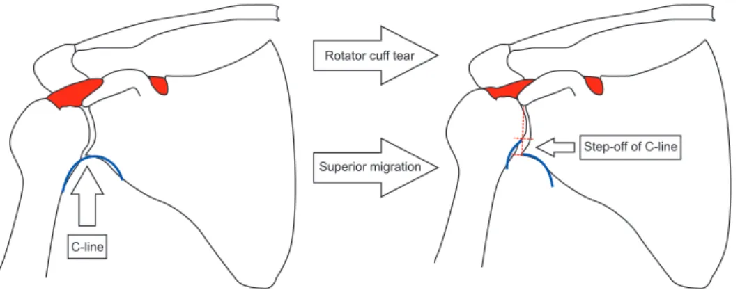
the inferior margin of the articular glenoid and then to the lateral border of the scapula, and coined it the term ‘C-line’ because the resulting line depicts the letter ‘C’. The superior migration of the humeral head triggered by a rotator cuff tear introduces a discontinuity in this C-line resulting in a break in the line and an elevation of the humeral end of C-line break—we described this as a step-off of the C-line (SOC) phenomenon (Fig. 2).
In this study, we investigated the clinical usefulness of SOC compared to that of the already established AHD as an indicator of superior migration of the humeral head.
Methods
Subjects of Study
In the control group, of the 120 patients who presented mild shoulder pain but did not show evidence for rotator cuff tears between January 2008 and July 2009, by taking an magnetic resonance imaging of the shoulder we excluded 62 patients who showed to have a rotator cuff tear, rotator cuff lesion, or bursitis (rotator cuff-related disease) to give a total of 58 patients (17–82
years) selected for subsequent plain radiography. For the patient group, we enrolled patients who had received either magnetic resonance angiography (MRA) of the shoulder or a rotator cuff repair for complete tears of intermediate size or larger between January 2005 and July 2009. We assessed the preoperative true antero-posterior (AP) radiographs of the 144 patients (range, 42–81 years). A diameter of 1 cm or larger was considered the minimum size for medium tears.
The control group comprised of 33 men and 25 women. The patient group comprised of 76 men and 68 women. The exclu- sion criteria in the patient group included a previous history of shoulder operation on the ipsilateral arm, inflammatory arthritis, glenohumeral arthritis, a pervious history of shoulder trauma, and radiographs derived from unstandardized X-rays. In the con- trol group, those with shoulder lesions such as multi-directional shoulder instability and recurrent shoulder dislocations or with a non-AP true image of the shoulder were excluded.
Experimental Approach
1) Method of radiological assessment
We took preoperative radiographs of the shoulder in the true anteroposterior views. Radiography was performed with the patients standing and with their arms in neutral position. The projection angle was at 45° and taken so that the humeral joint was not superimposed. To minimize the error in measurement between the actual length and the radiographically measured length, we used a sizing bar. Because on the radiographically mea- sured value was around 1.1 times longer than the actual length, we adjusted the sizing bar by taking into account this factor.
To measure the extent of the C-line break, first we drew a hypothetical, perpendicular line that intersects the line that con- nects the supraglenoid tubercle and the infraglenoid tubercle and the line that parallels the inferior margin of the infraglenoid tubercle. Then, we drew another line following the medial margin of the infraglenoid tubercle of the anatomic neck of the humerus. The SOC was measured as the shortest distance be- tween these two lines (Fig. 2). We used Philips digital diagnostic for plain radiography. To increase the accuracy of our results, we carried out an intra-observer study to estimate the extent of
8.41 mm
Fig. 1. Mearsurement of acromiohumeral distance.
C-line
Rotator cuff tear
Superior migration
Step-off of C-line
Fig. 2. Step-off of C-line.
the agreement between the measurements of SOC and of AHD taken by two examiners.
2) Statistical analysis
All statistical analyses were carried out by using the SAS pro- gram ver. 9.1 (SAS Institute, Cary, NC, USA). We used the fol- lowing non-parametric tests: the Pearson’s correlation test and the Spearman’s correlation test. A difference was considered as significant if p<0.05. We made a statistical analysis of the radio- graphic results of the AHD and SOC using the following non- parametric tests: the Mann-Whitney test and the Fisher’s exact test (SAS program ver. 9.1). Statistical significance was set to p<0.05. An receiver operating characteristic curve was used to calculate the diagnostic benchmarks of AHD and of SOC.
Results
We found that both the mean AHD and the mean SOC in
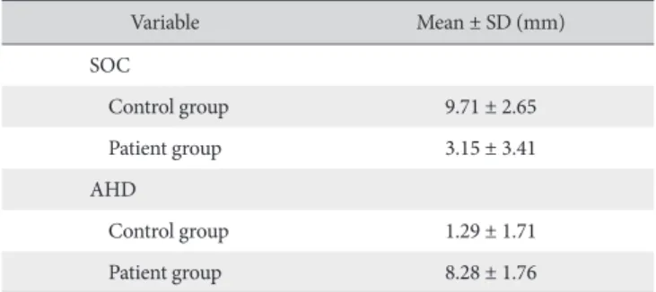
the patient group showed a statistically significant correlation with tear size (p<0.01); the larger the tear the greater the de- crease in the AHD and the greater the increase in SOC. In the control group (n=58), the mean AHD was 9.71 ± 2.65 mm and the mean SOC was 1.29 ± 1.71 mm (Table 1). In the pa- tient group (n=144), the mean AHD was 8.28 ± 1.76 mm, and the mean SOC was 3.15 ± 3.41 mm.
The mean SOCs according to tear-size groups were as fol- lows: the massive tear group (n=15) had a mean SOC of 5.38
± 3.59 mm; the large tear group (n=44), a mean SOC of 3.27
± 2.70 mm; the medium group (n=71), 2.93 ± 3.72 mm; and the small tear group (n=14), 1.44 ± 2.49 mm.
We compared the mean SOCs of the control group (n=58) and those of the patient group according to tear size. Although the SOCs between the control group and the small tear group did not show a statistically significant difference (p=1.000), we found that those of the massive, large, and the medium tear groups differed from the corresponding SOCs of the control group with statistical significance (massive tear group, p=0.001; large tear group, p=0.026; medium tear group, p=0.047) (Table 2).
Even though both the AHD and the SOC of the patient group was positively correlated with height, we found that these corre- lations were not statistically significant. We found that AHD was negatively correlated with age, but not significantly (p<0.725).
Conversely, we found that SOC was positively correlated with age with statistical significance (p<0.016), showing a correlation coefficient of 0.170.
In addition, we calculated the diagnostic benchmark of SOC as 3.135 mm, the sensitivity of which was 53.1% and the speci- ficity, 89.7%. The diagnostic benchmark of AHD was 4.200 mm, the sensitivity of which was 98.6% and the specificity, 5.2% (Fig.
3).
We found that the intraclass correlation coefficient of the mean AHDs was 0.834 mm, which indicates a statistically sig- Table 1. Comparison of the Mean AHD & SOC between Patient and Control
Groups
Variable Mean ± SD (mm)
SOC
Control group 9.71 ± 2.65
Patient group 3.15 ± 3.41
AHD
Control group 1.29 ± 1.71
Patient group 8.28 ± 1.76
AHD: acromiohumeral distance, SOC: step-off of the C-line, SD: standard deviation.
Table 2. Comparison of AHD and SOC between Control Group and Tear Size Groups
Variable Control Massive Large Medium Small AHD
Control - 0.001 0.004 0.042 0.716
Massive - 0.609 0.158 0.252
Large - 0.807 0.831
Medium - 0.995
Small -
SOC
Control - 0.000 0.026 0.047 1.000
Massive - 0.226 0.076 0.014
Large - 0.984 0.397
Medium - 0.567
Small -
AHD: acromiohumeral distance, SOC: step-off of the C-line.
0 1.0
0.8
0.6
0.4
0.2
0
Specificity
Sensitivity
0.2 0.4 0.6 1.0
ROC curve
0.8 AHD
SOC
Reference line
Fig. 3. Receiver operating characteristic (ROC) curve of acromiohumeral dis- tance (AHD) and step-off of the C-line (SOC).
nificant agreement of 83.4% of the measurements made by the two observers (p=0.000). The intraclass correlation coefficient of the mean SOCs was 0.906 mm, showing that the measure- ments made by the two observers matched with a high degree of agreement with statistical significance (p=0.000).
Discussion
We found that SOC, our novel indicator of superior migra- tion of the humeral head, showed a clinical usefulness in dif- ferentiating the presence of medium tears or larger tears from an absence of tears. But it was not useful in differentiating small tears. The SOC, which we measured using plain radiography, can be used as a measure of superior migration of the humeral head and shows a high specificity in differentiating cuff tears. We believe when SOC and AHD are used hand in hand, the effec- tiveness is synergistic, for AHD can differentiate cuff tears with high sensitivity.
The AHD is measured as the shortest distance between the proximal humeral head and the inferiolateral edge of the ante- rior acromion. Golding10) reported that an AHD range of 6–15 mm is normal in the absence of an acromial lesion and that of the available indicators of acromial lesions AHD is the most credible radiological marker.
The reduction in AHD is thought to be largely attributed to the proximal or the superior migration of the humeral head. A reduced AHD is also known to be associated with fatty infiltra- tion of the cuff or with cuff lesions such as tears. In addition, supraspinatus tendon tears are another cause of AHD reduction because they lead to deltoid muscle-dependent superior trac- tion of the humerus.2,11)
Weiner and Macnab11) suggested that an AHD of less than 6 mm can be a diagnostic marker of cuff tear. But radiographic findings from plain radiographs alone are inadequate to ob- jectively assess superior translation of the humeral head. For instance, the presence of acromial spurs influences radiological measurement, which undermines the credibility of the diagnos- tic marker. And removing the acromial spurs have shown to alter the preoperative AHD values leading to a poorly reproducible postoperative AHD value. Another shortcoming of AHD as a diagnostic marker of cuff tears is the discrepancies in what physi- cians used as the norm value of AHD, which comes as a result of the inter-observer variation in selecting the least distance between the medial margin of the proximal humeral head and the inferior margin of the acromion. Yet AHD is one of the most widely used to diagnose cuff tears through plain radiography.
Our aim was to devise a novel method that improves the accuracy of the diagnosis of cuff tears through indirect markers.
Several studies have investigated indirect markers that are indic- ative of cuff lesions. For instance, Krishnan et al.12) reported that a Gothic arch, an anatomical structure seen in a normal shoul-
der, plays an important role during the repair of proximal hu- meral fractures. The Gothic arch refers to the line that connects the lateral border of the scapula and the medial border of the humerus along the glenoid labrum on AP shoulder radiographs.
In addition, Keener et al.13) reported a computational measure- ment method to estimate humeral head migration by measuring the distance from the center of the humeral head to that of the glenoid; although accurate, the downside of this method was that a computer program is required.
Taking the concept based on the Gothic arch, we defined C-line as the line that forms from the medial margin of the proximal humeral head continuing up to the inferior margin of the articular glenoid and then to the lateral border of the scapula. The superior migration of the humeral head triggered by a rotator cuff tear introduces a discontinuity in this C-line;
we measured the corresponding break in the C-line. According to the measurements of SOC, we found that the extent of SOC was greater in patients with cuff tears than those without as with AHD. Further we found that extent of SOC was significantly cor- related to the severity of cuff tears. Further, we found that unlike the conservative diagnostic method of cuff tears, AHD, which is easily influenced by acromial deformity or by bony spurs, change in C-line occurred independently of the acromion. This means that less inter-observer variation would be seen in terms of SOC, and patients who have had acromioplasty may also be measured through SOC without being concerned with repro- ducibility. Taken together, we anticipate both the reproducibility and the reliability in SOC-dependent diagnosis of cuff tears may be enhanced through our novel approach.
Because AHD is highly influenced by variation in height, it may seem to fluctuate from the norm value between patients.
However, the extent of C-line break is independent of a person’s height, so it is deemed a more consistent marker than AHD.
Another advantage of SOC over AHD may be that although a broad range of the norm AHD requires that a bilateral measure- ment is made, because that of SOC is narrow, the variability of measurement is minimal and a single-side measurement is suffice to provide a relatively reliable indication of superior dis- placement. These make measuring SOC more time and cost effective than measuring AHD. In addition, because superior migration of the humeral head can be an indication of retears, SOC can also be used during postoperative follow-up examina- tions to predict retears in patients who have received a supraspi- natus tendon repair.
In their biomechanical study, Mura et al.14) found that in pa- tients with supraspinatus tendon tears alone a superior migration of the humeral head is not obvious but evident in those with combined tears of the supraspinatus and infrapinsatus tendon tears. In the same light, Keener et al.13) reported that combined tears of the supraspinatus and infrasinatus tendons (or infrasina- tus tendon tears) are more associated with superior migration of
the humeral head than supraspinatus tendon tears, especially those of small sizes, that occur alone. Through a cadaveric study, Mochizuki et al.15) investigated the anatomical characteristics of the supraspinatus and the infraspinatus tendons and of the humeral attachment sites of these tendons. Compared to pre- vious reports, their report showed that the attachment of the infraspinatus tendon tends to occur more broadly, that is more anterolateral to the greater tuberosity of the humeral head, and they suggested that this anatomical characteristic may lead to increased rate of combined tears that include the infraspina- tus tendon. When the radiological findings of 63 MRAs were analyzed to investigate the relationship between AHD and cuff lesions, Saupe et al.16) found that cuff tear size, infraspinatus atrophy, and fatty degeneration were factors with the largest im- pact on AHD reduction. In this present study, the patients with supraspinatus tears alone had only small tears, which meant that the superior migration of the humeral head was not prominent;
conversely, those with combined supraspinatus and infraspiantus tendon tears had intermediate- or larger-sized tears meaning that the superior migration of the humeral head in these patients was more prominent, and thus, for whom a diagnostic value of SOC could be attained.
The study is limited in that the degrees of tear size, of muscle atrophy, and of fatty degeneration of individual cuff muscles were not differentiated to take into account the impact of each cuff muscle within the whole unit, but rather the rotator cuff was analyzed as a collective unit. Thus, our study does not address the relative contributions of each muscle on the glenohumeral joint and on the migration of the humeral head but only the im- pact of the tear size of the overall cuff.
Conclusion
In sum, we found that a positive SOC sign determined through plain radiography is a strong predictor of rotator cuff tears of medium size and larger. We believe that the SOC test may be a more applicable approach to diagnosing cuff tears in the clinical setting than measuring ADH and may be used to dif- ferentially predict relatively large tears.
References
1. Bartolozzi A, Andreychik D, Ahmad S. Determinants of out- come in the treatment of rotator cuff disease. Clin Orthop Relat Res. 1994;(308):90-7.
2. Nové-Josserand L, Edwards TB, O’Connor DP, Walch G. The acromiohumeral and coracohumeral intervals are abnormal in rotator cuff tears with muscular fatty degeneration. Clin Or-
thop Relat Res. 2005;(433):90-6.
3. Goutallier D, Postel JM, Gleyze P, Leguilloux P, Van Driessche S.
Influence of cuff muscle fatty degeneration on anatomic and functional outcomes after simple suture of full-thickness tears.
J Shoulder Elbow Surg. 2003;12(6):550-4.
4. Kotzen LM. Roentgen diagnosis of rotator cuff tear. Report of 48 surgically proven cases. Am J Roentgenol Radium Ther Nucl Med. 1971;112(3):507-11.
5. Moosikasuwan JB, Miller TT, Burke BJ. Rotator cuff tears:
clinical, radiographic, and US findings. Radiographics.
2005;25(6):1591-607.
6. Nové-Josserand L, Lévigne C, Noël E, Walch G. The acromio- humeral interval. A study of the factors influencing its height.
Rev Chir Orthop Reparatrice Appar Mot. 1996;82(5):379-85.
7. Gruber G, Bernhardt GA, Clar H, Zacherl M, Glehr M, Wurnig C. Measurement of the acromiohumeral interval on standard- ized anteroposterior radiographs: a prospective study of ob- server variability. J Shoulder Elbow Surg. 2010;19(1):10-3.
8. Hébert LJ, Moffet H, Dufour M, Moisan C. Acromiohumeral distance in a seated position in persons with impingement syndrome. J Magn Reson Imaging. 2003;18(1):72-9.
9. Iannotti JP. Full-thickness rotator cuff tears: factors affecting sur- gical outcome. J Am Acad Orthop Surg. 1994;2(2):87-95.
10. Golding FC. The shoulder: the forgotten joint. Br J Radiol.
1962;35:149-58.
11. Weiner DS, Macnab I. Superior migration of the humeral head. A radiological aid in the diagnosis of tears of the rotator cuff. J Bone Joint Surg Br. 1970;52(3):524-7.
12. Krishnan SG, Bennion PW, Reineck JR, Burkhead WZ. Hemi- arthroplasty for proximal humeral fracture: restoration of the Gothic arch. Orthop Clin North Am. 2008;39:441-50.
13. Keener JD, Wei AS, Kim HM, Steger-May K, Yamaguchi K.
Proximal humeral migration in shoulders with symptomatic and asymptomatic rotator cuff tears. J Bone Joint Surg Am.
2009;91:1405-13.
14. Mura N, O’Driscoll SW, Zobitz ME, et al. The effect of infraspi- natus disruption on glenohumeral torque and superior migra- tion of the humeral head: a biomechanical study. J Shoulder Elbow Surg. 2003;12(2):179-84.
15. Mochizuki T, Sugaya H, Uomizu M, et al. Humeral insertion of the supraspinatus and infraspinatus. New anatomical findings regarding the footprint of the rotator cuff. J Bone Joint Surg Am. 2008;90(5):962-9.
16. Saupe N, Pfirrmann CW, Schmid MR, Jost B, Werner CM, Zanetti M. Association between rotator cuff abnormalities and reduced acromiohumeral distance. AJR Am J Roentgenol.
2006;187(2):376-82.