- 894 -
우관상동맥의 폐동맥 이상 기시증(ARCAPA)
최 에스터*․박 정 준*․윤 태 진*․김 영 휘**
고 재 곤**․박 인 숙**․서 동 만**
=Abstract=
Anomalous Origin of the Right Coronary Artery from the Pulmonary Artery
Esther Choi, M.D.
*, Jeong Jun Park, M.D.
*, Tae Jin Yoon, M.D.
*Young Hwoe Kim, M.D.
**, Jae Kon Ko, M.D.
**, In Sook Park, M.D.
**, Dong Man Seo, M.D.
*Anomalous origin of the right coronary artery from the pulmonary artery is a rare congenital anomaly that has generally been found incidentally during autopsy or surgery. Sudden death may occur without antecedent symptoms in apparently healthy, asymptomatic patients and hence operation is recommended when the lesion is recognized. As opposed to the more frequent anomalous origin of the left coronary artery from the pulmonary artery, only a few children with this anomaly have been reported to have undergone surgical treatment. This report describes a 2-year old patient whose diagnosis was made by echocardiography, confirmed by angiocardiography, and successfully corrected by reimplantation of the anomalous coronary artery into the aorta.
(Korean J Thorac Cardiovasc Surg 2002;35:894-7)
Key words : 1. ARCAPA syndrome 2. Pulmonary artery 3. Coronary artery
*
울산대학교 의과대학 서울아산병원 흉부외과
Department of Thoracic and Cardiovascular Surgery, Asan Medical Center, Ulsan University College of Medicine
**
울산대학교 의과대학 서울아산병원 소아심장과
Department of Pediatric Cardiology, Asan Medical Center, Ulsan University College of Medicine 논문접수일 : 2002년 7월 18일 심사통과일 : 2002년 9월 25일
책임저자 : 서동만(138-736) 서울시 송파구 풍납동 388-1, 서울아산병원 흉부외과. (Tel) 02-3010-3583, (Fax) 02-3010-6811 본 논문의 저작권 및 전자매체의 지적소유권은 대한흉부외과학회에 있다.
CASE
The patient's heart murmur was first noted at 3 months of age during hospital admission for operation of an anal fistula. At 2 years of age, she was referred to the Heart Institute at Asan Medical Center for evaluation. She was a well-looking child with normal vital signs for her age. Cardiac auscultation was typical for a large atrial septal defect (ASD) consisting of a
systolic ejection murmur and trace rumble. There was no continuous murmur. Her electrocardiogram was suggestive of right ventricular hypertrophy, and her chest radiograph showed mild cardiomegaly and increased vascularity. Two-dimensional echocardiography revealed a large secundum ASD, a large pulmonary artery, a slightly dilated left coronary artery with a 3.7mm orifice on the aorta(Fig. 1), and a right coronary artery with no luminal connection with the aorta(Fig.
대흉외지 서동만 외
2002;35:894-7 ARCAPA
- 895 -
Fig. 1. Parasternal short axis view of (Left) a slightly dilated left coronary artery (LCA) with a 3.7 mm orifice on the aorta, and(Right) a right coronary artery(RCA) with no luminal connection with the aorta. AO = aorta; PA = main pulmonary artery.
Fig. 2. Anomalous origin of the right coronary artery(RCA)
from the main pulmonary artery(PA). A) RCA has a luminal
connection to the PA but not to the aorta(AO). B) Color
Doppler echocardiography showing RCA flow entering the
main pulmonary artery. C) Color M-mode shows that this flow
occurs during diastole.
서동만 외 대흉외지
ARCAPA 2002;35:894-7
- 896 -
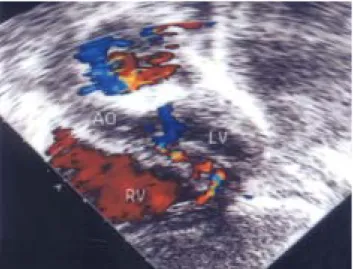
Fig. 3. Color flow mapping showing multiple tiny turbulet color flows in the ventricular srptum suggestive of intercoronary collateral flow during diastole. AO = aorta; RV, right ventricle; LV, left ventricle.
2). Using color Doppler echocardiography, the right coronary artery flow was seen entering the main pulmonary artery immediately above the pulmonary valve during diastole(Fig. 3). Multiple tiny turbulent color flows suggestive of coronary artery collateral vessels(intercoronary collateral anastomoses) were also seen during diastole near the apex of the ventricular septum(Fig. 4).
Cardiac catheterization demonstrated a single left coronary artery arising from the aorta with retrograde filling of a right coronary artery which drained into the main pulmonary artery(Fig. 5). Pulmonary artery pres- sures were normal(30/14 mmHg). Pressure in the aorta was 87/45 mmHg and there was no oxygen step-up in the main pulmonary artery. The Qp/Qs ratio was 2.3.
In the operating room, a dilated pulmonary artery was seen. Except for its origin, the right coronary artery had a normal distribution but was thin-walled and dilated. The anomaly was successfully corrected under cardiopulmonary bypass by a direct reimplantation of the anomalousright coronary artery along with a cuff of adjacent pulmonary artery into the aorta. Closure of the ASD was done through a right atriotomy. The patient's postoperative course was uncomplicated, and she was discharged on the seventh postoperative day. Six months after surgery, the patient was in good clinical condition with normal ventricular function.
DISCUSSION
Soloff1) noted four possible types of anomalous origin
Fig. 4. Preoperative selective coronary angiograms. A) In the
early phase of injection from the aortic root, only the left
coronary artery is opacified. B) At a later phase, retrograde
filling of the right coronary artery is seen draining into the
pulmonary artery. C) Right anterior oblique view shows
retrograde filling of the anomalous RCA from collaterals with
subsequent flow into the pulmonary artery.
대흉외지 서동만 외
2002;35:894-7 ARCAPA
- 897 -
중심 단어: 1. 관상동맥 이상 기시증 2. 폐동맥
3. 관상동맥
=국문초록=
우관상동맥의 폐동맥 이상 기시증(ARCAPA)은 매우 드문 선천성 심기형으로 증세가 없는 건강한 환자에 서 급사를 초래할 수 있으며 주로 부검시 혹은 수술 중 우연히 발견된다. 환아는 2세된 여아로 우관상동맥의 폐동맥 기시 이상을 심장초음파 검사로 진단하였고 관상동맥조영촬영으로 확진하였으며 이상 기시하는 우 관상동맥을 대동맥으로 성공적으로 전이하였다.
of the coronary arteries from the pulmonary artery: 1) origin of the right coronary artery from the pulmonary artery, 2) origin of the left coronary artery from the pulmonary artery, 3) origin of both coronary arteries from the pulmonary artery, and 4) origin of an accessory coronary artery from the pulmonary artery.
The most common of these is the anomalous origin of the left coronary artery, occurring in approximately 1 in 300,000 children. Origin of the right coronary artery from the pulmonary artery is extremely rare. In early case reports it was an incidental autopsy finding with no clear relation to clinical symptoms or death. In several subsequent reports it was an incidental finding at angiography or operation for other congenital heart disease2). Recently, origin of the right coronary artery from the pulmonary artery as an isolated congenital anomaly has been evaluated3).
This anomaly generally does not cause any typical clinical findings, often becoming an autoptic or surgical surprise after infancy or in adult age. However, some cases of cardiopulmonary arrests4) and sudden deaths have been reported in patients with this lesion5). As opposed to the more frequent anomalous origin of the left coronary artery from the pulmonary artery6,7), only a few children with this anomaly have been reported to have undergone successful surgical treatment. This report describes a 2-year old patient whose diagnosis was made by two-dimensional echocardiography, confirmed by angiocardiography, and successfully corrected at surgery.
The surgical reimplantation of the anomalous right coronary artery into the aorta in a child may be performed safely without complications. A surgical surprise can be avoided by keeping in mind the possibility of such malformations during careful pre- operative evaluation, especially when unusual diastolic flows are observed in the main pulmonary artery during echocardiography.
REFERENCES