1
T
he difficulty directly visualizing the coronary microvas- culature, as opposed to the epicardial coronary vessels, has made the diagnosis of coronary microvascular dysfunc- tion (CMD) in the catheterization laboratory challenging. The index of microcirculatory resistance (IMR) is a quantitative and reproducible measure of coronary microvascular func- tion, readily applicable in the cardiac catheterization labora- tory with a single pressure–temperature sensor guidewire.1,2 IMR has been increasingly applied to investigate the extentof CMD in various patient populations, such as those with ST-segment–elevation myocardial infarction, angina with or without obstructive coronary artery disease, and cardiac allograft vasculopathy after heart transplantation, establish- ing its role for assessing CMD in the cardiac catheterization laboratory.3–9
However, previous studies using IMR primarily investi- gated the presence or absence of CMD in only the left anterior descending coronary artery. Furthermore, little is known about Background—Difficulty directly visualizing the coronary microvasculature as opposed to the epicardial coronary artery makes its assessment challenging. The goal of this study is to measure the index of microcirculatory resistance (IMR) in all 3 major coronary vessels to identify the clinical and angiographic predictors of an abnormal IMR.
Methods and Results—Ninety-three patients who underwent coronary physiological assessment in all 3 major coronary vessels were prospectively enrolled (59.8±9.4 years with 77.4% men). IMR was corrected using Yong’s formula and coronary microvascular dysfunction (CMD) was defined using vessel-specific cutoffs. A global IMR was calculated as the sum of the IMR in all 3 major epicardial vessels. Angiographic epicardial disease severity was assessed with vessel-specific and overall SYNTAX score. Median IMR and fractional flow reserve was 17.2 (Q1–Q3: 13.3–22.9) and 0.92 (0.85–0.97). The majority of patients (59.1%) had no CMD, 23.7% had 1-vessel CMD, 14.0% had 2-vessel CMD, and 3.2% had 3-vessel CMD. CMD was observed at a similar rate in the territories supplied by all 3 major coronary vessels (left anterior descending coronary artery 28.0%, left circumflex artery 19.4%, and right coronary artery 23.7%;
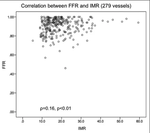
P=0.39). Fractional flow reserve had a weak, positive correlation with IMR (ρ=0.16; P<0.01). The SYNTAX score had no significant correlation with IMR, both at a patient level (ρ=−0.002; P=0.99) and a vessel-specific level (ρ=−0.06;
P=0.36). By multivariable ordinal logistic regression analysis, no variable was left as an independent predictor of an abnormal IMR.
Conclusions—Clinical factors and epicardial coronary disease severity are not predictors of the extent of CMD.
Clinical Trial Registration—URL: https://www.clinicaltrials.gov. Unique identifier: NCT01621438.
(Circ Cardiovasc Interv. 2017;10:e005445. DOI: 10.1161/CIRCINTERVENTIONS.117.005445.)
Key Words: coronary artery disease ◼ ischemia ◼ logistic model ◼ microcirculation ◼ observational study
© 2017 American Heart Association, Inc.
Circ Cardiovasc Interv is available at http://circinterventions.ahajournals.org DOI: 10.1161/CIRCINTERVENTIONS.117.005445 Received May 17, 2017; accepted October 23, 2017.
From the Division of Cardiovascular Medicine, Stanford University, CA (Y.K., W.F.F., T.N., D.-H.C.); Division of Cardiology, Department of Internal Medicine, Heart Vascular Stroke Institute, Samsung Medical Center, Seoul, Republic of Korea (J.M.L.); Department of Internal Medicine, Kyungpook National University Hospital, Daegu, Republic of Korea (J.H.L.); Department of Cardiology, Catharina Hospital Eindhoven, the Netherlands (F.M.Z.);
Department of Medicine, Seoul National University Hospital, Republic of Korea (J.-H.J., H.-J.L., B.-K.K.); Department of Medicine, Inje University Ilsan Paik Hospital, Goyang, Republic of Korea (J.-H.D.); Department of Medicine, Keimyung University Dongsan Medical Center, Daegu, Republic of Korea (C.-W.N.); Department of Cardiology, Ulsan University Hospital, University of Ulsan College of Medicine, Republic of Korea (E.-S.S.); and Institute of Aging, Seoul National University, Republic of Korea (B.-K.K.).
The Data Supplement is available at http://circinterventions.ahajournals.org/lookup/suppl/doi:10.1161/CIRCINTERVENTIONS.117.005445/-/DC1.
Correspondence to William F. Fearon, MD, Division of Cardiovascular Medicine, Stanford University, 300 Pasteur Dr, H2103, Stanford, CA 94305.
E-mail [email protected]
Three-Vessel Assessment of Coronary Microvascular Dysfunction in Patients With Clinical Suspicion of Ischemia
Prospective Observational Study With the Index of Microcirculatory Resistance
Yuhei Kobayashi, MD; Joo Myung Lee, MD; William F. Fearon, MD; Jang Hoon Lee, MD;
Takeshi Nishi, MD; Dong-Hyun Choi, MD; Frederik M. Zimmermann, MD; Ji-Hyun Jung, MD;
Hyun-Jung Lee, MD; Joon-Hyung Doh, MD; Chang-Wook Nam; MD;
Eun-Seok Shin, MD; Bon-Kwon Koo, MD
Downloaded from http://ahajournals.org by on February 24, 2019
whether clinical characteristics and epicardial coronary artery disease are predictive of the extent of microvascular dysfunction.
Accordingly, the primary goal of the present study is to investi- gate the prevalence of CMD in the myocardial regions supplied by each of the 3 major coronary vessels by measuring IMR and to investigate the predictive value of clinical characteristics and epicardial coronary artery disease on the extent of CMD.
Methods
Study Design and Patient PopulationThe 3V FFR-FRIENDS study (Three-Vessel Fractional Flow Reserve for the Assessment of Total Physiologic Atherosclerotic Burden and Its Clinical Impact in Patients With Coronary Artery Disease) is a multicenter, prospective, investigator-initiated observational study, assessing the 3-vessel coronary physiology in patients with clinical suspicion of ischemic heart disease.10 In patients ≥18 years who un- derwent invasive physiological assessment of coronary artery lesions for standard clinical indications, comprehensive coronary physiologi- cal assessment including fractional flow reserve (FFR) and IMR was performed in 3 major epicardial coronary arteries.
Patients were excluded if they had previous coronary artery bypass surgery, an extremely tortuous or calcified coronary artery, known severe left ventricular hypertrophy, left ventricular ejection fraction
<30%, inability to receive adenosine, or recent (within 3 weeks before cardiac catheterization) ST-segment–elevation myocardial infarction.
This study was approved by an institutional review committee from each participating site, and informed consent was obtained from all patients. The data, analytic methods, and study materials will not be made available to other researchers for purposes of reproducing the results or replicating the procedure.
Coronary Angiography and Coronary Physiological Measurements
Coronary angiography was performed in a standard fashion.
Angiographic views were obtained after the administration of
intracoronary nitrate (100 or 200 μg). Quantitative coronary angiog- raphy (QCA) was performed at each participating center with a con- tour-detection QCA system. Parameters including percent diameter stenosis, minimum lumen diameter, reference vessel size, and lesion length were reported.7
Invasive coronary physiological parameters were measured with a 0.014-inch pressure sensor guidewire and a console (PressureWire Certus or Aeris wire and the RadiAnalyzer console; then St. Jude Medical). After equalization to the guide catheter pressure with the sensor positioned at the ostium of the coronary artery, the pressure guidewire was advanced down the target coronary artery. With com- mercially available software, the shaft of the PressureWire can act as a proximal thermistor by detecting changes in temperature-dependent electric resistance. The sensor near the tip of the wire simultaneously measures pressure and temperature and can thereby act as a distal thermistor. The transit time of room-temperature saline injected down a coronary artery is then determined with a thermodilution technique.
Approximately 3 mL of room temperature saline was injected rapidly by hand into the target coronary artery, and the resting mean tran- sit time (Tmn) was obtained as an average of 3 injections. Thereafter, intravenous infusion of adenosine (140 μg/kg per minute) was ad- ministered to induce a steady state of maximal hyperemia, followed by 3 more injections of 3 mL of room temperature saline to calcu- late hyperemic Tmn. Simultaneous measurements of mean proximal coronary pressure (Pa, by guide catheter) and mean distal coronary pressure (Pd, by PressureWire) were also acquired in the resting and maximal hyperemic status. FFR was calculated by the ratio of Pd/Pa at hyperemia. IMR was calculated as Pd at hyperemia multiplied by hyperemic Tmn.1,2 All IMR values were also corrected by Yong for- mula (IMR=Pa×Tmn×([1.35×Pd/Pa] − 0.32)) to adjust for the influence of collateral flow.11 At the end of the study, the guidewire was pulled back to the guiding catheter, and the presence of pressure drift was assessed to ensure appropriate recordings. If significant drift (>0.05) was detected, repeated measurement was recommended.
We used the vessel-specific IMR cutoff values to diagnose CMD.
According to the previous publication by Lee et al,7 the cutoff values for an abnormal IMR were defined as >22, 24, and 28 U for left ante- rior descending coronary artery (LAD), left circumflex artery (LCx), and right coronary artery (RCA), respectively. In their study, an ab- normal IMR was defined as greater than the 75th percentile in each of the major coronary arteries. By using this method, the possible effect of left ventricular myocardial mass on IMR can be taken into account.12 In the current study, in an attempt to quantify the extent of CMD, the global IMR was further defined as the sum of IMR mea- sured in each of the 3 major coronary arteries. Because global IMR is the sum of IMR in all 3 myocardial territories and reflects the overall status of CMD, it is distinct from a vessel-specific IMR or a worst IMR among the 3 major coronary arteries.
Scoring Systems of Epicardial Coronary Artery Disease Status
Two representative scoring systems (SYNTAX score and Gensini score) were used to assess the severity of epicardial coronary artery disease.13–15 These scores were obtained for patient level (overall coronary artery tree) and for vessel-specific level. The SYNTAX score was calculated from the preprocedural angiogram in which each coronary lesion pro- ducing ≥50% diameter stenosis in vessels ≥1.5 mm by visual estima- tion was scored separately using the SYNTAX score algorithm from the website and added to obtain the overall SYNTAX score.13 Gensini14,15 score was calculated by taking severity of lesion narrowing, number of lesions, lesion location, and influence of collaterals into account.
End Points
The primary end point of this analysis was to assess the prevalence of CMD as determined by elevated IMR in each of the 3 major coronary arteries and to assess the correlation between epicardial coronary dis- ease and CMD determined by IMR. As described above, epicardial disease status was assessed using SYNTAX score, Gensini score, FFR, and percent diameter stenosis by QCA. Comparisons were
WHAT IS KNOWN
•
Difficulty directly visualizing the coronary micro- vasculature as opposed to the epicardial coronary artery makes its assessment challenging.•
The index of microcirculatory resistance is a quanti- tative and reproducible measure of coronary micro- vascular function, readily applicable in the cardiac catheterization laboratory.WHAT THE STUDY ADDS
•
By assessing the index of microcirculatory resis- tance in 3-vessel coronary territories, we found that the majority of patients (59.1%) had no coronary mi- crovascular dysfunction (CMD), 23.7% had 1-ves- sel CMD, 14.0% had 2-vessel CMD, and 3.2% had 3-vessel CMD.•
The rate of CMD was similar in the territories sub- tended by the 3 major epicardial vessels. Clinical factors and epicardial coronary disease severity are not predictive of CMD, suggesting the importance of measuring the index of microcirculatory resistance when CMD is suspected.Downloaded from http://ahajournals.org by on February 24, 2019
made on both a patient level and a vessel level if feasible. The sec- ondary end point was to determine correlates of the extent of CMD among clinical, angiographic, and physiological information using univariable and multivariable ordinal regression models.
Statistical Analysis
Categorical variables are presented as counts and percentages.
Pearson χ2 test was used for comparisons of 2 sets of categori- cal variables. Continuous variables are presented as mean±SD or median (Q1–Q3), as appropriate. Two sets of continuous variables were compared with Mann–Whitney U test. Kruskal–Wallis test was used to assess the difference in IMR and FFR between insti- tutions. Correlations between IMR and angiographic parameters were tested with Spearman correlation coefficient. Partial corre- lation accounting for the interrogated vessel was also performed between IMR and angiographic parameters. Logistic regression model was used to test whether the vessel territory is the inde- pendent predictor of the presence/absence of CMD after adjust- ing for patients’ identification. Variables listed in Table I in the Data Supplement, SYNTAX score, Gensini score, and number of positive FFR vessel per patient were tested in univariable ordinal logistic regression analysis. Variables with P<0.25 in univariable ordinal logistic regression analysis were included in multivariable ordinal logistic regression model to determine an independent pre- dictors of the extent of CMD.16 By using ordinal regression anal- ysis, the independent effect of variable on the severity of CMD (multiple outcome categories: no CMD, CMD in 1-vessel, CMD in 2-vessel, and CMD in 3-vessel territories) could be assessed.
The proportional odds assumption was assessed for the ordinal regression model by using the test for parallel lines. A P value of
<0.05 was considered statistically significant. All analyses were performed using SPSS version 21.
Results
A total of 93 patients and 279 vessels were included in this study from 4 Korean institutions. The patients’ clinical, laboratory, and medication characteristics are summarized in Table 1. The patients’ angiographic and physiologi- cal characteristics are summarized in Table 2. The mean age was 59.8±9.4 years with 77.4% men. Among all, 23 patients (24.7%) presented with acute coronary syn- drome. By quantitative coronary angiography, 42 patients (45.2%) had at least one epicardial coronary artery with significant disease defined as ≥50% diameter stenosis. The median numbers of vessels with ischemic FFR values was 0 (Q1–Q3: 0–1). The mean percent diameter stenosis by quantitative coronary angiography in the 279 vessels was 34.0% (24.3–46.6). The median IMR and FFR were 17.2 (13.3–22.9) and 0.92 (0.85–0.97), respectively. The median global IMR defined as the sum of IMR in all 3 major coro- nary vessels was 52.1 (45.7–67.7). The median global IMR was similar between patients presenting with acute coro- nary syndrome versus those without (54.1 [48.1–67.6] ver- sus 50.8 [45.4–67.7]; P=0.32).
Prevalence of CMD
The number of vessels with CMD is shown in Figure 1A. The majority of patients (n=55, 59.1%) had no CMD, 22 patients (23.7%) had CMD in 1 vessel, 13 patients (14.0%) had CMD in 2 vessels, and 3 patients (3.2%) had CMD in all 3 vessels.
The results were similar when the universal IMR cutoff of 25 was applied to all vessel territories. When the rate of CMD in each of the 3 vessels was compared between patients
presenting with acute coronary syndrome and without, it was similar (P=0.56; Figure I in the Data Supplement). The prevalence of CMD in each vessel is shown in Figure 1B.
The prevalence of CMD was similar between the 3 vessels (LAD versus LCx versus RCA: 28.0% versus 19.4% versus 23.7%; P=0.39). In the logistic regression model, the ves- sel territory (LAD, LCX, or RCA) was not the independent predictor of the presence/absence of CMD after adjusting for patients’ identification.
Correlations Between Epicardial Disease Severity and CMD
The SYNTAX score had no significant correlation with IMR both at the patient level (ρ=−0.002; P=0.99) and at the vessel level (ρ=−0.06; P=0.36). When IMR was compared between patients with a SYNTAX score=0 versus a SYN- TAX score>0, IMR at the patient level was similar (49.7 [43.0–58.1] versus 52.8 [46.5–68.2], P=0.39). Gensini score had no significant correlation with IMR at the patient level (ρ=−0.13− P=0.22), whereas it had a weak negative correla- tion at the vessel level (ρ=−0.13; P=0.03). FFR had a weak Table 1. Summarized Clinical, Laboratory, and Medication Characteristics
93 Patients Clinical characteristics
Age, y 59.8±9.4 Male, n (%) 72 (77.4) Diabetes mellitus, n (%) 29 (31.2) Hypertension, n (%) 55 (59.1) Hypercholesterolemia, n (%) 60 (64.5) Family history, n (%) 12 (12.9) Current smoker, n (%) 24 (25.8) Acute coronary syndrome, n (%) 23 (24.7) Laboratory data
Fasting blood glucose, mg/dL 101 (93–119) Hemoglobin A1c, % 6.1 (5.6–6.5) Total cholesterol, mg/dL 160 (133–189) High density lipoprotein, mg/dL 44 (37–53) Triglyceride, mg/dL 107 (83–159) Low density lipoprotein, mg/dL 89 (71–112) Medication
Aspirin, n (%) 74 (79.6) Angiotensin-converting enzyme inhibitor, n (%) 4 (4.3) Angiotensin receptor blocker, n (%) 33 (35.5) β-Blocker, n (%) 24 (25.8) Calcium channel blocker, n (%) 34 (36.6) Nitrate, n (%) 17 (18.3) Statin, n (%) 56 (60.2) Values are mean±SD, median (Q1–Q3), or n (%). For complete information, please see Table I in the Data Supplement.
Downloaded from http://ahajournals.org by on February 24, 2019
positive correlation with IMR at the vessel-level analysis (ρ=0.16; P<0.01; Figure 2). Percent diameter stenosis by QCA had no significant correlation with IMR at the vessel- level analysis (ρ=−0.06; P=0.35).
When partial correlation was performed to account for the interrogated vessel, none of the above parameters have significant correlation with IMR at the vessel-level analysis (ρ=−0.04; P=0.55 for SYNTAX score, ρ=−0.01; P=0.86 for Gensini score, ρ=0.07; P=0.28 for FFR, and ρ=−0.05; P=0.43 for percent diameter stenosis by QCA).
Predictors of CMD
When the IMR value was compared between the sexes, it was similar in all vessel territories (LAD P=0.33, LCx P=0.88, and RCA P=0.34, respectively). When the IMR value was compared between patients with and without diabetes melli- tus, it was significantly higher in patients with diabetes mel- litus in the LAD (19.5 [14.0–23.3] versus 15.2 [12.0–19.1];
P=0.048) but was similar in LCx and RCA (LCx P=0.22 and RCA P=0.38, respectively). When the IMR value was com- pared between patients with and without hypertension, it was similar in all vessel territories (LAD P=0.68, LCx P=0.71, and RCA P=0.25). When the IMR value was compared between patients with and without hypercholesterolemia, it tended to be lower in the LAD of patients with hypercholesterolemia (15.1 [11.9–21.3] versus 17.3 [14.2–23.3]; P=0.09), was sig- nificantly lower in the RCA in patients with hypercholesterol- emia (17.2 [12.8–24.6] versus 21.0 [17.9–30.5]; P=0.006) but was similar in LCx (P=0.70).
Univariable and multivariable ordinal logistic regression analysis is shown in Table 3. By univariable ordinal logistic regression analysis, hypercholesterolemia (odds ratio=0.37 [95% confidence interval, 0.16–0.86], P=0.02) and nitrate use (odds ratio=0.25 [0.07–0.93]; P=0.04) were significant predictors of CMD. Epicardial disease status was not a sig- nificant predictor of CMD. When variables with P<0.25 by univariable ordinal logistic regression analysis (hypercho- lesterolemia, β-blocker use, nitrate use, nicorandil use, high- density lipoprotein, low-density lipoprotein, and number of FFR-positive vessels) were tested in a multivariable model, Table 2. Angiographic and Physiological Characteristics
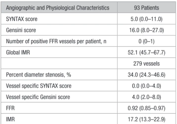
Angiographic and Physiological Characteristics 93 Patients
SYNTAX score 5.0 (0.0–11.0)
Gensini score 16.0 (8.0–27.0)
Number of positive FFR vessels per patient, n 0 (0–1)
Global IMR 52.1 (45.7–67.7)
279 vessels Percent diameter stenosis, % 34.0 (24.3–46.6) Vessel specific SYNTAX score 0.0 (0.0–4.0) Vessel specific Gensini score 4.0 (2.0–8.0)
FFR 0.92 (0.85–0.97)
IMR 17.2 (13.3–22.9)
Values are median (Q1–Q3) or n (%). FFR indicates fractional flow reserve;
and IMR, corrected index of microcirculatory resistance.
Figure 1. Prevalence of coronary microvascular dysfunction in three vessels and in each vessel. Majority of patients (59.1%) had no coro- nary microvascular dysfunction (CMD) in any vessels (A). Similar percentage of patients has CMD in each vessel territory (P=0.39; B). LAD indicates left anterior descending coronary artery; LCx, left circumflex artery; and RCA, right coronary artery.
Downloaded from http://ahajournals.org by on February 24, 2019
only hypercholesterolemia (odds ratio=0.35 [95% confidence interval, 0.14–0.91]; P=0.03) was left as an independent pre- dictor of CMD, but the proportional odds assumption was not met for this model (P<0.01). When hypercholesterolemia was excluded from the multivariable model, no variable remained significant. Similarly, when high-density lipoprotein and low- density lipoprotein were excluded from the multivariable model, no variable remained significant.
Discussion
The principal finding of this study is that the majority of patients with a clinical suspicion of ischemic heart disease do not have CMD in any epicardial vessel. When they do have CMD, in most cases, it is localized to one vessel, with an equal chance between the 3 vessels. Furthermore, clinical characteristics typically correlated with epicardial coronary artery disease are not predictive of the extent of CMD, mak- ing it challenging to predict CMD. These results suggest the usefulness of measuring IMR in the cardiac catheterization laboratory when CMD is suspected.
Vergallo et al17,18 showed an association between the Framingham risk score/presentation/chronic kidney disease and high-risk coronary plaque characteristics as assessed by 3-vessel optical coherence tomography, demonstrating the actual link between the clinical profile and epicardial coro- nary artery disease status. On the contrary, in this study, the Framingham risk score did not predict the severity of CMD as assessed by IMR. Clinical and laboratory characteristics also were not predictive of CMD, although they are typically cor- related with epicardial coronary artery disease development (note that in our model, the odds ratio of hypercholesterolemia was <1). These results may be because of the relatively small sample size of this study (279 vessels from 93 patients) and
thereby weaker statistical power, compared with the report by Lee et al7 (1452 vessels from 1092 patients), in which clini- cal characteristics such as history of myocardial infarction, sex, and high body mass index were predictive of elevated IMR. Or these results may be because of the different effect of clinical factors on the coronary microcirculation accord- ing to the distribution. Nevertheless, our results suggest that it would be challenging to predict CMD in a given patient using these clinical characteristics with C indexes of 0.67 (95%
confidence interval, 0.64–0.70).7 Furthermore, conventional noninvasive stress tests such as echocardiography and elec- trocardiography were shown to have no diagnostic value in identifying elevated IMR in patients presenting with angina and no obstructive coronary artery disease.19
In the cardiac catheterization laboratory, the severity of epicardial coronary artery disease assessed by the coronary angiogram (SYNTAX score, Gensini score, and percent diam- eter stenosis by QCA) or invasive coronary physiology (FFR) is not collinear with the development of CMD as determined by IMR. Rather, FFR is weakly but positively correlated with IMR, suggesting that microvascular disease influences FFR by blunting the vasodilator response. Therefore, the presence of significant epicardial disease is not necessarily related to the presence of CMD. Taken together, the current study suggests the difficulty predicting the presence and the extent of CMD without actually measuring IMR. In the recent article by Lee et al6 invasively investigating the left anterior descending cor- onary artery in patients with angina in the absence of obstruc- tive coronary artery disease, 20% of patients were found to have CMD as determined by elevated IMR. Identifying the cause of chest pain in patients with no obstructive epicardial coronary artery disease is a typical scenario where interven- tional cardiologists may consider measuring IMR.
Figure 2. Correlation between fractional flow reserve (FFR) and the index of micro- circulatory resistance (IMR). FFR had a weak positive correlation with IMR at the vessel level analysis (ρ=0.16; P<0.01).
Downloaded from http://ahajournals.org by on February 24, 2019
There are some limitations to our study. First, as discussed above, the relatively small sample size of this study may have resulted in low statistical power. However, this study is the first to describe invasive assessment of coronary microcircu- lation in all 3 major coronary vessels in nearly 100 patients.
Second, because the enrolled patients primarily had a low SYNTAX score, the relationship between epicardial coronary artery disease and CMD in severe epicardial disease is not fully known. Third, because the IMR cutoff in each vessel is based on the IMR international registry that included patients Table 3. Univariable and Multivariable Ordinal Logistic Regression Model to Predict CMD
Univariable* Multivariable†
Odds Ratio 95% CI P Value Odds Ratio 95% CI P Value
Age, y (per 10-y increase) 0.97 0.64–1.49 0.90 … … …
Male 0.65 0.26–1.65 0.37 … … …
Diabetes mellitus 1.29 0.55–3.03 0.55 … … …
Hypertension 0.69 0.31–1.56 0.38 … … …
Hypercholesterolemia 0.37 0.16–0.86 0.02 0.35 0.14–0.91 0.03
Family history 1.23 0.38–3.96 0.72 … … …
Current smoker 1.58 0.65–3.85 0.32 … … …
Previous myocardial infarction 1.86 0.52–6.73 0.34 … … …
Previous percutaneous coronary intervention 0.95 0.42–2.16 0.91 … … …
Acute coronary syndrome 1.29 0.52–3.22 0.58 … … …
Body mass index (per 1 kg/m2 increase) 1.00 0.87–1.15 0.97 … … …
Framingham risk score (per 10 U increase) 1.42 0.58–3.51 0.44 … … …
*Framingham predicted 10-y risk (per 10% increase) 1.15 0.77–1.73 0.49 … … …
Ejection fraction (per 10% increase) 1.05 0.60–1.83 0.86 … … …
Fasting blood glucose (per 10 mg/dL increase) 0.95 0.85–1.06 0.36 … … …
Hemoglobin A1c (per 1 mg/dL increase) 1.10 0.73–1.66 0.64 … … …
Total cholesterol (per 10 mg/dL increase) 1.04 0.94–1.15 0.48 … … …
High-density lipoprotein cholesterol (per 10 mg/dL increase) 1.24 0.90–1.71 0.19 1.33 0.93–1.89 0.12
Triglyceride (per 10 mg/dL increase) 1.01 0.96–1.06 0.63 … … …
Low-density lipoprotein cholesterol (per 10 mg/dL increase) 1.06 0.97–1.15 0.22 1.08 0.98–1.20 0.13
C-reactive protein (per 1 mg/dL increase) 0.53 0.09–2.99 0.47 … … …
Creatinine (per 1 mg/dL increase) 1.21 0.69–2.13 0.51 … … …
Creatine kinase myocardial band (per 1 IU/L increase) 1.00 1.00–1.01 0.65 … … …
Cardiac troponin I (per 0.01 ng/mL increase) 1.18 0.56–2.45 0.66 … … …
Aspirin 0.83 0.31–2.20 0.71 … … …
Angiotensin-converting enzyme inhibitor 0.42 0.04–4.35 0.46 … … …
Angiotensin receptor blocker 0.95 0.41–2.19 0.90 … … …
β-Blocker 1.85 0.76–4.51 0.17 1.90 0.69–5.24 0.21
Calcium channel blocker 1.06 0.46–2.43 0.89 … … …
Nitrate 0.25 0.07–0.93 0.04 0.33 0.08–1.38 0.13
Nicorandil 0.52 0.19–1.42 0.20 0.85 0.26–2.80 0.79
Statin 0.65 0.29–1.48 0.30 … … …
SYNTAX score (per 10 U increase) 0.93 0.52–1.68 0.82 … … …
Gensini score (per 10 U increase) 1.04 0.87–1.26 0.64 … … …
Number of positive FFR vessels per patient 0.65 0.34–1.27 0.21 0.76 0.37–1.57 0.46
CI indicates confidence interval; CMD, coronary microvascular dysfunction; and FFR, fractional flow reserve.
*Ten-year risk of cardiovascular disease.
†When hypercholesterolemia was excluded from the multivariable model, no variable remained significant. Similarly, when high-density lipoprotein and low-density lipoprotein were excluded from the multivariable model, no variable remained significant.
Downloaded from http://ahajournals.org by on February 24, 2019
in this study, this needs to be replicated in other cohorts.7 Fourth, we used corrected IMR values in all analyses instead of uncorrected IMR. The results would be similar if we used uncorrected IMR given a close relationship between the 2 values (r2=0.90; P<0.001; y=1.02x+0.96). Fifth, it is possible that CMD overlaps territories supplied by 2 major coronary vessels and cannot be detected by evaluating vessel-specific IMR. Sixth, we did not collect angiographic data such as Duke Scores and Myocardial Jeopardy Index to account for the amount of subtended left ventricular mass. Finally, inva- sive coronary physiological assessments were performed at a single time point without clinical follow-up, and hence, we do not know the rate at which epicardial and microvascular disease progressed or the prognostic importance of the find- ings in this study.
Conclusions
Clinical characteristics typically associated with epicardial coronary artery disease development and the severity of epi- cardial coronary artery disease are not predictive of the extent of CMD. This may be particularly true in patients with angina who are referred to the cardiac catheterization laboratory but found to have no obstructive coronary artery disease.
Sources of Funding
This study was supported by the unrestricted research grant from St.
Jude Medical.
Disclosures
Dr Fearon receives institutional research support from St. Jude Medical. Dr Koo receives institutional research grant from St. Jude Medical. The other authors report no conflicts of interest.
References
1. Fearon WF, Balsam LB, Farouque HM, Caffarelli AD, Robbins RC, Fitzgerald PJ, Yock PG, Yeung AC. Novel index for invasively assessing the coronary microcirculation. Circulation. 2003;107:3129–3132. doi:
10.1161/01.CIR.0000080700.98607.D1.
2. Kobayashi Y, Fearon WF. Invasive coronary microcirculation assess- ment–current status of index of microcirculatory resistance. Circ J.
2014;78:1021–1028.
3. Fearon WF, Low AF, Yong AS, McGeoch R, Berry C, Shah MG, Ho MY, Kim HS, Loh JP, Oldroyd KG. Prognostic value of the index of microcirculatory resistance measured after primary percutaneous coro- nary intervention. Circulation. 2013;127:2436–2441. doi: 10.1161/
CIRCULATIONAHA.112.000298.
4. McGeoch R, Watkins S, Berry C, Steedman T, Davie A, Byrne J, Hillis S, Lindsay M, Robb S, Dargie H, Oldroyd K. The index of microcircu- latory resistance measured acutely predicts the extent and severity of myocardial infarction in patients with ST-segment elevation myocardial infarction. JACC Cardiovasc Interv. 2010;3:715–722. doi: 10.1016/j.
jcin.2010.04.009.
5. De Maria GL, Cuculi F, Patel N, Dawkins S, Fahrni G, Kassimis G, Choudhury RP, Forfar JC, Prendergast BD, Channon KM, Kharbanda RK, Banning AP. How does coronary stent implantation impact on the status of the microcirculation during primary percutaneous coronary
intervention in patients with ST-elevation myocardial infarction? Eur Heart J. 2015;36:3165–3177. doi: 10.1093/eurheartj/ehv353.
6. Lee BK, Lim HS, Fearon WF, Yong AS, Yamada R, Tanaka S, Lee DP, Yeung AC, Tremmel JA. Invasive evaluation of patients with an- gina in the absence of obstructive coronary artery disease. Circulation.
2015;131:1054–1060. doi: 10.1161/CIRCULATIONAHA.114.012636.
7. Lee JM, Layland J, Jung JH, Lee HJ, Echavarria-Pinto M, Watkins S, Yong AS, Doh JH, Nam CW, Shin ES, Koo BK, Ng MK, Escaned J, Fearon WF, Oldroyd KG. Integrated physiologic assessment of isch- emic heart disease in real-world practice using index of microcircula- tory resistance and fractional flow reserve: insights from the International Index of Microcirculatory Resistance Registry. Circ Cardiovasc Interv.
2015;8:e002857. doi: 10.1161/CIRCINTERVENTIONS.115.002857.
8. Kobayashi Y, Fearon WF, Honda Y, Tanaka S, Pargaonkar V, Fitzgerald PJ, Lee DP, Stefanick M, Yeung AC, Tremmel JA. Effect of sex differences on invasive measures of coronary microvascular dysfunction in patients with angina in the absence of obstructive coronary artery disease. JACC Cardiovasc Interv. 2015;8:1433–1441. doi: 10.1016/j.jcin.2015.03.045.
9. Yang HM, Khush K, Luikart H, Okada K, Lim HS, Kobayashi Y, Honda Y, Yeung AC, Valantine H, Fearon WF. Invasive assessment of coronary physiology predicts late mortality after heart transplantation. Circulation.
2016;133:1945–1950. doi: 10.1161/CIRCULATIONAHA.115.018741.
10. Lee JM, Koo BK, Shin ES, Nam CW, Doh JH, Hwang D, Park J, Kim KJ, Zhang J, Hu X, Wang J, Ahn C, Ye F, Chen S, Yang J, Chen J, Tanaka N, Yokoi H, Matsuo H, Takashima H, Shiono Y, Akasaka T. Clinical implica- tions of three-vessel fractional flow reserve measurement in patients with coronary artery disease [published online ahead of print August 19, 2017].
Eur Heart J. doi: 10.1093/eurheartj/ehx458.
11. Yong AS, Layland J, Fearon WF, Ho M, Shah MG, Daniels D, Whitbourn R, Macisaac A, Kritharides L, Wilson A, Ng MK. Calculation of the in- dex of microcirculatory resistance without coronary wedge pressure mea- surement in the presence of epicardial stenosis. JACC Cardiovasc Interv.
2013;6:53–58. doi: 10.1016/j.jcin.2012.08.019.
12. Echavarría-Pinto M, van de Hoef TP, Nijjer S, Gonzalo N, Nombela- Franco L, Ibañez B, Sen S, Petraco R, Jimenez-Quevedo P, Nuñez-Gil IJ, Cerrato E, Salinas P, Quirós A, Garcia-Garcia HM, Fernandez-Ortiz A, Macaya C, Davies J, Piek J, Escaned J. Influence of the amount of myo- cardium subtended to a coronary stenosis on the index of microcirculatory resistance. Implications for the invasive assessment of microcirculatory function in ischaemic heart disease. EuroIntervention. 2017;13:944–952.
doi: 10.4244/EIJ-D-16-00525.
13. Serruys PW, Onuma Y, Garg S, Sarno G, van den Brand M, Kappetein AP, Van Dyck N, Mack M, Holmes D, Feldman T, Morice MC, Colombo A, Bass E, Leadley K, Dawkins KD, van Es GA, Morel MA, Mohr FW. Assessment of the SYNTAX score in the Syntax study. EuroIntervention. 2009;5:50–56.
14. Gensini GG. A more meaningful scoring system for determining the severity of coronary heart disease. Am J Cardiol. 1983;51:606.
15. Gensini GG. Coronary Arteriography. Mount Kisco, NY: Futura Pub, Co;
1975.
16. Mccullagh P. Regression-models for ordinal data. J R Stat Soc Series B.
1980;42:109–142.
17. Vergallo R, Xing L, Minami Y, Soeda T, Ong DS, Gao L, Lee H, Guagliumi G, Biasucci LM, Crea F, Yu B, Uemura S, O’Donnell CJ, Jang IK. Associations between the Framingham Risk Score and coronary plaque characteristics as assessed by three-vessel optical coherence tomography. Coron Artery Dis.
2016;27:460–466. doi: 10.1097/MCA.0000000000000383.
18. Vergallo R, Uemura S, Soeda T, Minami Y, Cho JM, Ong DS, Aguirre AD, Gao L, Biasucci LM, Crea F, Yu B, Lee H, Kim CJ, Jang IK. Prevalence and predictors of multiple coronary plaque ruptures: in vivo 3-vessel opti- cal coherence tomography imaging dtudy. Arterioscler Thromb Vasc Biol.
2016;36:2229–2238. doi: 10.1161/ATVBAHA.116.307891.
19. Pargaonkar V, Khandelwal A, Kobayashi Y, Tanaka S, Mathur MB, Froelicher V, Yeung A, Tremmel J. The diagnostic value of stress echocar- diography and electrocardiography in identifying occult coronary abnor- malities in patients with angina and no obstructive coronary artery disease.
J Am Coll Cardiol. 2015;65:A1623–A1623.
Downloaded from http://ahajournals.org by on February 24, 2019