7
흰쥐 왼쪽관상동맥의 분지 양상에 관한 해부학적 연구
안동춘1·김인식*
전북대학교 수의과대학, 1강원대학교 수의학부 (게재승인: 2007년 1월 9일)
An anatomical study on the branching patterns of left coronary artery in the rats
Dong-Choon Ahn
1, In-Shik Kim*
College of Veterinary Medicine, Chonbuk National University, Jeonju 561-756, Korea
1School of Veterinary Medicine, Kangwon National University, Chuncheon 200-701, Korea (Accepted: January 9, 2007)
Abstract : The left main descending artery (LMDA) of left coronary artery (LCA) in rats runs around the left side of conus arteriosus after arising from the aortic sinus and descends to the apex of heart with branching several branches into the wall of left ventricle (LV). The ligation site of LMDA for myocardial infarction (MI) is the 2~4 mm from LCA origin, between the pulmonary trunk and left auricle. The characteristics that rat heart has no interventricular groove on the surface and its coronary arteries run intramyocardially with branching several branches give the difficulty in surgery for MI which resulted in expected size. This study was aimed to elucidate the branching patterns of the left coronary artery for analysis of MI size and for giving the basic data to producing small MI intentionally in 2 male species that are widely used, Sprague-Dowley (SD) and Wistar-Kyoto (WKY), in the world. Red latex casting was followed by the microdissection in 27 and 28 hearts of SD and WKY male rats, respectively. The branching patterns of LMDA were classified into 3 major types and others based on the left ventricular branches (L). The Type I, Type II, Type III and others are shown in 55.6%, 22.2%, 14.8%, and 7.4% in SD, 60.7%, 10.7%, 7.1%, and 21.5% in WKY, respectively. The branching number of the first left ventricular branch (L1) that are distribute the upper one third of LV was 1.2~1.5, and its branching sites were ranging 0.9~2.1 ᒠfrom LCA origin. L2, the second left ventricular branch distributing middle one third of LV, was the number of 1.2~1.4 and branching out ranging 5.1~5.7 mm. L3, the third left ventricular branch of LMDA distributing lower one third of LV, was the number of 1~1.5 and branching out ranging 7.0~9.3 mm from LCA origin. The common branch of L1 and L2 was branched from LMDA with the number of 1.1, and its site was located in the distance of mean of 1.5 mm and 2.8 mm in SD and WKY, respectively. The common branch of L2 and L3 was branched from LMDA with the number of 1, and its site was located in the distance of mean of 7.2 mm and 2.9 mm in SD and WKY, respectively. The right ventricular branches (R) of LMDA were short and branched in irregularly compared with L. The number of 1~4 of R were branched from LMDA. With regarding to the distribution area of L and the ligation site for MI, moderate MI (25~35% of LV) might be resulted in 70.4% and 60.7% in SD and WKY rats. Small MI might be produced intentionally if the ligation would be located at the 4~6 mm from LCA origin in the left side of LMDA. These data wold be helpful to expect the size of MI and to reproduce of small MI, intentionally, in rat hearts.
Key words : branching pattern, heart, left coronary artery, myocardial infarction, rat
*Corresponding author: In-Shik Kim
College of Veterinary Medicine, Chonbuk National University, Jeonju 561-756, Korea [Tel: +82-63-270-2556, Fax: +82-63-270-3780, E-mail: [email protected]]
서 론
설치류심장은다른동물과달리심실사이고랑이뚜 렷하지않으며관상동맥주행도큰포유동물과다른주 행을 보인다
[1-4, 10, 25].
흰쥐 왼쪽관상동맥(left coronary artery; LCA)
주행을보면이는곳에서몇밀리 미터(mm)
이내는심장바깥막밑층을주행(subepicardial
running)
하면서동맥원뿔곁에있지만이후심장근육층내주행
(intramyocardial running)
을하면서심장끝(apex of heart)
을향하여왼쪽심실(left ventricle; LV)
앞쪽을달리면서분지한다
[1, 7, 8, 14]. LCA
의분지는대개있 을경우중격가지(septal branch)
가첫분지이고,
이어서 동맥원뿔가지(conual branch)
가나오며이후LV
와왼쪽심방
(left atrium)
에분포하는가지가나온다[2, 4, 7, 8,
14].
다른동물처럼왼쪽방실사이고랑,
즉,
왼쪽관상고랑을따라달리는휘돌이가지
(circumflex branch)
는없으며[7],
동아래심실사이가지(subsinusal interventricular branch)
도없다
[2, 14].
Dbalý
와공동연구자들[7, 8]
은흰쥐관상동맥분지유형을나누고그가지수를조사하였으며
, LCA
의분지유형은크게
I
형과Y
형으로나눌수있다고하였다. I
형은LCA
가심장끝을 향해달리면서심실사이중격과동맥원뿔
, LV
벽에가지를내는유형이라고하였고, Y
형은시작부분은
I
형과같으나LV
앞쪽벽,
즉,
심방귀면(atrial surface)
에이르러굵기가거의비슷한두가지로나뉘어심장끝에이르는유형이라고하였으며
, LCA
에 서분지하여심실벽에분포하는심실가지들(ventricular branches)
은LV
앞쪽과뒤쪽벽(anterior and posterior wall)
을따라달리는데그수는
2~10
이며대개3
이라고하였다.
한편
Johns
와Olson [18]
이실험동물을대상으로LCA
를결찰
(
結紮)
하여심근경색(myocardial infarction; MI)
을유발한모델을제시한이후
,
생쥐MI
모델과더불어 흰쥐MI
모델은만성심부전을비롯한심장질환동물모델로널리쓰이고있다
[6, 15, 21-23, 26, 27].
흰쥐심장관상동맥은측부순환이거의없어
MI
유발이쉽고 실험비용이적게들며,
표본크기를크게할수있고,
살아있는상태로장시간관찰할수있는점
,
사람만성 심부전의임상적특징과비슷하다는장점을지니고있 다[15, 27].
주로사용하는품종은Sprague-Dowley(SD)
흰쥐와
Wistar
흰쥐로수컷을대부분사용하고있다[15,
19, 22, 27]. LCA
결찰위치는폐동맥과왼쪽심방귀모서리사이이며
,
이부위를결찰하여생긴MI
크기는LV
의4~46%
정도로다양하지만[19, 23],
혈동력학적 변화(hemodynamic changes)
는MI
크기에따라달라지고
[22, 23], 20%
이하의크기인경우심장기능의변화가적은것으로알려져있어
[12]
서로다른크기로구분해야만하는어려움이있다
[21, 23].
따라서일정한
MI
크기를지닌모델을많이얻는것은동물실험에서매우중요한일이다
.
그러나일정한MI
크기를지닌모델을구한다는것은쉬운일이아니다
.
흰쥐
LCA
는심장근육층내주행을하므로 수술시잘보이지않는특징이있고
,
가지분지위치의개체별[6],
품종별
[19]
차이도있어서MI
유발성공률이100%
에이르기어렵다
[6].
그리고수술후사망률이15~50%
에이르러
MI
크기가35%
이상인 흰쥐는수술후6
개월생존율이
50%
이하에이른다[6, 12].
이러한문제는설치류인생쥐에서도보이지만이를 해결하기위하여최근
Ahn
등[3]
은LCA
의분지양상과 그위치를해부학적으로구명하여원하는MI
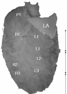
크기를얻Fig. 1. The each numbers of ventricular branches of left coronary artery in a SD rat. The left ventricle was divided into 3 area on auricular surface. The numbering of left ventricular branches were based on the area of distribution.
There are no branches that run in left coronary groove (atrioventricular groove). LA: left auricle of left atrium, PT:
pulmonary trunk, R1, R2, R3: right ventricular branches,
L1, L2, L3: left ventricular branches from the left main
descending artery, respectively. The oval ring marks the site
of ligation for myocardial infarction. The arrows of both
direction represent the each segment of left ventricle. View
of auricular surface (Surgical view of thoracotomy for the
myocardial infarction).
을수있었으며
,
그동안실험자료에서제외하였던중등도크기에서도혈동력학적변화가있음을제시한바
있다
.
이에저자는MI
모델로널리쓰이는SD
흰쥐와Wistar
흰쥐에서LCA
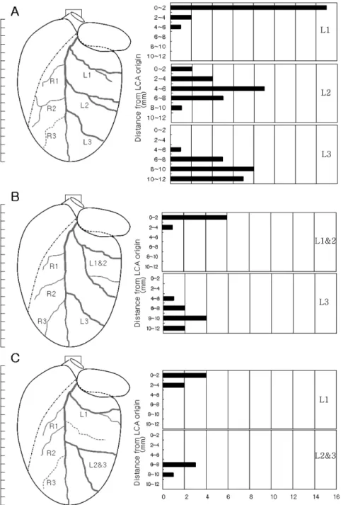
분지양상을해부학적으로구명Fig. 2. The types (type I: A, type II: B, and type III: C) of branching patterns (left) and corresponding graphs of frequency distribution of LV branches of left coronary artery (right) in SD male rats. The Type I has L1, L2 and L3, separately.
The common branch of L1 and L2 originates from the left main descending artery in Type II, and the common branch
of L2 and L3 in Type III. R1~R3, the 1st~3rd right ventricular branches. Dotted lines represent the branches observed
only in some rats. Scale bar, 1 mm. The numbers of left side of the graph represent the distance from the LCA origin
where the left ventricular branches were divided from the LMDA. The number of branches were shown under the graph.
하여일정한크기
,
원하는크기의MI
창출에도움이되고자본연구를시도하였다
.
재료 및 방법
실험동물은체중이
300~350 g
인SD
와Wistar-Kyoto (WKY)
흰쥐수컷을각각27
마리와28
마리사용하였다.
실험동물을
National Institutes of Health(NIH)
지침과Ahn
등
[3]
의방법에따라펜토바비탈(65 mg/kg, I.P)
마취후 배대동맥을통해헤파린을함유한인산완충용액(pH 7.4)
으로관상동맥을관류세척하고붉은색라텍스를주입 하였다
.
대동맥궁(aortic arch)
을결찰한다음심장을적출하여
10%
중성포르말린액에2~3
일이상고정하였다
.
심장절반을담을수있고눈금을새긴판위에심 장을놓고해부현미경을사용하여미세해부를 한다음(Fig. 1), MI
를위한수술면인심방귀면을위로가도록하였다
. LCA
모식도는모눈종이에작성하였고,
각분지위치는
LCA
이는곳에서직선으로내려계측하고심장평균크기에맞추어환산하였다
.
그리고왼쪽심실가지가나오는빈도를조사하고
2 mm
간격으로구분하여그수를그래프로표시하였다
(Fig. 2, 3) [3].
LCA
가지의명명은왼쪽대동맥동에서시작하여심장끝을 향하는 굵은 가지는 왼쪽큰내림가지
(left main descending artery; LMDA)
라고 하였고, LMDA
에서오 른쪽심실(right ventricle; RV)
쪽으로 분지하는 가지는LCA
의 오른쪽심실가지(right ventricular branches of
LCA; R),
왼쪽으로분지하여왼쪽모서리와심방면(atrial
surface)
으로달려분포하는가지는LCA
의왼쪽심실가지
(left ventricular branches of LCA)
라고하였다.
왼쪽심 방귀(left auricle)
아래로LV
를3
등분하였을때위쪽1/
3
에분포하며왼쪽심실사이고랑(
관상고랑)
과떨어져서평행하게달리는가지를첫째왼쪽심실가지
(the first left ventricular branch; L1),
가운데1/3
에분포하는가지를둘째왼쪽심실가지
(the second left ventricular branch; L2),
아래쪽
1/3
에분포하는 가지를 셋째왼쪽심실가지(the third left ventricular branch; L3)
라고하였다. R
은그길이가매우짧고분지위치가다양하여결찰하는대상 이될수없고그수와분포영역이일정하지않으나
,
그길이가
4 mm
이상인것만나타냈으며,
위쪽에서나오는순서에따라번호를붙였다
(Fig. 1).
한편심장끝에서3
mm
이내에분지하는가지는구분하지않았다.
LCA
의심실가지분지유형은왼쪽심실가지를기준으 로나누었으며, I
형(Type I)
은심실세구역에분포하는
L1, L2, L3
가각각분지하는유형이다(Fig. 2A, 3A).
II
형(Type II)
은L1
과L2
가공통가지로분지하지만심 실의위쪽1/3
과중간1/3
부분에분포하며, L3
는별도로분지하는유형이다
(Fig. 2B, 3B). III
형(Type III)
은L1
이분지한후L2
와L3
이공통가지로분지하는유형이며
(Fig. 2C, 3C),
기타유형은위의분류에따르지않는것이었다
.
본연구는관상동맥결찰을하여
MI
을유발하는모 델에서MI
크기의다양성을분석함과아울러적정크기의
MI
유발부위를제시하고자하였고,
중격가지와동 맥원뿔가지는LCA
에서나오는경우가일정하지는않고 일부흰쥐에서만분지하므로[7, 8, 14] LCA
의분지유형에서는제외하였다
.
결 과
심장 크기와 분지 유형
흰쥐심장에라텍스를주입하고고정한심장크기는 길이
13.8
±1.2 mm,
폭은10.0
±0.9 mm
이었다. LMDA
와
LMDA
에서분지하는가지는왼쪽심실벽에서심장근육층내주행을하고있었다
. L
은몇가닥이나오며서로일정한간격을유지하면서관상고랑과평행하게 달려
LV
앞쪽에서왼쪽모서리를지나심방면에가서그치는모습을보였다
.
심실가지중왼쪽관상고랑내에위치하는것을없었다
. L1
에서는중간에왼쪽심방으로 분포하는작은가지가나오기도하였다.
LCA
분지유형은SD
흰쥐에서I
형을보이는 것은15
마리로55.6%, II
형은6
마리로22.2%, III
형은4
마리로
14.8%
였으며, LMDA
와비슷한큰가지가왼쪽심방귀밑에서갈라져심실에분포하되심장끝에이르지
는않는예로 기타유형은
2
마리(7.4%)
에서나타났다(Table 1, Fig. 2). WKY
흰쥐에서는SD
와크게다르지않았다
. I
형은17
마리로60.7%, II
형은3
마리로10.7%, III
형은2
마리로7.1%,
기타유형은6
마리로21.5%
로SD
보다조금높게나타났다(Table 1, Fig. 3).
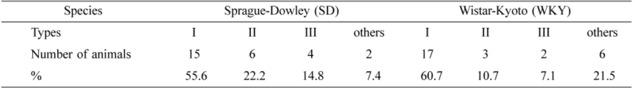
Table 1. The types of branching patterns of left coronary artery in the male hearts of the 2 species of rats
Species Sprague-Dowley (SD) Wistar-Kyoto (WKY)
Types I II III others I II III others
Number of animals 15 6 4 2 17 3 2 6
% 55.6 22.2 14.8 7.4 60.7 10.7 7.1 21.5
LCA의 왼쪽심실가지 분지 위치와 수
1) SD
흰쥐SD
흰쥐의I
형에서L1
의수는18
로평균1.2
이었고,
LCA
이는곳부터2 mm
이내에서 나오는것이15
가지(branches)
로가장많았으며분지하는지점의평균거리는
LCA
이는곳부터1.3
±1.1 mm
이었다. L2
의수는21 Fig. 3. The types (type I: A, type II: B, and type III: C) of branching patterns (left) and corresponding graphs of frequency distribution of LV branches of left coronary artery (right) in WKY male rats. The Type I has L1, L2 and L3, separately.
The common branch of L1 and L2 originates from the left main descending artery in Type II, and the common branch
of L2 and L3 in Type III. R1~R3, the 1st~3rd right ventricular branches. Dotted lines represent the branches observed
only in some rats. Scale bar, 1 mm. The numbers of left side of the graph represent the distance from the LCA origin
where the left ventricular branches were divided from the LMDA. The number of branches were shown under the graph.
(
평균1.4)
이었고, LCA
의이는곳부터4~6 mm
에서나 오는것이9
가지로많았으며분기하는지점의평균거리는
5.1
±1.7 mm
이었다. L3
의수는21(
평균1.4)
이었고
, 8~10 mm
에서기시하는것이8
가지로가장많이나타났으며이들의기시부위거리는평균
8.8
±1.5 mm
이었다
(Table 2, Fig. 2).
II
형에서L1
과L2
공통가지(L1 & 2)
의수는7
로평균1.2
이었고LCA
이는곳부터2 mm
이내에서분지하는것이
6
으로대부분을차지하였으며분지지점의평균거 리는1.5
±0.8 mm
이었다.
독립적인L1
도일부분지하였 으며그수는3(
평균0.2)
이었다. L3
은9
가지(branches)
가분지하여평균
1.5
였으며8~10 mm
지점에서분지하는가지 수는
4
로가장 많았고 그평균거리는8.6
±1.7 mm
이었다.
III
형에서L1
의수는6(
평균1.5)
이었고, 2 mm
이내 에서나오는것이4
으로많았으며,
이는곳의평균거리 는1.6
±0.9 mm
이었다. L2
와L3
의공통가지(L2 & 3)
수는
4(
평균1)
이었고6~8 mm
에서3
가지가분지하였으며 이는곳의평균거리는7.2
±1.5 mm
이었다.
독립적인L2
와
L3
이분지하는것이있었는데각각2
예와1
예에서나타났다
.
2) WKY
흰쥐WKY
흰쥐의I
형에서L1
의수는26
으로평균1.5
이었고
, LCA
이는곳부터2 mm
이내에서나오는것이15
가지(branches)
로가장많았으며분지하는지점의평균거리는
LCA
이는곳에서2.1
±1.5 mm
이었다. L2
의수는
21(
평균1.2)
이었고, LCA
의이는곳부터2~4 mm
에서나오는것이
9
가지, 4~6 mm
에서나오는것이8
가 지로 많았으며 분기하는 지점의 평균 거리는5.8
±1.6 mm
이었다. L3
의수는19(
평균1.1)
이었고, 8~10 mm
에서기시하는것이
11
가지로가장많이나타났으며이 들의기시부위거리는평균9.3
±1.2 mm
이었다(Table 3, Fig. 3).
II
형에서L1
과L2
공통가지(L1 & 2)
의수는3
으로평균
1
이었고LCA
이는곳부터2 mm
이내에서분지하는것이
2, 4~6 mm
에서나오는것이1
이었다.
이들의분지 지점의평균거리는2.8
±2.8 mm
이었다.
독립적으로분 지하여 분포하는L1
의 수는2
이었다. L3
은3
가지(branches)
가분지하여평균1
이었으며8~10 mm
지점에 서2
가지가분지하였고그평균거리는7.9
±2.3 mm
이 었다.
III
형에서L1
의수는3(
평균1.5)
이었고, 2 mm
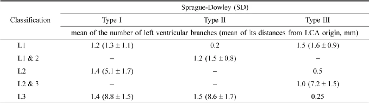
이내Table 2. The numbers of the left ventricular branches of LCA and the their mean distances from LCA origin on its types in the hearts of male SD rats
Classification Sprague-Dowley (SD)
Type I Type II Type III
mean of the number of left ventricular branches (mean of its distances from LCA origin, mm)
L1 1.2 (1.3
±1.1) 0.2 1.5 (1.6
±0.9)
L1 & 2
−1.2 (1.5
±0.8)
−L2 1.4 (5.1
±1.7)
−0.5
L2 & 3
− −1.0 (7.2
±1.5)
L3 1.4 (8.8
±1.5) 1.5 (8.6
±1.7) 0.25
Table 3. The numbers of the left ventricular branches of LCA and the their mean distances from LCA origin on its types in the hearts of male WKY rats
Classification Wistar Kyoto (WKY)
Type I Type II Type III
mean of the number of left ventricular branches (mean of its distances from LCA origin, mm)
L1 1.5 (2.1
±1.5) 0.7 1.5 (0.9
±0.6)
L1 & 2
−1.0 (2.8
±2.8)
−L2 1.2 (5.8
±1.6)
−0.5
L2 & 3
− −1.0 (2.9
±1.0)
L3 1.1 (9.3
±1.2) 1.0 (7.9
±2.3) 1 (9.3
±1.3)
에서모두분지하고 있었으며이는곳의평균거리는
0.9
±0.6 mm
이었다. L2
와L3
의공통가지(L2 & 3)
수는2 (
평균1)
이었고2~4 mm
에서분지하여이는곳의평균 거리는2.9
±1.0 mm
으로그위치는매우높았다.
독립 적인L2
와L3
가분지하는것이있었는데각각1
예와2
예에서나타났다
.
R 분지 수와 개체 수
LCA
의R
는개체별분지수가 다양하였고 그수는1~5
였다(
평균2.0~3.0)(Table 4, Fig. 2, 3). L
과마찬가지 로R
도심장근육층내주행을하고있었고분지하는곳의위치도다양하였으며
,
왼쪽심실가지에비해그길이는짧았다
(Fig. 1).
이들중일부는오른쪽심실까지이르는경우도있었다
.
고 찰
흰쥐는생쥐
,
햄스터,
기니픽,
토끼등과더불어MI
을 유발하여실험에쓰이는동물이다.
값이싸고,
다루기쉬우며
,
심장관상동맥은측부순환이거의없어MI
유발 이쉽고,
실험표본크기를크게할수있고,
살아있는상 태로장시간관찰할수있는점,
사람만성심부전의임상적특징과비슷하다는장점을지니고있어널리쓰이 고있다
[15, 27].
주로사용하는품종은SD
흰쥐와Wistar
흰쥐로수컷을 대부분사용하고있다
[15, 19, 22, 27].
LCA
결찰위치는폐동맥과왼쪽심방귀모서리사이로LCA
이는곳에서2~4 mm
정도떨어진곳이다.
그러나 흰쥐는LCA
가심장근육층내주행을하므로[4, 7, 8, 14]
개흉술(thoracotomy)
을할때관상동맥이지나는곳 을확인하기어려우며LCA
을결찰하여생긴MI
크기 는LV
의4~46%
정도로다양하다[23].
대개MI
크기는25~35%
로 생기고 있으나 혈동력학적 변화(hemod-
ynamic changes)
는MI
크기에따라 달라지고[22, 23],
20%
이하의크기인경우심장기능의변화가적은것으로알려져있어
[12] 20%
이하로생긴개체는실험군에서제외하고있는실정이다
.
그리고결찰이확실히되 었는지확인하는방법은LV
의색깔변화를통하여이루어지고있으며
100%
성공이어렵다[6].
심실에분포하는
LCA
의가지가분지하는데있어개체별[6],
품종별
[19]
차이도있음에도이에대한명확한해부학적연구가선행되지않은채실험을시도하고있어많은수
의동물이필요하다
[6, 21].
더구나실험방법상차이는있으나수술후사망률이대개
15~50%
에이르며, MI
크기가
35%
이상인흰쥐는수술후6
개월생존율이50%
이하에이르므로[6, 12]
혈동력학적변화를보이는일정한크기의
MI
를지닌동물을창출해내기가어려운실정이다
.
따라서관상동맥에 대한해부학적정보를바탕으로 일정한
MI
크기를지닌모델을많이얻는것은동물윤리적인면과실험정확도에서매우중요한일이다
.
본연구는
LCA
결찰과MI
크기에대한정보를제공할목적으로 가장널리쓰이고있는
SD
와WKY
흰쥐의
LCA
분지중심실가지들에대한해부학적연구를시도하였고
LCA
에서분지하는중격가지와동맥원뿔가지는제외하였다
.
중격가지와동맥원뿔가지는MI
를만 들기위한결찰대상이아니며LCA
에서반드시분지하 지않고단지일부에서만분지하기때문이다[7, 8, 14, 19].
흰쥐심장은심방귀면
(auricular surface)
과심방면(atrial
surface)
에서외형상심실사이중격위치를나타내는심실사이고랑이뚜렷하지않다
.
그리고생쥐,
햄스터,
들쥐와같은다른설치류와마찬가지로관상동맥은심장 근육층내주행
(intramyocardial course)
을한다[4, 7, 8,
14]. LCA
주행은이는곳에서몇밀리미터(mm)
이내는심장바깥막밑층을주행
(subepicardial running)
하면서동 맥원뿔곁에있지만 이후심장근육층내주행(intram- yocardial running)
을하면서심장끝(apex of heart)
을향하여
LV
앞쪽을달린다[1, 7, 8, 14].
흰쥐에서이가지 는LV
앞쪽,
즉,
심방귀면에서심실에분포하는여러가 지를내는데그수는2~10
으로대략3
이다[8].
본연구에서
LCA
의가지들(branches)
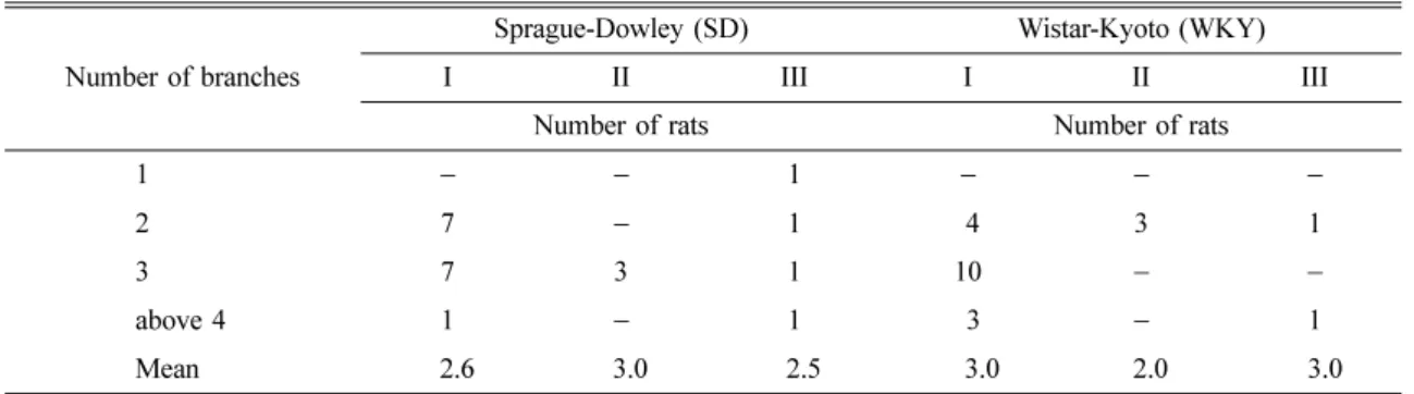
에대한명명은Table 4. The number of right ventricular branches of LCA on its types in SD and WKY male rats Number of branches
Sprague-Dowley (SD) Wistar-Kyoto (WKY)
I II III I II III
Number of rats Number of rats
1
− −1
− − −2 7
−1 4 3 1
3 7 3 1 10
− −above 4 1
−1 3
−1
Mean 2.6 3.0 2.5 3.0 2.0 3.0
흰쥐가 네발동물이므로
Nomina Anatomica Veterinaria
(NAV) [17]
를원칙적으로따랐으며NAV
에없는것은Nomina Anatomica (NA) [11]
에따랐다.
그러나LCA
에서 분지하는중격가지(septal branch)
와동맥원뿔가지(conual branch)
를제외하고,
설치류에서LV
벽(wall)
에분포하는가지는그주행이심실사이고랑에위치하는것이아니
고
,
또분지하여나온가지들의분포가다르므로[3, 5,
9, 10, 16, 24, 25] NAV
에서사용하는원뿔곁심실사이가 지(paraconal interventricular branch), N.A
에서사용하는 앞쪽내림가지(anterior descending branch)
를사용할 수없었다
.
또한휘돌이가지(circumflex branch)
라는용어를 사용할수없었다.
원뿔곁심실사이가지는NA
의앞쪽내 림가지에해당하지만이용어는심방귀면에서심실사이 고랑,
즉,
원뿔곁심실사이고랑이뚜렷하고 이고랑에LCA
의가지가위치하면서주행하는동물에서적용할 수있는용어이기때문이다.
또한원뿔곁심실사이가지와갈라지는첫째큰가지에휘돌이가지라는용어도적 합하지않다
.
그것은흰쥐에서처음으로갈라져나와LV
외측벽에분포하는가지가관상고랑에위치하지않고
[8],
이고랑과어느정도평행하게비스듬히달려심방면에그치며
[14],
동아래심실사이가지로서그치는것이없기
[2, 14]
때문에이용어는적합하지않다고생각한다
.
한편본연구에서관찰한결과흰쥐LCA
의분지는 심장끝을향해가는가지가가장굵고이보다굵기가가 는가지들이나와왼쪽과오른쪽에분포하고있었다.
특히왼쪽으로분지하여가는가지들은서로일정한간격 을유지하면서관상고랑과평행하게달려
LV
앞쪽에서왼쪽모서리를지나심방면에가서그치는모습을보 였으며이는
Icardo
와Colvee [16], Ahn
등[3]
이관찰한생쥐의
LCA
분지모습과유사하였다.
따라서본연구에서는
LCA
의가지중심장끝을향해가면서심실에분지하는이가지의이름은
Ahn
등[3]
이사용한
LMDA
라고하였고,
휘돌이가지라는용어대신
L1
라고하였다.
또이와평행하게달리는가지들을분포영역에따라
L2, L3
라고하였다.
이러한명명은관 상동맥분지유형을분류하는데도매우도움이되고MI
크기를예측하거나분석하는데도유용하다
[3]. LMDA
에서오른쪽으로분지하는가지는
LCA
의R
라고하였 다.
이가지는그길이가왼쪽심실가지에비해짧았으며LMDA
에서분지하는위치가다양하였고,
그가지수는1~4
로다양하였다.
흥미로운것은R
중에서RV
까지분 포하는경우가있었다.
이것은RV
까지분포하는가지를낸다는보고
[14]
와일치하는것이다.
이런경우에LCA
를결찰하면
RV
벽까지MI
가일어날수있다.
실제로 몇몇예에서는RV
벽이반흔조직으로남아있는경우를볼수있다
.
설치류에서
LMDA
에대한명칭은아직도연구자에따라달라서혼란스럽다
. Ahmed
등[2]
은흰쥐와기니픽에대한연구에서앞쪽심실사이가지
(anterior interven- tricular branch),
김등[1], Chimenti
등[6], Liu
등[19]
을 포함한 많은 연구자들은 왼쪽앞쪽내림가지
(left anterior descending branch), Durán
등[9]
은쥐과에속 하는11
종의 쥐에서 둔각모서리가지(obtuse marginal branch),
배쪽심실가지(ventral ventricular branch), Had i- selimovi
등[13]
은가축과다람쥐를비롯한야생동물에 서왼쪽심실사이가지(left interventricular branch), Icardo
와
Colvee [16]
는스위스백색생쥐(Swiss albino mouse)
에 서LCA
큰줄기(main coronary trunk), Sans-Coma
등[24, 25]
은시리안햄스터와안경겨울잠쥐에서둔각모서리가지
(obtuse marginal branch),
배쪽심실사이가지(ventral interventricular branch)
로부르고있고,
북미산비버를연 구한Bisaillon [1981]
은NVA
에따라원뿔곁심실사이가지
(paraconal interventricular branch)
라고하고있다. L1
에대한명칭도휘돌이가지
(circumflex branch) [1, 2, 5, 9, 10, 14, 19, 24, 25]
로부르고있다.
심실가지에대해서도
Dbalý [7]
는얕은가지또는벽쪽가지(superficial or parietal branch)
라고하고있다.
그러나대부분연구자들 은뚜렷하지않은심실사이고랑이존재한다고가정하여 명명하고있거나동물해부학용어를찾지않고사람해 부학에서사용하는용어를그대로사용하고있는바이 에대한정확한명칭도정립되어야할것이다.
흰쥐관상동맥유형에대한해부학적분석은
Dbalý
와공동연구자들
[7, 8]
이Wistar
흰쥐로처음시행하였다.
이들은흰쥐관상동맥분지유형을나누고그가지수
를조사하였으며
, LCA
의분지유형은크게I
형과Y
형으로나눌수있다고하였다
. I
형은LCA
가심장끝을 향해달리면서심실사이중격과동맥원뿔, LV
벽에가지를내는유형이라고하였고
, Y
형은시작부분은I
형과 같으나LV
앞쪽벽,
즉,
심방귀면에이르러굵기가거의비슷한두가지로나뉘어심장끝에이르는유형이라고 하였다
.
흰쥐태자에서I
형은76%, Y
형은24%
이며[8],
성숙한흰쥐에서
I
형은69%, Y
형은31%
라고하였다[7].
그리고LCA
에서분지하여심실벽에분포하는심실가지들
(ventricular branches)
은LV
앞쪽과 뒤쪽벽(anterior and posterior wall)
을따라달리는데 그수는2~10
이며대개3
이라고하였다.
본실험에서I
형, III
형은
Dbalý
와공동연구자들이분류한I
형과 비슷하고,
L1 & 2
의공통가지가크게분지하는II
형과큰가지가둘로나뉘어심실에분포하는기타유형을
Y
형으로볼 수있다.
본실험결과SD
에서I
형, III
형은70.4%, II
형과기타유형은