PCR-RFLP를 이용한 국내 분포 씨스트선충 4종의 동정*
1)
고형래
**․강헌일
***․박은형
**․김은화
**․이재국
****Identification of Four Cyst Nematodes using PCR-RFLP in Korea
Ko, Hyoung-Rai․Kang, Heonil․Park, Eun-Hyoung․Kim, Eun-Hwa․Lee, Jae-Kook
To identify four cyst nematodes (Heterodera schachtii, H. trifolii, H. glycines, H.
sojae) that are economically important plant-parasitic nematodes in Korea, restric- tion fragment length polymorphism (RFLP) by 8 endonucleases (PstI, VspI, AlwI, RsaI, MvaI, EcoRI, Eco72I, Hinf I) was performed based on sequence difference of mitochondrial DNA cytochrome c oxidase subunit I (COI) gene. As a result, species-specific DNA band patterns by RsaI endonuclease were observed in H.
schachtii. The specific patterns was in H. trifolii by 3 endonucleases (VspI, AlwI, Hinf I), and was in H. glycines by Hinf I. While, H. sojae was not digested by 4 endonuclease (VspI, AlwI, RsaI, Hinf I). This study showed that four cyst nemato- des could be distinguished using RFLP by 4 endonucleases (RsaI, VspI, AlwI, Hinf I) based on the sequence difference of COI gene.
Key words : cyst nematode, identification, PCR, RFLP
Ⅰ. 서 론
실과 같이 가느다랗고 긴 원통형의 모양을 하고 있는 선충은 크기가 수 ㎛ 정도로 매우 작아 19세기까지 존재 여부를 잘 알지 못했으나(Dropkin, 1980), 현미경의 성능 발달과 함께 선충의 형태적 관찰이 용이해져 20세기 이후 수많은 연구자들에 의해 활발히 연구되어 왔 다. 선충은 바다, 민물, 토양, 동물 등 다양한 환경에 서식하고 있는데 이러한 서식 환경에
****
이 연구의 수행은 농촌진흥청 국립농업과학원 기관고유 연구사업(PJ01342805)의 지원으로 이루어
졌다.
****
농촌진흥청 국립농업과학원 작물보호과
****
부산대학교 식물생명과학과
****
Corresponding author, 국립농업과학원 작물보호과 농업연구사([email protected])
따라 해양 선충, 동물기생선충, 식물기생선충, 자유생활 토양선충 등으로 구분된다. 농업적 으로 중요시 되고 있는 식물기생선충은 전 세계적으로 150속 3,000종 이상이 보고되었으며, 약 50속 150종 이상의 식물기생선충이 농산물의 수량 감소, 품질 저하 등의 피해를 통해 경 제적으로 큰 손실을 야기하고 있다(Lamberti et al., 2007).
우리나라 농업에 있어 중요한 식물기생선충으로는 뿌리혹선충(Meloidogyne spp.), 뿌리썩 이선충(Pratylenchus spp.), 씨스트선충(Heterodera spp.) 등이 잘 알려져 있으며, 뿌리혹선충 은 과채류 시설재배지에서(Kim, 2001), 뿌리썩이선충은 딸기 등 과채류와 화훼류 재배지에 서 주로 문제를 일으키고 있다(Lee et al., 2008; Ko et al., 2016). 최근에는 밭작물에 씨스트 선충이 다수 검출되어 문제 선충으로 급부상하여 작물 생산에 큰 차질을 빚고 있다(Kang et al., 2016; Kwon et al., 2018). 특히 콩, 배추 등 밭작물은 2종 이상의 씨스트선충이 피해를 주거나 피해를 주지 않는 선충이 혼재되어 있어 신속한 방제 의사결정에 어려움을 겪고 있 다(Kang et al., 2016; Mwamula et al., 2018). 또한, 비기주작물 윤작, 저항성 품종 개발 및 이 용, 살선충성 녹비작물 재배 등을 통한 선충 방제 시에도 종에 따라 방제 효과가 다르게 나 타날 수 있기 때문에 방제를 이행하기에 앞서 선충의 종 동정은 매우 중요하다고 할 수 있 다(Cook and Noel, 2002).
최근 밭작물에 문제되고 있는 선충인 씨스트선충의 종 동정은 광학현미경(light micros- copy), 주사전자현미경(scanning electron microscopy), 투과전자현미경(transmission electron microscopy) 등을 이용한 형태적 특성, 등전점 전기영동(isoelctric focusing) 기법과 같은 단 백질 기반의 생화학적 특성, 중합효소연쇄반응(polymerase chain reaction; PCR), 제한효소절 편길이다형성(restriction fragment length polymorphism; RFLP), 유전자증폭산물길이다형성 (amplified fragment length polymorphism), DNA 바코드 등 분자생물학적 특성들이 이용되고 있다(Subbotin et al., 2010a). 근래에는 분자생물학의 발전에 힘입어 신속 진단이 가능한 실 시간 PCR (real-time PCR)이 활발히 연구되고 있지만(Braun-Kiewnick and Kiewnick, 2018), 고가의 장비임에 따라 아직 보편화되어 있지 않아 기존의 종 동정 기술들이 많이 이용되고 있다. 특히, RFLP 기법은 이미 보편화되어 있는 PCR 장비만 있으면 비교적 쉽고 빠르게 종 동정이 가능하고, 자매종(sibling species)을 구별할 수 있는 기술로 아직까지도 신종 또는 발생 보고 시 많이 이용되고 있다(Subbotin, 2015; Feng et al., 2018). 씨스트선충의 RFLP 연 구 초기에는 ITS (internal transcribed spacer) 영역의 유전자를 대상으로 주로 연구되었다 (Subbotin et al., 2000; Amiri et al., 2002). 그러나 최근에는 씨스트선충 DNA 바코드 마커로 미토콘드리아 DNA cytochrome c oxidase subunit I (COI) 유전자가 주로 이용되고 있으며 (Subbotin et al., 2018), COI 유전자에 대한 RFLP 연구는 거의 수행되지 않았다. 따라서, 본 연구는 콩과 배추 밭에 2종 이상의 씨스트선충이 혼재되어 있을 시 COI 유전자를 대상으 로 RFLP 분석을 통해 종을 동정함으로써 신속한 방제 의사 결정을 위한 기술을 제공하고 자 수행하였다.
Ⅱ. 재료 및 방법
1. 씨스트선충 감염 토양 채취 및 씨스트 분리공시선충을 확보하기 위해 2017년부터 2018년까지 태백 3점, 정선 3점, 세종 1점, 천안 1 점, 서산 2점, 태안 1점, 무안 1점의 씨스트선충 감염 토양시료를 채취하였다(Table 1). 채취 한 토양 시료는 가로 21 cm, 세로 27 cm 크기의 지퍼백에 담아 수분이 증발하는 것을 방지 하였고 8℃ 저온창고에 보관하면서 수시로 꺼내어 분석에 이용하였다.
토양 내 씨스트(cyst)는 체법을 이용하여 추출하였다(Barker, 1985). 씨스트 추출을 위해 토양 시료를 골고루 잘 섞은 다음 100 ml 플라스틱 비커를 이용하여 100 cm3 토양을 정량 하였다. 정량한 토양을 약 7 L의 수돗물이 들어있는 플라스틱 물통(8 L)에 넣어 토양현탁액 을 만들고 20 mesh와 60 mesh체에 순서대로 거른 다음 60 mesh체 위에 남은 물질을 한데로 모으고 수돗물을 이용하여 50 ml 비커에 옮겨 담았다. 옮겨 담은 용액을 150 mm 크기의 필 터페이퍼로 거르고 실체현미경(×50)으로 관찰하면서 핀셋으로 씨스트만 골라내어 직경 2 cm 크기의 micro-sieve에 보관하였다.
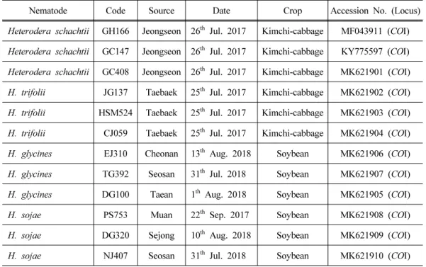
Table 1. The nematode samples used in this study
Nematode Code Source Date Crop Accession No. (Locus)
Heterodera schachtii GH166 Jeongseon 26
thJul. 2017 Kimchi-cabbage MF043911 (COI)
Heterodera schachtii GC147 Jeongseon 26
thJul. 2017 Kimchi-cabbage KY775597 (COI)
Heterodera schachtii GC408 Jeongseon 26
thJul. 2017 Kimchi-cabbage MK621901 (COI)
H. trifolii JG137 Taebaek 25
thJul. 2017 Kimchi-cabbage MK621902 (COI)
H. trifolii HSM524 Taebaek 25
thJul. 2017 Kimchi-cabbage MK621903 (COI)
H. trifolii CJ059 Taebaek 25
thJul. 2017 Kimchi-cabbage MK621904 (COI)
H. glycines EJ310 Cheonan 13
thAug. 2018 Soybean MK621906 (COI)
H. glycines TG392 Seosan 31
thJul. 2018 Soybean MK621907 (COI)
H. glycines DG100 Taean 1
thAug. 2018 Soybean MK621905 (COI)
H. sojae PS753 Muan 22
thSep. 2017 Soybean MK621908 (COI)
H. sojae DG320 Sejong 10
thAug. 2018 Soybean MK621909 (COI)
H. sojae NJ407 Seosan 31
thJul. 2018 Soybean MK621910 (COI)
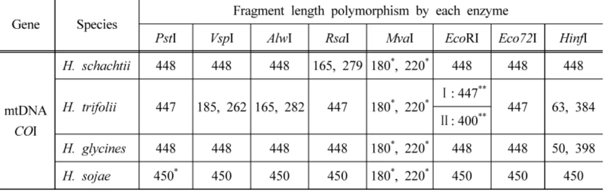
Table 2. The restriction fragment length of the four cyst nematodes by 8 eodonucleases based on the sequence difference of cytochrome c oxidase subunit I (COI) gene
Gene Species
Fragment length polymorphism by each enzyme
PstI VspI AlwI RsaI MvaI EcoRI Eco72I HinfI
mtDNA COI
H. schachtii 448 448 448 165, 279 180
*, 220
*448 448 448
H. trifolii 447 185, 262 165, 282 447 180
*, 220
*Ⅰ: 447
**447 63, 384
Ⅱ: 400
**H. glycines 448 448 448 448 180
*, 220
*448 448 50, 398 H. sojae 450
*450 450 450 180
*, 220
*450 450 450
**Approximate length of DNA band
**Type of the fragment length polymorphism in Heterodera trifolii
2. DNA 추출
씨스트선충의 DNA는 Iwahori 등(2000)의 방법을 응용하여 추출하였다. 슬라이드글라스 에 10 ㎕의 3차 증류수를 떨어뜨린 다음, 골라낸 단일 씨스트를 슬라이드 글라스 위의 증류 수에 옮겨 세척하였다. 낚싯줄을 이용하여 세척한 씨스트를 증류수가 없는 표면으로 옮기 고 핀셋을 이용하여 필터페이퍼(2 mm × 2 mm)로 껍질을 터뜨려 씨스트 내부의 알과 유충 을 슬라이드글라스 표면으로 노출시켰다. 노출된 알과 유충은 새로운 필터페이퍼(2 mm × 2 mm)로 문질러 체벽을 파쇄시키고, 이 때 사용한 필터페이퍼 칩을 30 ㎕의 DNA 추출 용액 (2 M KCl, 10 mM Tris-HCl, 1 M MgCl2, 10% [w/v] Triton-X 100, 20.6 mg/ml Proteinase K)이 들어있는 0.2 ml PCR tube에 침지하였다. 이를 Thermal cycler (PTC-200; MJ Research, Alameda, CA, USA)에 넣고 60℃에서 30분, 95℃에서 10분간 Lysis하여 DNA를 추출하였다.
3. 중합효소연쇄반응
12개 포장에서 각각 검출된 씨스트로부터 추출한 genomic DNA를 주형으로 PCR을 수행 하였다. 뿌리혹선충 미토콘드리아 DNA cytochrome c oxidase subunit I (COI) 유전자 부위의 증폭에는 forward primer JB3 (5′-TTTTTTGGGCATCCTGAGGTTTAT-3′)와 reverse primer JB5 (5′-AGCACCTAAACTTAAAACATAATGAAAATG-3′) 세트를 이용하였다(Derycke et al., 2005). PCR은 PCR premix (Ready-2×-Go with Taqplus; Nanohelix™, Daejeon, Korea) 15 ㎕가 담긴 0.2 ml PCR tube에 template DNA 3 ㎕ (1.03~52.32 ng/㎕), 10 pmol forward primer 1 ㎕, 10 pmol reverse primer 1 ㎕, triple distilled water 30 ㎕를 넣어 총 50 ㎕의 반응액을 만든 다 음, thermal cycler (PTC-200; MJ Research, Alameda, CA, USA)를 이용하여 denaturation 94℃
에서 30초, annealing 52℃에서 30초, extension 72℃에서 45초의 조건으로 수행하였다. PCR 증폭산물은 전기영동기(Mupid eXu; Advance, Tokyo, Japan)를 이용하여 1% agarose gel, 1 × TAE buffer (0.045 M Trisborate, 0.001 M EDTA), 100 V, 25 mA 조건에서 25분간 전기영동 하고 UV장치(MLB-21; Maestrogen, Hsinchu, Taiwan)에서 증폭 여부를 확인하였다.
4. 제한효소절편길이다형성 분석
국내 분포하는 씨스트선충 4종(H. schachtii, H. trifolii, H. glycines, H. sojae)의 미토콘드리 아 DNA COI 유전자 부위 PCR 산물의 제한효소절편길이다형성(RFLP)을 분석하기 위해 Webcutter 2.0 (http://rna.lundberg.gu.se/cutter2/)를 이용하여 8종의 제한효소 PstI (Takara, Otsu, Japan), VspI (Thermo Scientific, Carlsbad, CA, USA), AlwI (Thermo Scientific, Carlsbad, CA, USA), RsaI (Thermo Scientific, Carlsbad, CA, USA), MvaI (Thermo Scientific, Carlsbad, CA, USA), EcoRI (Takara, Otsu, Japan), Eco72I (Thermo Scientific, Carlsbad, CA, USA), HinfI (Takara, Otsu, Japan)을 선발하였다(Subbotin et al., 2010b). 씨스트선충 4종의 각각 3개체군 (총 12개체군)의 PCR 산물 2 ㎕와 triple distilled water 6 ㎕, 제한효소 10 unit, 제한효소와 동봉된 10 × Buffer 1 ㎕를 0.2 ml PCR tube에 넣어 반응물을 조제하였으며, 37℃ 조건에서 overnight 하였다. RFLP 결과는 전기영동장치(Mupid-eXu; Advanced, Tokyo, Japan)에서 3%
agarose gel, 100 V, 25 mA 조건으로 40분간 전기영동하고, UV장치(UVCI-1100; Major science, New Taipei City, Taiwan)에서 각 제한효소별 RFLP 양상을 확인하였다.
Ⅲ. 결 과
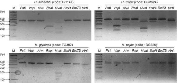
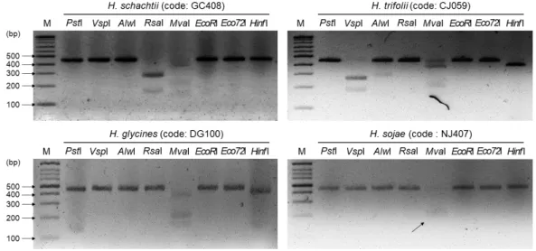
1. 씨스트선충 4종의 PCR 및 제한효소절편길이다형성국내 분포하는 씨스트선충 4종(사탕무씨스트선충, 클로버씨스트선충, 콩씨스트선충, 반 짝이콩씨스트선충) 12개체군의 미토콘드리아 DNA COI 유전자에 대한 PCR 결과 4종 모두 약 450 bp의 PCR 산물을 확인하였다(자료 미제시). 이때 생성된 씨스트선충 4종 12개체군 의 COI 유전자 PCR 산물에 8종류(PstI, VspI, AlwI, RsaI, MvaI, EcoRI, Eco72I, Hinf I)의 제한 효소를 처리한 결과는 Fig. 1~3과 같다. 사탕무씨스트선충(Heterodera schachtii)의 COI 유전 자 PCR 산물에 6종(PstI, VspI, AlwI, EcoRI, Eco72I, Hinf I)의 제한효소 처리 시 448 bp의 단 일 DNA 밴드가 관찰되었고, RsaI 처리 시 165와 279 bp, MvaI 처리 시 약 180과 220 bp의 DNA 밴드가 관찰되었다. 콩씨스트선충(H. glycines)은 Hinf I 처리 시 398 bp, MvaI 처리 시 약 180과 220 bp의 주요 밴드가 관찰되었고, 나머지 6종류의 제한효소 처리 시 448 bp의 단
일 DNA 밴드가 관찰되었다. 반짝이콩씨스트선충(H. sojae)은 MvaI 처리 시 PS753 개체군에 서는 약 180과 220 bp의 주요 밴드가 관찰되었고(Fig. 1), DG320과 NJ407 개체군에서는 약 220 bp의 주요 밴드만 관찰되었다(Fig. 2, 3). 나머지 7종류의 제한효소 처리 시에는 약 450 bp의 DNA 밴드가 관찰되었다. 클로버씨스트선충(H. trifolii)은 JG137 개체군의 COI 유전자
Fig. 1. Restriction fragment length polymorphism of four cyst nematodes population (GH166, JG137, NJ310 and PS753) by 8 endonuclease.
Fig. 2. Restriction fragment length polymorphism of four cyst nematodes population (GC147, HSM524, TG392 and DG320) by 8 endonuclease. Black arrow represented weakened DNA band.
Fig. 3. Restriction fragment length polymorphism of four cyst nematodes population (GC408, CJ059, DG100 and NJ407) by 8 endonuclease. Black arrow represented weakened DNA band.
PCR 산물에 PstI, RsaI, Eco72I 처리 시 447 bp, VspI 처리 시 185와 262 bp, AlwI 처리 시 282 bp, MvaI 처리 시 약 180과 220 bp, Hinf I 처리 시 384 bp의 주요 DNA 밴드가 각각 관 찰되었다. 반면 EcoRI 처리 시에는 JG137, CJ059 개체군에서는 447 bp의 DNA 밴드(type I), HSM524 개체군에서는 약 400 bp의 주요 DNA 밴드(type II)가 관찰되어 2가지 유형의 RFLP 양상을 보이는 것으로 나타났다(Fig. 2, 3).
Ⅳ. 고 찰
씨스트선충의 종 동정 연구에는 ITS rRNA 영역이 주로 이용되어 왔으며, 2000년대 초반 많은 연구자들이 씨스트선충을 대상으로 ITS 영역에 대한 PCR-RFLP 연구를 수행하였다 (Subbotin et al., 2000; Amiri et al., 2002). 그러나 최근에는 COI 유전자가 다양한 생물종의 DNA 바코드 연구에 주로 이용되고 있으며(Powers et al., 2018; Subbotin et al., 2018), 국제 바코드 컨소시움(Consortium for the Barcode of Life)에서도 COI 유전자를 DNA 바코드 마커 로 권장한 바 있다(Blok and Powers, 2009). ITS rRNA는 유전적으로 잘 보존된 영역이긴 하 지만 Heterodera 속의 경우 약 1,020~1,030 bp의 염기서열을 가지는 것으로 알려져 있으며 (Sharma, 1998), 선행 연구나 Webcutter 2.0 웹사이트(http://rna.lundberg.gu.se/cutter2/)를 이용 하여 비교하였을 때 상대적으로 짧은 염기서열을 가지는 COI 유전자 부위(약 450 bp)에 비
해 제한효소 인식 부위가 많았다(supplementary Fig. 1). 따라서 COI 유전자 PCR 산물을 이 용하면 상대적으로 단순한 RFLP 양상을 보일 것으로 기대하였으며, 실험 결과 예상했던 COI 유전자의 RFLP 양상은 ITS 영역의 RFLP 양상보다 비교적 단순하였다(Fig. 1; Subbotin et al., 2000; Amiri et al., 2002).
한편, 일부 제한효소 처리 시 제한효소 인식부위가 없음에도 DNA 단편이 생기는 흥미로 운 결과가 도출되었다. Ko 등(2017)의 연구에서 사탕무씨스트선충과 콩씨스트선충의 COI 유전자 PCR 산물에 MvaI 제한효소를 처리하면 제한효소 인식부위(5′- CC↓WGG - 3′/ 3′- GGW↓CC - 5′)가 없어 448 bp의 단일 DNA 밴드가 관찰되는 것으로 나타났다. 제한효소와 동봉된 매뉴얼(Thermo Scientific Corp.)과 Webcutter 2.0 웹사이트를 이용하여 분석하여도 제 한효소 인식부위가 없는 것으로 나타났다(supplementary Fig. 1). 반면, 본 연구에서는 사탕 무씨스트선충, 클로버씨스트선충, 콩씨스트선충과 반짝이콩씨스트선충 4종 모두 MvaI 처리 시 약 180과 220 bp 2개의 주요 DNA 밴드가 확인되었다. 클로버씨스트선충도 EcoRI 처리 시 JG137과 CJ059 개체군은 447 bp의 단일 DNA 밴드(type I), HSM524 개체군은 약 400 bp 의 DNA 밴드(type II)로 2가지 유형의 DNA 밴드 양상이 관찰되었다. 마찬가지로 제한효소 인식부위(5′ - G↓AATTC - 3′ / 3′ - CTTAA↓G - 5′)가 클로버씨스트선충 모든 개체군의 COI 유전자 염기서열에 없었음에도 2가지 유형의 RFLP 양상을 보였다. 이러한 연구 결과는 MvaI, EcoRI의 제한효소 인식서열을 가지는 자매종(sibling species)이 존재할 때 나타날 수 있는 결과로 판단된다(Subbotin et al., 2015). 하지만 제한효소 처리 시간에 따라 RFLP 결과 의 차이가 있을 수 있으므로 이에 대한 추가적인 연구가 수행되어야 정확한 해석이 가능할 것으로 판단된다. 한편, 반짝이콩씨스트선충의 경우 COI 유전자 PCR 산물에 MvaI 제한효 소 처리 시 PS753 개체군은 약 180과 220 bp의 주요 DNA 밴드가, DG320, NJ407 개체군은 약 220 bp의 DNA 밴드가 관찰되었다. 이는 PCR 증폭산물의 농도 차이에 의한 결과로 판 단되나, COI 유전자에 MvaI 제한효소의 인식서열이 없어 정확한 원인 분석이 어려워 추가 실험 분석이 필요하다. 하지만 본 연구의 목적은 국내 분포하는 씨스트선충 4종(사탕무씨 스트선충, 클로버씨스트선충, 콩씨스트선충, 반짝이콩씨스트선충)의 동정이므로 혼동을 줄 수 있는 MvaI이나 EcoRI 제한효소를 제외하고 종을 동정하는 것이 좋을 것으로 판단된다.
따라서, 각 종별로 특이적인 RFLP 양상을 보인 VspI, AlwI, RsaI, Hinf I 4종류의 제한효소를 국내 분포하는 씨스트선충 4종의 COI 유전자 PCR 산물에 처리하면 각각의 종 구분이 가능 할 것으로 판단된다. 한편, 토양이나 식물체로부터 추출한 씨스트를 수일 간 냉장고나 상온 에 방치하는 경우 씨스트 표면에 다양한 미생물이 증식되고 이들의 DNA가 함께 추출되어 PCR 시 증폭될 수 있다. 이러한 PCR 산물 시료를 RFLP에 이용한다면 특정 종이 가지는 DNA 밴드 패턴이 나오지 않아 종 동정시 오류를 범할 수 있다. 따라서 본 연구에서 개발 된 PCR-RFLP를 이용할 시에는 토양으로부터 1~2일 이내 추출하여 미생물이 증식되지 않 은 깨끗한 상태의 씨스트를 이용해야 실험적 오류로 발생할 수 있는 오동정을 피할 수 있
을 것으로 판단된다.
[Submitted, May. 10, 2019 ; Revised, July. 31, 2019 ; Accepted, August. 6, 2019]
References
1. Amiri, S., S. A. Subbotin, and M. Moens. 2002. Identification of the beet cyst nematode Heterodera schachtii by PCR. Eur. J. Plant Pathol. 108: 497-506.
2. Barker, K. R. 1985. Nematode extraction and bioassay.: Barker, K. R. C. C. Carter, J. N.
Sasser (eds). An advanced treatise on Meloidogyne Volume II: Methodology, North Carolina State University Graphics, US. pp. 19-35.
3. Blok, C. V. and T. O. Powers. 2009. Biochemical and molecular identification.: Perry R. N.
M. Moens, J. L. Starr (eds). Root-knot nematodes, eds. CABI International, UK. pp. 98-118.
4. Braun-Kiewnick, A. and Kiewnick, S. 2018. Real-time PCR, a great tool for fast identifi- cation, sensitive detection and quantification of important plant-parasitic nematodes. Eur. J.
Plant Pathol. 152(2): 271-283.
5. Cook, R. and G. R. Noel. 2002. Cyst nematodes: Globodera and Heterodera species.: Starr, J. L. R. Cook, J. Bridge (eds). Plant resistance to parasitic nematodes, CABI International, US. pp. 71-105.
6. Derycke, S., T. Remerie, A. Vierstraete, T. Backeljau, J. Vanfleteren, M. Vincx, and T.
Moens. 2005. Mitochondrial DNA variation and cryptic speciation within the free-living marine nematode Pellioditis marina. Mar. Ecol. Prog. Ser. 300: 91-103.
7. Dropkin, V. H. 1980. Introduction.: Dropkin, V. H. (eds). Introduction to plant nematology, A Wiley-Interscience Publication, US. pp. 1-5.
8. Feng, Y., D. Wang, D. Xiao, T. J. Pereira, Y. Xuan, Y. Wang, X. Liu, L. Chen, Y. Duan, and X. Zhu. 2018. Cactodera chenopodiae (Nematoda: Heteroderidae), a new species of cyst nematode parasitizing common lambsquarter (Chenopodium album) in Liaoning, China.
Zootaxa 4407(3): 361-375.
9. Iwahori, H., N. Kanzaki, and K. Futai. 2000. A simple, polymerase chain reaction fragment length polymorphism-aided diagnosis method for pine wilt disease. Forest Pathol. 30:
157-164.
10. Kang, H., G. Eun, J. Ha, Y. Kim, N. Park, D. Kim, and I. Choi. 2016. New cyst nematode,
Heterodera sojae n. sp. (Nematoda: Heteroderidae) from soybean in Korea. J. Nematol.
48(4): 280-289.
11. Kim, D. G. 2001. Occurrence of root-knot nematodes on fruit vegetables under greenhouse conditions in Korea. Res. Plant Dis. 7(2): 69-79.
12. Ko, H. R., E. H. Kim, S. J. Kim, J. K. Lee, and W. H. Lee. 2017. Rapid methods to distinguish Heterodera schachtii from Heterodera glycines using PCR technique. Res. Plant Dis. 23(3): 241-248.
13. Ko, H. R., M. A. Lee, E. H. Kim, S. J. Kim, and J. K. Lee. 2016. Incidence of major plant-parasitic nematodes in main producing areas of strawberry in Korea. Res. Plant Dis.
22(4): 249-256.
14. Kwon, S. B., D. G. Park, H. S. Won, Y. G. Moon, J. H. Lee, Y. B. Kim, B. G. Choi, H.
T. Seo, H. R. Ko, J. K. Lee, and D. W. Lee. 2018. Spread of cyst nematodes in highland chinese cabbage field in Gangwon-do. Korean J. Appl. Entomol. 57(4): 339-345.
15. Lamberti, F., N. Greco, and A. Troccoli. 2007. Control and management of plant-parasitic nematodes.: Saxena G. K. G. Mukerji (eds). Management of nematode and insect-borne plant diseases, The Haworth Press, US. pp. 107-164.
16. Lee, J. K., B. Y. Park, D. R. Choi, and J. W. Heo. 2008. Damage and occurrence of Pratylenchus vulnus and Pratylenchus sp. on spray chrysanthemum, Dendranthema grandi- flourm Kitamura. Korean J. Appl. Entomol. 47(4): 473-478.
17. Mwamula, A. O., H. R. Ko, Y. Kim, Y. H. Kim, J. K. Lee, and D. W. Lee. 2018.
Morphological and molecular characterization of Heterodera schachtii and the newly recorded cyst nematode, H. trifolii associated with Chinese cabbage in Korea. Plant Pathol.
J. 34(4): 297-307.
18. Powers, T., T. Harris, R. Giggins, P. Mullin, and K. Powers. 2018. Discovery and identifi- cation of Meloidogyne species using COI DNA barcoding. J. Nematol. 50(3): 399-412.
19. Sharma, S. B. 1998. The cyst nematodes. Kluwer academic publishers, Dordrecht, The Netherlands. p. 452.
20. Subbotin, S. A. 2015. Heterodera sturhani sp. n. from China, a new species of the Heterodera avenae species complex (Tylenchida: Heteroderidae). Russ. J. Nematol. 23(2):
145-152.
21. Subbotin, S. A., F. Toumi, I. H. Elekcioglu, L. Waeyenberge, and Z. T. Maafi. 2018. DNA barcoding, phylogeny and phylogeodraphy of the cyst nematode species of the avenae group from the genus Heterodera (Tylenchida: Heteroderidae). Nematology 20(7): 671-702.
22. Subbotin, S. A., M. Mundo-Ocampo, and J. G. Baldwin. 2010a. Biology and evolution.:
Subbotin, S. A., M. Mundo-Ocampo, J. G. Baldwin (eds). Systematics of cyst nematodes (Nematoda: Heteroderinae) 8A, Koninklijke Brill NV, Netherlands. pp. 43-70.
23. Subbotin, S. A., M. Mundo-Ocampo, and J. G. Baldwin. 2010b. Identification of Heterodera species.: Subbotin, S. A., M. Mundo-Ocampo, J. G. Baldwin (eds). Systematics of cyst nematodes (Nematoda: Heteroderinae) 8B, Koninklijke Brill NV, Netherlands. pp. 9-34.
24. Subbotin, S. A., L. Waeyenberge, and M. Moens. 2000. Identification of cyst forming nema- todes of the genus Heterodera (Nematoda: Heteroderidae) based on the ribosomal DNA- RFLP. Nematology 2(2): 153-164.