*Received: August 3, 2013 / Revised: August 28, 2013 Accepted: August 29, 2013
†Corresponding author: Kyungsoo Chang. Department of Clinical Laboratory Science, College of Health Sciences, Catholic University of Pusan, Busan 609‐757, Korea.
Tel: +82‐51‐510‐0565, Fax: +82‐51‐510‐0568 e-mail: [email protected]
ⒸThe Korean Society for Biomedical Laboratory Sciences. All rights reserved.
J. Exp. Biomed. Sci. 2013, 19(3): 213~223 pISSN : 1738-3226
Molecular Epidemiology of Cryptococcus neoformans/Cryptococcus gattii Complex Isolates from Pigeon Droppings in Korea
Kyungsoo Chang†
Department of Clinical Laboratory Science, College of Health Sciences, Catholic University of Pusan, Busan 609‐757, Korea
The objectives of this study are to develop a molecular diagnosis to differentiate serotypes and mating‐types of C.
neoformans/C. gattii complex isolates from pigeon droppings in Korea and to elucidate molecular epidemiology of the isolates. Phenotypes and genotypes of C. neoformans/C. gattii complex isolates were identified by biochemical properties and PCR using specific CNLAC1 gene, respectively. To classify serotypes and mating‐types of C. neoformans/C. gattii complex isolates, the five reference strains and thirty‐three isolates in Korea were investigated by restriction fragment length polymorphism (RFLP) analysis using CNLAC1 gene for varieties, by random amplified polymorphic DNA (RAPD) for serotyping, and by PCR using specific primer sets for mating typing. All isolates in Korea were belonged to C. neoformans var. grubii (serotype A) by RFLP and RAPD patterns which showed high sensitivity and specificity. Therefore, RFLP and RFLP were available to differentiate varieties and serotypes of C. neoformans. Amplification patterns of the five reference strains by specific PCR for mating typing were differentiable, and all isolates were classified into MATα. All C. neoformans environmental isolates in Korea were Cr. neoformans serotype A and MATα which is a more virulent pathogen. This study suggests that RFLP and RAPD are rapid and correct molecular diagnosis tools for epidemiology of C. neoformans/C. gattii complex isolates.
Key Words: C. neoformans/C. gattii complex, Pigeon droppings, Molecular epidemiology, RFLP, RAPD
INTRODUCTION
The encapsulated yeast Cryptococcus neoformans/
Cryptococcus gattii (C. neoformans/C. gattii) species complex is a basidiomycetous dimorphic organism and are found worldwidely, because its natural habitats are pigeon droppings and contaminated soil. Its inhalation from an environmental source may cause pulmonary and central nervous system infection in susceptible human and animal hosts (Vidotto et al., 2006; Matsumoto et al., 2007; Ribeiro
and Ngamskulrungroj, 2008). This opportunistic fungal pathogen is responsible for life‐threatening infections, particularly in immunocompromised patients. Cryptococcosis has been responsible for great morbidity and mortality rates among patients with AIDS. The incidence of cryptococcal infection has increased in recent years as a result of a large increase in AIDS patients, population aging and the expanded use of immunosuppressive drugs for cancer treatment and organ transplantation (Ohkusu et al., 2002;
Vidotto et al., 2006; Matsumoto et al., 2007; Ribeiro and Ngamskulrungroj, 2008).
Cryptococcus species complex have been divided into five distinct serotypes, A, B, C, D, and A/D type according to the immunologic properties (antigenic determinants) of their capsular polysaccharides, which are related to pathogenicity. Cryptococcus species complex have been divided into two species (C. neoformans and
Original Article
C. gattii) regard to biochemically, genetically, ecologically, and epidemiologically. Also, C. neoformans has been divided into two varieties (C. neoformans var.
grubii and C. neoformans var. neoformans). C.
neoformans var. neoformans corresponds to serotypes D and A/D, C. neoformans var. grubii corresponds to serotype A, whereas C. gattii corresponds to serotypes B and C. Some epidemiologic properties such as geographic distribution have been associated with serotypes. C.
neoformans var. grubii (serotype A) and C. neoformans var. neoformans (serotype D and A/D) has a worldwide distribution in pigeon droppings and decaying wood. C.
gattii (serotype B and C) has a more limited global distribution, occurring in tropical and subtropical regions.
It has been shown to have a specific ecological association with a number of Eucalyptus species trees in Australia (Baró et al., 1999; Franzot et al., 1999; Kwon‐Chung and Varma, 2006; Okabayashi et al., 2006; Enache‐Angoulvant et al., 2007; Frasés et al., 2009; MacDougall et al., 2011).
Several serotyping methods have been used in analysis of clinical and environmental isolates of Cryptococcus species complex. For classification of the two species of C. neoformans, canavanine‐glycine‐bromthymol blue (CGB) agar test and disk test of D‐proline on carbon base agar are required. CGB agar test is performed to determine the use of glycine as the source of carbon. D‐proline assimilation test is performed to determine the use of D‐
proline as the source of nitrogen (Kwon‐Chung et al., 1982;
Baró et al., 1999; Franzot et al., 1999; Kwon‐Chung and Varma, 2006). In addition, Cryptocheck agglutination test (Iatron Laboratories Inc., Tokyo, Japan) had been used as the most common serotyping method (Baró et al., 1999;
Nakamura, 2001; Ohkusu et al., 2002; Frasés et al., 2009).
This method cannot be available anymore because they are not made in Japan anymore (Enache‐Angoulvant et al., 2007).
Mating type is well known as a virulence factor of Cryptococcus species complex. Two mating types MATa and MATα are existed in Cryptococcus species complex.
The mating system is controlled by single‐locus with two‐
idiomorphic alleles. When crossed on an appropriate medium, equal numbers offspring of the a and α mating
types are produced (Halliday et al., 1999; Chaturvedi et al., 2000; Ohkusu et al., 2002; Litvintseva et al., 2011).
However, according to the survey results of C.
neoformans var. neoformans isolates from environmental (mostly pigeon droppings) and clinical sources have shown that the MATα is an approximately 30 to 40 times more frequent than the MATa. Also, the MATα strain was significantly more virulent then the MATa strain when injected intravenously into the murine model (Kwon‐Chung et al., 1992; Halliday et al., 1999; Ohkusu et al., 2002; Okabayashi et al., 2006).
Many researchers is already known that the importance of Cryptococcus species complex affecting the worldwide public health. Thus, ecological, epidemiological, human infectious mechanism, and pathogenicity studies are actively underway on the global scale. Nevertheless, domestic studies are concentrated on medical cases, other studies are few. As a result, there is a paucity of important ecological and epidemiological information for Cryptococcus species complex, in Korea. Therefore, ecological and epidemiological studies on Cryptococcus species complex are essential to understand this fungus in order to improve diagnosis and therapy against their infections.
In this study, we attempted to differentiate serotypes of Cryptococcus species complex isolates from environmental and clinical sources by using molecular biological methods.
Restriction fragment length polymorphism (RFLP) and random amplified polymorphic DNA (RAPD) methods were performed in this study. CNLAC1 gene was encoding laccase to produce melanin pigment and then selected for RFLP. A phenoloxidase called laccase is the crucial cooper‐
containing enzyme responsible for the conversion of diphenolic compounds to dopaquinone, followed by their polymerization to melanin (Buchanan and Murphy, 1998).
Additionally, we investigated the mating types of Cryptococcus species complex isolates from Korea by PCR using specific primer sets. Molecular methods using RAPD and RFLP might be useful for rapid differentiation of serotypes and mating types of Cryptococcus species complex, as well as providing important epidemiological and ecological information.
MATERIALS AND METHODS Cryptococcus species complex strains and other yeast‐
like fungi
In previous study, thirty one environmental strains of Cryptococcus species complex were isolated from pigeon droppings which were collected from public places (such as parks and under the bridges) in Korea from February 2010 to March 2011 (Chae et al., 2012). Briefly, some parts of each sample were homogenized as much as possible. Homogeneous samples were suspended in sterilized phosphate buffered saline (PBS) at a ratio of 1:5 by vortex, and centrifuged at 500× g for 5 min. One hundred micro‐liter of aliquot from supernatant was inoculated onto sunflower seed (Helianthus annus) agar plate containing 0.1% creatinine and 40 ml chloramphenicol (50 μg/ml) for detection of the typical brown pigmentation due to the activity of phenoloxidase of Cryptococcus species complex. The plates were incubated in the dark at 25℃ under a humid condition.
After 5~10 days, brown colonies by melanin pigment production of Cryptococcus species complex were subcultured on sunflower seed agar (SSA) and Sabouraud dextrose agar (SDA) plates at 25℃ under a humid condition for the isolation and maintenance of pure colony (Chae et al., 2012). Two clinical isolates of Cryptococcus species complex were provided from general hospitals in Busan from February 2010 to March 2011. One identified clinical strain of C. neoformans var.
grubii was provided from Dr. Hwang (Catholic University of Pusan, Busan), and used as a reference strain for serotype A of C. neoformans.
Non C. neoformans yeast like‐fungi isolates:
Cryptococcus laurentii (C. laurentii), Cryptococcus uniguttulatus (C. uniguttulatus), Candida zeylanoides (C.
zeylanoides), Candida guilliermondii (C. guilliermondii), Candida sake (C. sake), Rhodotorula mucilaginosa (R.
mucilaginosa), Rhodotorula glutinis (R. glutinis), Malassezia furfur (M. furfur) and Stephanoscus ciferrii (S. ciferrii) isolated from pigeon droppings were used in this study. All isolates were identified by using the Vitek2 ID‐YST card (Biomerieux, France) and
genotyping in previous studies (Chae et al., 2012).
Candida albicans (C. albicans) clinical isolates were provided from general hospitals in Busan.
All reference strains of C. neoformans and C. albicans were purchased from American Type Culture Collection (ATCC). Serotype D (ATCC 66031) and serotype A/D (ATCC 48184) of C. neoformans var. neoformans were used as reference strain. Serotype B (ATCC MYA‐4560) and serotype C (ATCC 32608) of C. gattii was used as reference strain. C. albicans (ATCC 10231) was used as reference strain.
Differentiation of species of Cryptococcus species complex by biochemical tests
All of pure brown colonies were identified as Cryptococcus species complex according to standard biochemical tests (Chae et al., 2012). Briefly, pure brown colonies of isolates on SSA were stained by india ink and nigrosine stains, and observed by light microscopy examination to detect capsule formation. Additionally, we performed specific biochemical tests such as urease activity test on Cristensen’s urea broth, nitrate reduction test, and the growth ability test at 37℃ for identification of Cryptococcus species complex. Final identification of isolates was performed using Vitek2 ID‐YST card (Biomerieux, France).
Species of Cryptococcus species complex isolates were determined by CGB agar test and disk test of D‐proline on carbon base agar. CGB agar test was performed based on the method described by Kwon‐Chung et al., 1982.
Thirty three isolates were cultured on CGB agar, and incubated in the dark at 25℃ under a humid condition for 5 days. Positive reaction, a color change from greenish yellow to cobalt blue indicated that variety of isolate is C. gattii (serotype B and C). Negative reaction, no color change indicated that variety of isolate is C.
neoformans var. grubii (serotype A) or C. neoformans var. neoformans (serotype D and A/D). Disk test of D‐
proline on carbon base agar was performed based on the method described by Mukamurangwa et al., 1995.
Growth around the disk as positive reaction indicated that variety of isolate is C. gattii (serotype B and C). No
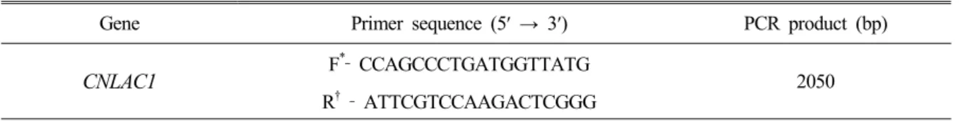
Gene Primer sequence (5′ → 3′) PCR product (bp)
CNLAC1 F
*‐ CCAGCCCTGATGGTTATG R† ‐ ATTCGTCCAAGACTCGGG 2050
*Forward primer.
†Reverse primer.
Table 1. CNLAC1 primer sequences for serotyping of Cryptococcus species complex growth around the disk as negative reaction indicated that
variety of isolate is C. neoformans var. grubii (serotype A) or C. neoformans var. neoformans (serotype D and A/D).
Extraction of genomic DNA from Cryptococcus species complex
Genomic DNA of Cryptococcus species complex isolates was extracted based on the method described by Yamamoto et al., 1995. Briefly, each of isolates was cultured on SDB for 48h at 25℃. One milliliter cultivated broth was moved to a new tube and centrifuged at 6,500×
g for 1 min. Sample pellet was suspended 500 µl of extraction buffer (100 mM Tris HCl (pH 9.0), 40 mM EDTA), and mixed after addition of 100 µl of 10%
sodium dodecyl sulfate (SDS) and 300 µl of 99% benzyl chloride. The mixture was homogenized, and incubated at 50℃ for 30 min with gentle shaking. After centrifugation at 14,200× g, at 4℃ for 10 min, the supernatant was transferred to a new tube. After addition of 1/10 volume of 3 M sodium acetate and equal volume of isopropanol of the supernatant, the tube was slightly vortexed, and stored at ‐70℃ for 1 h. The DNA was precipitated after centrifugation at 14,200× g, at 4℃ for 20 min, and the supernatant was removed. The pellet was washed with 500 µl of ethanol and centrifuged at 14,200× g at 4℃ for 5 min, and after that the supernatant was removed. The pellet was dried in room temperature for 10 min. The pellet was resuspended in 50 µl of distilled water (D.W).
Polymerase chain reaction (PCR) and Restriction fragment length polymorphism (RFLP) with CNLAC1 gene
Genotyping of isolates from pigeon droppings was carried out using CNLAC1 gene sequencing with CNLAC1 unique oligonucleotide primer described by
previous study (Chae et al., 2012).
For RFLP analysis, new CNLAC1 primer pair was designed by the Biotoolkit320 program. The sequence of CNLAC1 primer pair is described in Table 1. RFLP analysis of CNLAC1 gene was performed after checking the amplified CNLAC1 gene of Cryptococcus species complex isolates. PCR was performed using the AccuPower PCR Premix Kit (Bioneer, Seoul, Korea). A total volume of 20 µl of reaction mixture was contained within each 1 µl of primer (10 pM/ µl), and 2 µl of template DNA (1 µg/µl), and 16 μl of D.W. Amplification of CNLAC1 gene was carried out using the following conditions: initial denaturation at 95℃ for 5 min; 35 cycles of 95℃ for 1 min, 58℃ for 1 min, and 72℃ for 90 sec;
and a final extension at 72℃, 7 min. PCR products were analyzed by electrophoresis with 1.2% agarose gels containing ethidium bromide (EtBr), and visualized under UN light.
For RFLP patterns analysis of CNLAC1 gene, one restriction enzyme which can be classified into three varieties were chosen by using the NEBcutter V2.0 program (http://tools.neb.com/NEBcutter2/index.php):
ApaLI (Enzynomics, Seoul, Korea). The restriction enzyme reaction was conditioned with 10× Buffer, 10U restriction enzyme, including 2 µl of PCR product and D.W. to become 20 µl. After six hours of digestion at 37℃, electrophoresis was carried out on 2.0% agarose gels containing EtBr (10 µg/ml) in 0.5× Tris‐bortate‐
EDTA (TBE) buffer at 100V for 80 min. RFLP patterns of isolates and reference strains of Cryptococcus species complex were visualized under UV light.
Random amplified polymorphic DNA (RAPD) analysis
Six different primers for RAPD were designed as
previously described by Koeleman et al., 1998. PCR was performed using the AccuPower PCR Premix Kit. A total volume of 20 µl of reaction mixture was contained within each 1 µl of RAPD primer (25 pM/µl), and 2 µl of template DNA (1 µg/µl), and 17 μl of D.W. PCR was carried out using the following conditions: initial denaturation at 95℃ for 5 min; 45 cycles of 94℃ for 1 min, 36℃ for 1 min, and 72℃ for 2 min. PCR products were separated by electrophoresis on 2.0% agarose gels containing EtBr (10 μg/ml) in 0.5× TBE buffer at 100V for 90 min, and visualized under UV light.
Determination of mating type of Cryptococcus species complex isolates
Mating type determination for Cryptococcus species complex isolates were performed by PCR using two unique oligonucleotide primer pairs, which are specific for mating type a and α. The primer pairs were designed by Chaturvedi et al., 2000. The sequence and expected size of PCR products are described in Table 1. PCR was performed using the AccuPower PCR Premix Kit. A total volume of 20 µl of reaction mixture was contained within each 1 µl of primer (10 pM/µl), and 2 µl of template DNA (1 µg/µl), and 16 μl of D.W. Amplification of primer pairs were carried out using the following conditions: initial denaturation at 95℃ for 3 min; 30 cycles of 94℃ for 1 min, 58℃ for 1 min, and 72℃ for 1 min; and a final extension at 72℃, 7 min. PCR products were separated by electrophoresis on 2.0% agarose gels containing EtBr (10 µg/ml) in 0.5× TBE buffer at 100V for 90 min, and visualized under UV light.
RESULTS
Differentiation of species of Cryptococcus species complex by biochemical properties
Thirty three Cryptococcus species complex environmental and clinical isolates were identified by various biochemical properties. All Cryptococcus species complex isolates were shown typical brown colonies on SSA by phenoloxidase activity, and identical positive results for capsule formation,
urease activity, nitrate reduction, and growth ability at 37℃. Finally, they were confirmed as Cryptococcus species complex by biochemical properties using Vitek2 ID YST system.
For classification of species for Cryptococcus species complex isolates, CGB agar test and disk test of D‐proline on carbon base agar were performed. As a result, thirty three Cryptococcus species complex isolates and reference strains (C. neoformans var. neoformans (ATCC 66031 and 48184) and C. neoformans var. grubii) were not detected color changes on CGB agar. Whereas, reference strains of C. gattii (ATCC MYA‐4560 and 32608) were shown color changes from greenish yellow to cobalt blue on CGB agar.
In D‐proline disk test on carbon base agar, thirty three Cryptococcus species complex isolates and reference strains (C. neoformans var. neoformans (ATCC 66031 and 48184) and C. neoformans var. grubii) were not detected strong growth around the D‐proline disk. Whereas, C.
gattii (ATCC MYA‐4560 and 32608) were shown strong growth around the D‐proline disk. Therefore, varieties of thirty three Cryptococcus species complex environmental and clinical isolates were confirmed as C. neoformans var.
grubii (serotype A) or C. neoformans var. neoformans (serotype D and A/D).
Determination of varieties of Cryptococcus species complex isolates by PCR‐RFLP patterns
RFLP was performed to differentiate species and varieties of Cryptococcus species complex isolates. The specificity of CNLAC1 gene for detection of Cryptococcus species complex isolates have been reported in our previous study (Chae et al., 2012). The approximately 2000 bp of amplified fragments were detected in thirty three Cryptococcus species complex isolates and reference strains by PCR using CNLAC1 primer pair. However, it was not amplified in other yeast isolates. In addition, CNLAC1 gene was detected in five serotypes of Cryptococcus species complex, but not differentiated into their serotypes.
For determination of classification of Cryptococcus species complex, RFLP analysis was performed by using one restriction enzyme. As a result, two species and
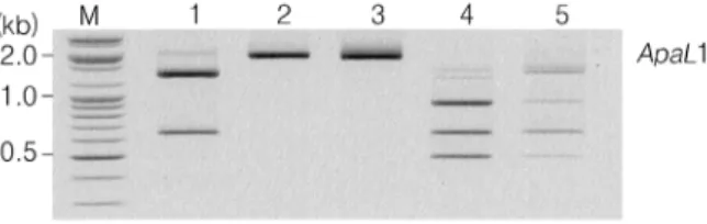
Fig. 1. Restriction patterns of the amplified CNLAC1 gene from Cryptococcus species complex isolates and reference strains by using ApaL1. Lanes: M. 100 bp molecular marker (Bioneer, Seoul), 1. reference strain of C. neoformans var.
grubii (serotype A), 2. reference strain of C. gattii (serotype B, ATCC MYA‐4560), 3. reference strain of C. gattii (serotype C, ATCC 32608), 4. reference strain of C. neoformans var.
neoformans (serotype D, ATCC 66031), 5. reference strain of C. neoformans var. neoformans (serotype A/D, ATCC 48184).
varieties of reference strains, clinical and environmental isolates were classified through different RFLP patterns (Fig. 1). Digestion of the CNLAC1 gene by ApaLI resulted in 3 different RFLP patterns. Serotypes B and C strains (ATCC MYA‐4560 and ATCC 32608: C.
neoformans var. gattii) were not digested by ApaLI.
Serotype A strains (thirty‐ three environmental and clinical isolates: C. neoformans var. grubii) were digested into two different size fragments, approximately between 650 bp to 1,400 bp by ApaLI. In addition, serotypes D and A/D strains (ATCC 66031 and ATCC 48184: C. neoformans var. neoformans) were digested into three different size fragments, approximately 500 bp, 700 bp and 900 bp by ApaLI. Therefore, two species and varieties of Cryptococcus species complex were determined by ApaLI.
Analysis of PCR‐RFLP by using one restriction enzyme is one of useful diagnostic molecular methods to differentiate species and varieties of Cryptococcus species complex.
Determination of serotypes for Cryptococcus species complex isolates by RAPD patterns
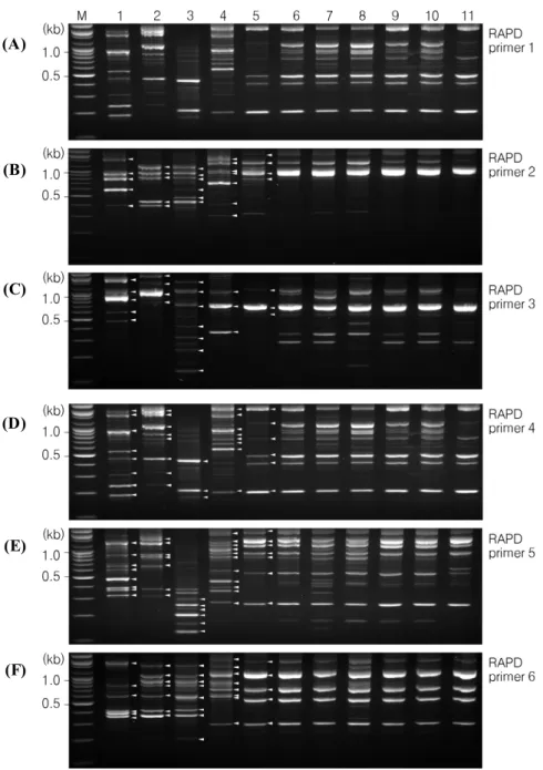
The RAPD patterns of five reference strains and thirty three isolates of Cryptococcus species complex were obtained from the six different RAPD primers (Fig. 2).
The specific RAPD patterns were detected in five reference strains of Cryptococcus species complex by serotype. Especially, RAPD patterns of thirty four clinical and environmental isolates were identical. RAPD primer
1; mainly 5 bands of 500, 650, 750, 1,400, and 1,600 bp were detected in all environmental and clinical isolates (Fig. 2A). RAPD primer 2; mainly 4 bands of 800, 910, 1,300, and 2,100 bp were detected in environmental and clinical isolates (Fig. 2B). RAPD primer 3; mainly 3 bands of 580, 660, and 1,200 bp were detected in environmental and clinical isolates (Fig. 2C). RAPD primer 4; mainly 6 bands of 210, 410, 500, 750, 900, 1,100, and 2,100 bp were detected in environmental and clinical isolates (Fig. 2D). RAPD primer 5; mainly 7 bands of 270, 550, 900, 1,000, 1,200, 1,500, and 2,100 bp were detected in environmental and clinical isolates (Fig. 2E). RAPD primer 6; mainly 5 bands of 300, 520, 700, 1,100 and 2,000 bp were detected in environmental and clinical isolates (Fig. 2F). RAPD patterns of Cryptococcus species complex reference strains were different from thirty three environmental and clinical isolates. RAPD patterns between Cryptococcus species complex reference strains were different from each other by serotype. In other words, RAPD patterns are one of useful diagnostic molecular methods for serotyping of Cryptococcus species complex.
Specificity of RAPD patterns to identify Cryptococcus species complex from other yeast like‐fungi
RAPD patterns between serotypes of Cryptococcus species complex were specific. Especially, RAPD patterns by using primers 5 were more specific than others. All of the yeast like‐fungi isolates obtained from the environmental and clinical sources gave different band patterns after RAPD‐
PCR with primer 5 (Fig. 3). RAPD primer 5 was able to produce unique different RAPD patterns from Cryptococcus species complex. Consequently, analysis of RAPD patterns by using RAPD primer 5 is one of useful diagnostic molecular methods for identification of Cryptococcus species complex from other yeast like‐fungi.
Determination of mating types for C. neoformans isolates by PCR
For determination of mating types for C. neoformans isolates, PCR was performed by using specific two primer pairs. As a result, only MATα specific primer pair
Fig. 2. RAPD patterns of Cryptococcus species complex isolates and reference strains by serotypes. A: RAPD profiles by PCR using RAPD primer 1, B: RAPD profiles by PCR using RAPD primer 2, C: RAPD profiles by PCR using RAPD primer 3, D: RAPD profiles by PCR using RAPD primer 4, E: RAPD profiles by PCR using RAPD primer 5, F: RAPD profiles by PCR using RAPD primer 6.
Lanes: M. 100 bp molecular marker (Bioneer, Seoul), 1. reference strain of C. gattii (serotype B, ATCC MYA 4560), 2.
reference strain of C. gattii (serotype C, ATCC 32608), 3. reference strain of C. neoformans var. neoformans (serotype D, ATCC 66031), 4. reference strain of C. neoformans var. neoformans (serotype A/D, ATCC 48184), 5. reference strain of C.
neoformans var. grubii (serotype A), 6‐9. environmental isolates of C. neoformans (serotype A), 10‐11. clinical isolates of C. neoformans (serotype A).
was amplified specific fragments of 101 bp in all isolates (Fig. 4). Therefore, thirty‐four C. neoformans isolates from environmental and clinical sources were MATα (33/33, 100%).
DISCUSSION
Cryptococcus species complex are opportunistic fungal pathogens that are prevalent worldwide and have been identified from various environmental sources, particularly (A)
(B)
(C)
(D)
(E)
(F)
pigeon droppings. These pathogens cause life‐threatening infection of the central nervous system in mainly immunodeficiency hosts (Chen et al., 1996; Ohkusu et al., 2002; Vidotto et al., 2006). Especially, C. neoformans var.
grubii (serotype A) and C. neoformans var. neoformans (serotype D and A/D) are responsible for the most cases of cryptococcosis in immunocompromised patients.
Whereas C. gattii (serotype B and C) has been associated with infections in subjects with a normal immunologic status in British Columbia (Canada) and Pacific Northwest (United States America) (Centers for Disease Control and Prevention. 2010; Litvintseva et al., 2011; MacDougall et al., 2011). The frequency of severe systemic mycosis has increased in the last few decades.
Serotyping of Cryptococcus species complex isolates has been useful for explanation of the epidemiologic, pathogenic and clinical information associated with this fungal pathogen. The mating types of isolates are important for understanding their ecology and virulence.
In addition, serotypes and mating types of Cryptococcus species complex isolates are essential to rapid diagnosis and accurate treatment against the attacks of this fungal pathogen.
Currently, the prevalent laboratory investigation methods for serotypes and mating types of C. neoformans are limited to biochemical tests and immunodiagnosis.
Cryptocheck agglutination test kit (Iatron Laboratories Inc., Tokyo, Japan) had been used as most common serotyping method (Baró et al., 1999; Nakamura, 2001;
Ohkusu et al., 2002; Enache‐Angoulvant et al., 2007;
Frasés et al., 2009). However it is no longer available.
Therefore, new methods need to be developed for the typing of Cryptococcus species complex strains. Molecular methods have high specificity and sensitivity, and their potential to overcome the limitations of conventional methods. They are being used as methods of identification, typing and epidemiology studies on the global scale.
Nested PCR assay using ITS rDNA was very useful molecular method for detection Cr. neoformans in CSF (Mitchell et al., 1994; Rappelli et al., 1998; Sidrim et al., 2010). ITS region has been the most frequently used for yeast genotyping depending on nucleotide sequences. PCR
‐RFLP method classified serotypes of Cryptococcus species complex by using CAP59 gene which is necessary for capsule formation (Enache‐Angoulvant et al., 2007;
Sidrim et al., 2010). However, it was not a specific gene for the identification of Cryptococcus species complex.
ITS region and CAP59 gene were the common genes in yeasts. Serotypes of C. neoformans and C. gattii were grouped into 8 molecular types (VNI, VNII, VNIII, VNIV, VGI, VGII, VGIII, and VGIV) by PCR fingerprinting using M13 sequences in the earlier studies (Meyer et al., 2003; Matsumoto et al., 2007; Sidrim et al., 2010).
However, there still is not exactly correlation between serotype and molecular type for Cr. gattii.
This study demonstrated the usefulness of PCR‐RFLP and RAPD methods to differentiate varieties and serotypes of Cryptococcus species complex isolates from environmental and clinical sources. There are many advantages using our methods compared to using conventional methods. We found and demonstrated that CNLAC1 gene was a specific gene of Cryptococcus species complex strains. In addition, CNLAC1 gene was capable to detect all serotypes of Cryptococcus species complex (Chae et al., 2012). The PCR‐RFLP method by using CNLAC1 gene was demonstrated to be specific method for rapid identification and detection of two species of Cryptococcus species complex (Fig. 1). Also, one restriction enzyme, ApaL1 was suitable enzyme for differentiate varieties of C. neoformans. PCR‐RAPD method was also useful method for detection of five serotypes of Cryptococcus species complex (Fig. 2). Six different RAPD primers were successful in differentiating Cryptococcus species complex serotypes. Specific RAPD band patterns were visualized easily by serotype.
Especially, RAPD primer 5 was more specific than others. RAPD primer 5 was able to produce different RAPD patterns from Cryptococcus species complex by serotype. In addition, the non‐Cryptococcus species complex isolates obtained from the environmental and clinical sources gave different band patterns from Cryptococcus species complex (Fig. 3). Therefore, these results have proved that the RFLP and RAPD methods can save a lot of time for serotyping Cryptococcus
Fig. 3. RAPD patterns of non‐Cryptococcus species complex yeast like‐fungi isolates by PCR using RAPD primer 5. Lanes:
M. 100 bp molecular marker (Bioneer, Seoul), 1. C. laurentii, 2. C. uniguttulatus, 3. C. zeylanoides, 4. C. guilliermondii, 5. C. sake, 6. R. mucilaginosa, 7. R. glutinis, 8. M. furfur, 9. S. cifferrii.
Fig. 4. Mating types of C. neoformans isolates by PCR. Lanes: M. 100 bp molecular marker (Bioneer, Seoul), 1. reference strain of C. neoformans var. neoformans (MATα, ATCC 66031), 2‐4. environmental isolates of C. neoformans, lane 5. clinical isolate of C. neoformans.
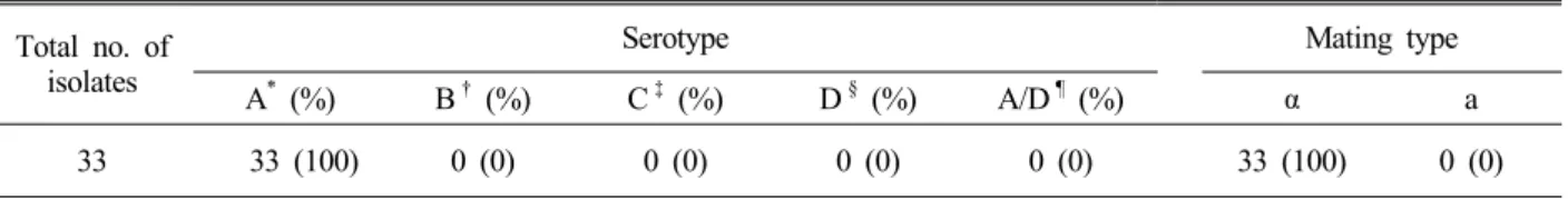
Total no. of isolates
Serotype Mating type
A* (%) B† (%) C‡ (%) D§ (%) A/D¶ (%) α a
33 33 (100) 0 (0) 0 (0) 0 (0) 0 (0) 33 (100) 0 (0)
*Serotype A (C. neoformans var. grubii)
†Serotype B (C. gattii)
‡Serotype C (C. gattii)
§Serotype D (C. neoformans var. neoformans)
¶Serotype A/D (C. neoformans var. neoformans)
Table 2. Characteristics of environmental and clinical isolates of Cryptococcus species complex in Korea species complex environmental and clinical isolates
without serological and biochemical tests.
The environmental and clinical isolates in Busan, Korea were reported to be serotype A and mating type α in previous study (Hwang, 2002; Hwang, 2010). Our results showed that all Cryptococcus species complex isolates from environmental and clinical sources in Korea between 2010 and 2011 were belonged to C. neoformans var.
grubii (serotype A) by analysis of RAPD and RFLP patterns (33/33, 100%). Other serotypes were not identified in this study. Mating type α‐specific primer was used in this study and amplified a 101 bp MATα fragment in all environmental and clinical isolates (33/33, 100%), showing that all Cryptococcus species complex isolates
were MATα (Fig. 4). Information about the mating types of Cryptococcus species complex isolates from Korea is important for an understanding of their ecology, epidemiology and virulence. These results suggest that the pathogen of animal and human cryptococcosis cases in Korea might be only C. neoformans serotype A and MAT α (Table 2).
In conclusion, the molecular diagnostic methods by PCR‐RFLP and RAPD were rapid molecular tools for serotyping Cryptococcus species complex isolates. It provides more rapid and reliable method than other conventional methods. In addition, our results obtained from in this study will contribute to the epidemiological and ecological researches.
Acknowledgments
This study was supported by a grant (Z‐AD20‐2011‐12‐
01) from Animal, Plant & Fisheries Quarantine and Inspection Agency (QIA), Ministry of Food, Agriculture, Forestry and Fisheries, Republic of Korea in 2011.
REFERENCES
Baró T, Torres‐Rodríguez JM, Morera Y, Alía C, López O, Méndez R. Serotyping of Cryptococcus neoformans isolates from clinical and environmental sources in Spain.
J Clin Microbiol. 1999. 37: 1170‐1172.
Buchanan KL, Murphy JW. What makes Cryptococcus neoformans a pathogen? Emerg Infect Dis. 1998. 4: 71‐83.
Centers for Disease Control and Prevention (CDC). Emergence of Cryptococcus gattii‐‐Pacific Northwest, 2004‐2010.
MMWR Morb Mortal Wkly Rep. 2010. 59: 865‐868.
Chae HS, Jang GE, Kim NH, Son HR, Lee JH, Kim SH, Park GN, Jo HJ, Kim JT, Chang KS. Classification of Cryptococcus neoformans and yeast‐like fungus isolates from pigeon droppings by colony phenotyping and ITS genotyping and their seasonal variations in Korea. Avian Dis. 2012. 56: 58‐64.
Chae HS, Park GN, Kim SH, Jo HJ, Kim JT, Jeoung HY, An DJ, Kim NH, Shin BW, Kang YI, Chang KS. Rapid direct identification of Cryptococcus neoformans from pigeon droppings by nested polymerase chain reaction using CNLAC1 gene. Poul Sci. 2012. 91: 1983‐1989.
Chaturvedi S, Rodeghier B, Fan J, McClelland CM, Wickes BL, Chaturvedi V. Direct PCR of Cryptococcus neoformans MATalpha and MATa pheromones to determine mating type, ploidy, and variety: a tool for epidemiological and molecular pathogenesis studies. J Clin Microbiol. 2000. 38:
2007‐2009.
Chen LC, Blank ES, Casadevall A. Extracellular proteinase activity of Cryptococcus neoformans. Clin Diagn Lab Immunol. 1996. 3: 570‐574.
Enache‐Angoulvant A, Chandenier J, Symoens F, Lacube P, Bolognini J, Douchet C, Poirot JL, Hennequin C. Molecular identification of Cryptococcus neoformans serotypes. J Clin Microbiol. 2007. 45: 1261‐1265.
Franzot SP, Salkin IF, Casadevall A. Cryptococcus neoformans var. grubii: separate varietal status for Cryptococcus neoformans serotype A isolates. J Clin Microbiol. 1999.
37: 838‐840.
Frasés S, Ferrer C, Sánchez M, Colom‐Valiente MF. Molecular epidemiology of isolates of the Cryptococcus neoformans species complex from Spain. Rev Iberoam Micol. 2009.
26: 112‐117.
Halliday CL, Bui T, Krockenberger M, Malik R, Ellis DH, Carter DA. Presence of alpha and a mating types in environmental and clinical collections of Cryptococcus neoformans var. gattii strains from Australia. J Clin Microbiol. 1999. 37: 2920‐2926.
Hwang SM. Serotyping of Cryptococcus neoformans strains isolated in Korea. J Microbiol. 2002. 40: 166‐169.
Hwang SM. Molecular Characterization of Clinical and Environmental Strains of Cryptococcus neoformans Isolated from Busan, Korea. J of Bacteriology and Virology. 2010. 40: 91‐98.
Koeleman JG, Stoof J, Biesmans DJ, Savelkoul PH, Vandenbroucke‐Grauls CM. Comparison of amplified ribosomal DNA restriction analysis, random amplified polymorphic DNA analysis, and amplified fragment length polymorphism fingerprinting for identification of Acinetobacter genomic species and typing of Acinetobacter baumannii. J Clin Microbiol. 1998. 36: 2522‐2529.
Kwon‐Chung KJ, Polacheck I, Bennett JE. Improved diagnostic medium for separation of Cryptococcus neoformans var. neoformans (serotypes A and D) and Cryptococcus neoformans var. gattii (serotypes B and C).
J Clin Microbiol. 1982. 15: 535‐537.
Kwon‐Chung KJ, Edman JC, Wickes BL. Genetic association of mating types and virulence in Cryptococcus neoformans.
Infect Immun. 1992. 60: 602‐605.
Kwon‐Chung KJ, Varma A. Do major species concepts support one, two or more species within Cryptococcus neoformans?
FEMS Yeast Res. 2006. 6: 574‐587.
Litvintseva AP, Carbone I, Rossouw J, Thakur R, Govender NP, Mitchell TG. Evidence that the human pathogenic fungus Cryptococcus neoformans var. grubii may have evolved in Africa. PLoS One. 2011. 6: e19688.
MacDougall L, Fyfe M, Romney M, Starr M, Galanis E. Risk factors for Cryptococcus gattii infection, British Columbia, Canada. Emerg Infect Dis. 2011. 17: 193‐199.
Matsumoto MT, Fusco‐Almeida AM, Baeza LC, Melhem Mde S, Medes‐Giannini MJ. Genotyping, serotyping and determination of mating‐type of Cryptococcus neoformans clinical isolates from São Paulo State, Brazil. Rev Inst Med
Trop Sao Paulo. 2007. 49: 41‐47.
Meyer W, Castañeda A, Jackson S, Huynh M, Castañeda E, IberoAmerican Cryptococcal Study Group. Molecular typing of IberoAmerican Cryptococcus neoformans isolates. Emerg Infect Dis. 2003. 9: 189‐195.
Mitchell TG, Freedman EZ, White TJ, Taylor JW. Unique oligonucleotide primers in PCR for identification of Cryptococcus neoformans. J Clin Microbiol. 1994. 32: 253‐ 255.
Mukamurangwa P, Raes‐Wuytack C, De Vroey C.
Cryptococcus neoformans var. gattii can be separated from var. neoformans by its ability to assimilate D‐tryptophan.
J Med Vet Mycol. 1995. 33: 419‐420.
Nakamura Y. Molecular analyses of the serotype of Cryptococcus neoformans. Nihon Ishinkin Gakkai Zasshi.
2001. 42: 69‐74.
Ohkusu M, Tangonan N, Takeo K, Kishida E, Ohkubo M, Aoki S, Nakamura K, Fujii T, Siqueira IC, Maciel EA, Sakabe S, Almeida GM, Heins‐Vaccari EM, Lacaz Cda S.
Serotype, mating type and ploidy of Cryptococcus neoformans strains isolated from patients in Brazil. Rev Inst Med Trop Sao Paulo. 2002. 44: 299‐302.
Okabayashi K, Kano R, Watanabe T, Hasegawa A. Serotypes and mating types of clinical isolates from feline
cryptococcosis in Japan. J Vet Med Sci. 2006. 68: 91‐94.
Rappelli P, Are R, Casu G, Fiori PL, Cappuccinelli P, Aceti A. Development of a nested PCR for detection of Cryptococcus neoformans in cerebrospinal fluid. J Clin Microbiol. 1998. 36: 3438‐3440.
Ribeiro MA, Ngamskulrungroj P. Molecular characterization of environmental Cryptococcus neoformans isolated in Vitoria, ES, Brazil. Rev Inst Med Trop Sao Paulo. 2008.
50: 315‐320.
Sidrim JJ, Costa AK, Cordeiro RA, Brilhante RS, Moura FE, Castelo‐Branco DS, Neto MP, Rocha MF. Molecular methods for the diagnosis and characterization of Cryptococcus: a review. Can J Microbiol. 2010. 56: 445‐458.
Vidotto V, Ito‐Kuwa S, Nakamura K, Aoki S, Melhem M, Fukushima K, Bollo E. Extracellular enzymatic activities in Cryptococcus neoformans strains isolated from AIDS patients in different countries. Rev Iberoam Micol. 2006.
23: 216‐220.
Yamamoto Y, Kohno S, Koga H, Kakeya H, Tomono K, Kaku M, Yamazaki T, Arisawa M, Kara K. Random amplified polymorphic DNA analysis of clinically and environmentally isolated Cryptococcus neoformans in Nagasaki. J Clin Microbiol. 1995. 33: 3328‐3332.