Original Articles Korean Circulation J 1998;;;;28((((4))))::::620-625
상심실성 빈맥환자에서 관상정맥동의 형태비교
계명대학교 의과대학 내과학교실
현대우·김윤년·박소영·한성욱·허승호·김기식·김권배
Coronary Sinus Morphology in Patients with Supraventricular Tachycardia
Dae-Woo Hyun, MD, Yoon-Nyun Kim, MD, So-Young Park, MD, Seung-Ho Hur, MD, Seong-Wook Han, MD, Kee-Sik Kim, MD and Kwon-Bae Kim, MD
Division of Cardiology, Department of Internal Medicine, School of Medicine Keimyung University, Taegu, Korea
ABSTRACT
Background:Coronary sinus catheterization is important in electrophysiological studies. However the mor- phologic feature of the coronary sinus and its significance in patients with supraventricular tachycardia (SVT) have not been determined. During diagnostic electrophysiological studies, coronary sinus catheterization was easier in patients with atrioventricular nodal reentry tachycardia (AVNRT) than in patients with atrioventricular reentry tachycardia (AVRT). Therefore, we studied coronary sinus morphology in patients with SVT and compared AVNRT and AVRT patients. Methods:The size and shape of the coronary sinus were measured in 13 patients who underwent retrograde coronary sinus venogram during electrophysiologic study between May and June 1996. The diagnosis was 7 cases of AVNRT, 2 of Wolff-Parkinson-White syndrome and 4 of concealed bypass tracts (mean age, 40 years:male vs female, 1:1.2). Results:The mean coronary sinus ostial diameter was 10.4±2.0 mm;for AVNRT, it was 11.4±2.2 mm, and for AVRT it was 9.3±1.0 mm in left anterior oblique projection (p=0.031). The mean coronary sinus-to-spine angle was 82.6±17.4°:AVNRT 95.4±24.4° and AVRT 67.7±15.2° in anterior posterior projection (p=0.035).
Conclusion:The coronary sinus ostial diameter of AVNRT patients was significantly larger than that of AVRT patients. This finding may have important implications for arrythmia pathogenesis in such patients.
(((
(Korean Circulation J 1998;28((((4)))):620-625)))) KEY WORDS:Coronary sinus·AVNRT·AVRT.
서 론
관상정맥동 도자는 상심실성 빈맥환자의 전기생리 적 심장검사에 중요한 역할을 한다. 관상정맥동 도자
는 WPW증후군(Wolff-Parkinson-White syndrome) 환자에서 후중격영역과 좌측방실환에서의 전기활성도 에 대한 정보를 제공하고, 방실결절회귀성 빈맥환자에 서 느린 방실결절 경로의 정확한 선정을 위한 해부학적 지침을 제공한다.1)2) 부회로와 느린 방실결절 경로의 정확한 위치 선정은 시술시간을 단축시켜 주고 환자나 검사자에 대한 방사선 노출을 감소시켜준다.3)4)
그러나 상심실성 빈맥환자에서 관상정맥동의 형태에 대해서는 잘 알려져 있지 않다. 또한 방실결절회귀성 논문접수일:1997년 11월 17일
심사완료일:1998년 4월 27일
교신저자:김윤년, 700-712 대구광역시 중구 동산동 194 계명대학교 의과대학 내과학교실
전화:(053) 250-7432・전송:(053) 250-7434 E-mail:[email protected]
빈맥환자에서 방실회귀성 빈맥환자보다 쉽게 도자가 관상정맥동으로 위치함을 경험할 수 있다. 이에 연구자 는 상심실성 빈맥환자에서 관상정맥동의 형태와 방실 결절회귀성 빈맥과 방실회귀성 빈맥환자에서 관상정맥 동의 형태 차이에 대해 비교 분석하였다.
대상 및 방법
대 상
1996년 5~6월동안 계명의대 순환기내과에서 전기 생리 심장검사를 시행한 환자중 13명의 상심실성 빈맥 환자를 대상으로 하였고 이들은 방실결절회귀성 빈맥 7명, 불현성 우회로성 빈맥 4명, WPW증후군 2명이었 다. 이들의 평균연령은 40±17(20~61)세였고 남녀 비는 1:1.2(6명:7명)였다. 이들 모두 도자절제술을 시행하였고 불현성 우회로성 빈맥 1명을 제외하고는 모두 도자절제를 성공하였다(Table 1).
방 법
대상환자에서 전기생리검사중에 좌측쇄골하정맥접근 으로 7F Daig Coronary Sinus Catheter(모델명, Daig ResponseTM 401132)를 투시영상하에 관상정맥동에 위치케 하였다. 혈관조영은 앙와위에서 중호기에 수동 으로 조영제를 주입하여 좌전측면과 전후면 두방면에 서 활동혈관필름에 기록후 분석을 시행하였다.
관상정맥동구 직경은 조영제가 흘러나오는 외곽선과 관상정맥동이 우심방으로 들어오는 장축에서 측정하였 으며 각각 5, 10, 20, 30, 40 mm거리의 간격으로 관상 정맥동의 직경을 측정하였다. 관상정맥동 각분지의 수 와 직경, 관상정맥동구와의 거리를 측정하여 기록하였 다. 모든 거리와 직경의 측정은 7F Daig Coronary Sinus Catheter의 실제직경을 혈관조영필름상에 관상 정맥동구가 가장잘 보이는 정지영상상에 배율로 환산 하여 측정하였다.
관상정맥동구의 각도는 좌전측면에서는 척추체전연 과 관상정맥동구사이의 각도를 측정하였고 전후면에서 는 척추체중심선과 관상정맥동구사이의 각도를 측정하 였다. 관상정맥동분지의 각도는 분지에서의 관상정맥동 수직선과 분지사이의 각도를 측정하였다(Figs. 1, 2 and 3). 통계처리는 Window용 SPSS(statistical pa-
Table 1. Characteristics of study patients
AVNRT AVRT(CBT+WPW) Total
Sex(M:F) 4:3 2:4 6:7
Age 42±19 36±16 40±17 AVNRT:Atrioventricular nodal reentry tachycardia AVRT:Atrioventricular reentry tachycardia CBT:Concealed bypass tract
WPW:Wolff-Parkinson-White syndrome
Fig. 1. Schematic diagram of coronary sinus-spine an- gle measurement at anterior posterior(A) and left an- terior oblique(B) projections. (1):angle of coronary sinus-spine, (2):angle of branch, (3):diameter of coronary sinus ostium.
Fig. 2. Left anterior oblique projection of coronary sinus angiogram in a patient with atrioventricular nodal re- entry tachycardia.
Fig. 3. Anterior posterior projection of coronary sinus angiogram in a patient with atrioventricular reentry ta- chycardia.
ckage for social science)통계처리 프로그램을 이용하 였고 두군사이의 평균을 비교하기 위하여 independent sample t-test를 시행하였고 p값이 0.05이하일 때 통 계적 유의성을 두었다.
결 과
관상정맥동의 직경
좌전측면(LAO projection)에서 관상정맥동구의 평 균직경은 10.4±2.0 mm이고 방실결절회귀성 빈맥은 11.4±2.2 mm, 방실회귀성 빈맥은 9.3±1.0 mm로 통계학적 유의한 차이가 있었다(p=0.046). 관상정맥 동구 40 mm거리에서 측정한 관상정맥동의 평균직경 은 4.9±0.7 mm이고 방실결절회귀성 빈맥은 5.4±
0.7 mm, 방실회귀성 빈맥은 4.3±0.8 mm로 통계적 유의한 차이가 있었다(p=0.031)(Fig. 4).
전후면(AP projection)에서 관상정맥동구의 평균직 경은 11.3±2.3 mm이고 방실결절회귀성 빈맥은 12.3
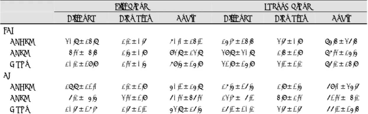
±2.1 mm, 방실회귀성 빈맥은 10.1±2.1 mm였으며 통계학적 유의한 차이는 없었다. 관상정맥동 40 mm거 리에서 측정한 관상정맥동의 평균직경은 5.4±1.9 mm 이고 방실결절회귀성 빈맥은 6.5±1.7 mm이고 방실회 귀성 빈맥은 4.1±1.0 mm로 통계적 유의한 차이가 있 었다(p=0.011)(Table 2).
관상정맥동의 분지
좌전측면에서 관상정맥동의 평균분지수는 1.9±1.3 개였으며 첫 번째 분지까지의 평균거리, 직경, 각도는 20.2±19.9 mm, 2.3±0.5 mm, 79.5±15.9°였다. 첫 번째 분지까지 평균거리에서 방실결절회귀성 빈맥은 30.8±24.8 mm, 방실회귀성 빈맥 4.3±4.4 mm로 통 계적 유의한 차이가 있었다(p=0.047). 두 번째 분지 까지의 평균거리, 직경, 각도는 31.9±15.9 mm, 3.1±
1.2 mm, 86.2±24.9°였으며 방실결절회귀성 빈맥과 방실회귀성 빈맥사이의 유의한 차이는 없었다.
전후면에서 관상정맥동의 평균분지수는 1.9±1.0개 였으며 첫 번째 분지까지의 평균거리, 직경, 각도는 20.6±17.7 mm, 2.6±1.1 mm, 53.8±26.5°였다. 두 번째 분지까지의 평균거리, 직경, 각도는 26.1±10.2 mm, 3.6±1.6 mm, 66.1±15.4°였으며 방실결절회귀 성 빈맥과 방실회귀성 빈맥사이에는 통계적 유의한 차 이는 없었다.
관상정맥동구와 척추와의 각도
좌전측면에서 평균각도는 84.1±13.1°이고 전후면
Table 2. Mean measurements of AVNRT and AVRT patients in two agiographic projections of the coronary sinus
Ostium 5 mm 10 mm 20 mm 30 mm 40 mm
LAO
AVNRT 11.4±2.2* 10.5±1.9 8.4±1.5 6.3±1.5 6.0±0.9 5.4±0.7*
AVRT 9.3±1.0* 8.1±1.1 7.1±1.2 5.8±0.7 5.5±0.7 4.3±0.8*
Mean 10.4±2.0 9.4±2.0 7.8±1.5 6.1±1.2 5.8±0.9 4.9±0.9
AP
AVNRT 12.3±2.1 10.4±2.0 8.5±1.4 7.9±1.2 7.1±1.3 6.5±1.7*
AVRT 10.1±2.1 8.3±2.2 7.3±1.7 6.4±2.5 5.3±1.8 4.1±1.0*
Mean 11.3±2.3 9.4±2.3 7.9±1.6 7.2±2.0 6.3±1.7 5.4±1.9 AVNRT:atrioventricular nodal reentry tachycardia AVRT:atrioventricular reentry tachycardia
LAO:left anterior oblique projection AP:anteroposterior projection Results are mean±SD expressed in min. *p<0.05
Fig. 4. Diagram comparing the mean measurement of the left anterior oblique projection of the coronary sinus for patients with atrioventricular nodal reentry tachyc- ardia (AV NRT) and atrioventricular reentry tachycar- ida (AVRT).
에서는 82.6±17.4°였으며, 전후면에서 방실결절회귀 성 빈맥 95.4±24.4° 방실회귀성 빈맥 67.7±15.2°로 통계적 유의한 차이가 있었다(p=0.035)(Table 4).
고 안
방실결절
방실결절은 1906년 Tawara5)에 의해 처음 발견되 었고 Keith와 Flack6)에 의해 치밀결절(true or com- pact node)이라 명명되었다. 방실결절은 관상정맥동구 의 앞쪽과 삼첨판막 중격소엽륜(septal leaflet an- nulus)기저부의 심방중격 심내막아래에 존재하고 있으 며 이 부위에 대한 연구가 많이 이루어지고 있다.7-9) 최근에는 치밀결절로 입력(input)되는 두 가지의 기본 적인 경로가 있다고 널리 수용되고 있으며 첫 번째 경 로는 관상정맥동과 떨어져있고 전난와연(anterior li- mbus of the fossa ovalis)에서 치밀결절로 들어가는 빠른경로(fast pathway)이다. 두 번째 경로는 관상정 맥동구 후하방에서 들어가는 느린경로(slow pathway)
이다.10-13) 경피적 도자절제술의 발달로 방실결절기능
변형(AV node modification)과 우회로절제를 비침습 적 방법으로 치료하게 되었으며 외과적 수술방법의 개 선으로 약물치료와 도자절제가 실패한 상심실성 빈맥 환자의 치료율을 개선시키고 있다.14)
관상정맥동의 확장과 방실결절회귀성 빈맥과의 관계 방실결절회귀성 빈맥환자에서 느린경로 도자절제시 치밀결절을 구성하고 있는 해부학적 형태와 결절주위 심방조직의 역할에 대해 새로운 연구와 논쟁이 이루어 지고 있다. 이러한 느린경로는 전기생리학적인 검사에 서 관상정맥동주위로 많은 다양성이 있다. 느린경로의 도자절제는 대개 관상정맥동구의 앞쪽, 삼첨판막 중 겹소엽륜 직상방에서 이루어지나 어떤 환자에서는 관 상정맥동 하후방에서 성공적으로 도자절제가 가능하 며 이러한 점은 느린경로가 Koch삼각(Koch’ trian- gle)기저부에서 관상정맥동 주위로 넓게 존재하고 있 다는 것을 의미한다.15)16)
심실성 빈맥은 만성 심부전환자에서 자주 관찰되
며17)18) 이러한 심실성 빈맥의 정도와 빈도는 심근 기
능부전(myocardial dysfunction)의 정도와 비례한
다.19)20) 이러한 점은 여러 연구에서 만성 심부전과 같
이 심근의 신장이나 확장(stretch or dilatation)은 활 동전위기간(action potential duration), 불응기(refr- actoriness), 전도속도(conduction velocity)에 영향을 미치는 것으로 알려졌다.21-23)
Michael JR22)은 토끼 심장에서 급성 심실확장(ac- ute ventricular dilatation)이 심장 전기 생리에 미치는 Table 3. Mean measurements of AVNRT and AVRT patients in two angiographic projections of the coronary sinus venous tributaries
First branch Second branch
Distance Diameter Angle Distance Diameter Angle
LAO
AVNRT 30.8±24.8* 2.2±0.6 70.0±24.1 25.7±14.4 3.6±0.9 85.4±36.4 AVRT 4.3± 4.4* 2.5±0.9 93.8±23.8 39.8±30.8 2.4±1.9 87.3±25.5 Mean 20.2±19.9 2.3±0.5 79.5±15.9 31.9±15.9 3.1±1.2 86.2±24.9
AP
AVNRT 28.8±21.0 2.2±1.9 50.1±25.8 27.5±16.5 2.9±1.5 69.0±35.6 AVRT 6.2± 5.5 3.3±1.9 60.3±46.3 23.7± 6.1 4.9±2.3 61.3± 4.2 Mean 20.6±17.7 2.6±1.1 53.8±26.5 26.1±10.2 3.6±1.6 66.1±15.4 AVNRT:atrioventricular nodal reentry tachycardia AVRT:atrioventricular reentry tachycardia
LAO:left anterior oblique projection AP:anteroposterior projection Results are mean±SD expressed in mm or degree. *p<0.05
Table 4. Relation of the coronary sinus ostium to the spine
LAO AP
AVNRT 86.1± 9.3 95.4±24.4*
AVRT 81.7±29.0 67.7±15.2*
Mean 84.1±13.1 82.6±17.4 AVNRT:atrioventricular nodal reentry tachycardia AVRT:atrioventricular reentry tachycadia LAO:left anterior oblique projection AP:anteroposterior projection
Results are mean±SD expressed in degree
*:p<0.05
영향에 대한 연구를 하였다. 이 연구에서 좌심실의 급 성확장은 심외막 조율역치(epicardial pacing thre- shold), 전도속도에는 영향이 없고 효과적 불응기(ef- fective refractory period, ERP)가 감소하는 것을 알 게 되었다. 또한 좌심실 급성 확장에서 심실조율(ve- ntricular pacing)시 심실성 부정맥이 유도되는 빈도가 더 많았다. 그러나 Michael RR23)은 쥐심장 전기생리 검사에서 푸르키니에 섬유속(Purkinje fiber bundle)이 1.5배까지 신장(stretch)하면 전기자극 전도속도가 점 점 빨라지고 1.5배 이상으로 신장하면 전도속도가 느 려지며 안정전위(resting potential)와 활동전위폭(ac- tion potential amplitude)이 감소 하였다. 이러한 이 유는 푸르키니에 섬유속이 1.5배 팽창시 푸르키니에 섬유와 교원질(collagen)비가 증가하고 1.5배 이상으 로 팽창하면 세포간 열(cleft)이 감소하고 세포내 부 종이 생겼기 때문이라 하였다. 이와 같이 푸르키니에 섬유속의 구조적 변화가 전도속도를 변화시키는 것을 알게 되었다.
전형적인 회귀성 빈맥은 한 방향의 차단(unidirec- tional bock)이 존재하고 느린경로부위가 있으며 느린 경로는 전도속도에 필수적으로 관여하지는 않는다. 그 러나 관상정맥동구의 확장이 정상 심방조직을 신장 또 는 확장시키므로써 구조적 변화를 일으켜 전도속도가 느려지고 불응기에도 영향을 주어 느린경로 부위가 실 제로 작용하는 영역을 형성할 수 있겠다. 이러한 것이 방실결절 주위로 부정맥을 유발 가능한 한 원인이 되고 관상정맥동구의 확장이 방실결절회귀성 빈맥의 병인 될 수도 있으리라고 생각된다.
관상정맥동의 형태이상
관상정맥동은 태생 7~8주경 좌측 동각근위부(pr- oximal left sinus horn)와 정맥동 횡측부근에서 발생한 다. 관상정맥동 형태이상은 이런 태생기에 발생부전에 의해 일어난다.24)25)
관상정맥동의 형태이상과 심장질환과의 연관성에 대 해서 아직 논란이 많으며 Mantini E25)등은 관상정맥동 의 형태이상에 대해 관상정맥동확장, 관상정맥동결여, 우심방 관상정맥동구의 폐쇄, 관상정맥동의 발육부전등 으로 분류하여 다른 심장기형에 동반되어 나타남을 기 술하였고 Chiang CE3)등에 의하면 모가난형(angula-
tion), 협착(narrowing), 누(fist-ula), 게실(diverti- culum)등으로 분류하였으며 심실상성빈맥에도 관상정 맥동의 이상이 발견되며 이중 대부분은 모가난형이라 하였다.
상기의 분류에서와 같이 관상정맥동의 모가난형이 형태이상의 대부분을 차지하므로 본연구에서도 관상 정맥동의 각도를 척추를 기준으로 비교 분석하였다.
척추와 관상정맥동사이의 각도는 횡격막의 위치에 따 라 달라질 수 있으므로 체형, 호흡시기, 심장의 크기 등에 따라 달라질 수 있다. 본연구에서 대부분의 환자 에서 이러한 변수의 효과를 최소화 하기위하여 앙와 위에 호기중에 측정하였으며 관상정맥동 형태이상은 발견되지 않았다.
요 약
연구배경:
관상정맥동은 전기생리학적 심장검사에서 절제도자 의 위치선정에 중요한 역할을 하나 그 형태에 대해서는 잘 알려져 있지 않다. 또한 도자절제시 방실결절회귀성 빈맥환자에서 방실회귀성 빈맥환자보다 쉽게 도자가 관상정맥동으로 위치함을 경험할 수 있다. 이에 상심실 성 빈맥환자의 관상정맥동의 형태와 방실결절회귀성 빈맥과 방실회귀성 빈맥환자에서 형태 차이에 대해 비 교 분석하였다.
방 법:
1996년 5~6월까지 전기생리 심장검사를 시행한 환 자중 13명을 대상으로 하여 관상정맥동을 직접 조영하 여 관상정맥동 형태분석을 시행하였다. 방실결절회귀성 빈맥 7명, 잠재성 우회로성 빈맥 4명, WPW증후군 2명 이었고 이들의 평균연령은 40세 남녀 비는 1:1.2 였다.
결 과:
좌전측면에서 관상정맥동구의 평균직경은 10.4±2.0 mm였고 방실결절회귀성 빈맥환자에서 11.4±2.2 mm, 방실회귀성 빈맥환자에서 9.3±1.0 mm로 측정되었다 (p=0.0031). 전후면에서 관상정맥동구와 척추와의 각 도는 평균 82.6±12.4 였으며 방실결절회귀성 빈맥 95.4±24.4, 방실회귀성 빈맥 67.7±15.2로 측정되었 다(p=0.035).
결 론:
방실결절회귀성 빈맥환자가 방실회귀성 빈맥환자보
다 관상정맥동의 직경이 더 크며 이러한 관상정맥동의 형태가 상심실성빈맥의 병인과 관련이 있으리라 생각 되며 이에 대해서는 향후 더 세밀한 연구가 필요하리라 생각된다.
중심 단어:관상정맥동・방실결절회귀성 빈맥・방실회 귀성 빈맥.
REFERENCES
1) Davis LM, Byth K, Lau KC, Uther JB, Richards DAB, Ross DL. Accuracy of various methods of localization of the orifice of the coronary sinus at electrophysiologic st- udy. Am J Cardiol 1992;70:343-6.
2) Jackman WM, Wang X, Friday KJ, Roman CA, Mou- lton KP, Beckman KJ, McClelland JH, Twidale N, Haz- litt A, Prior MI, Margolis PD, Calame JD, Overholt ED, Lazzara R. Catheter ablation of accessory atrioventricular pathway (Wolff-Parkinson-White syndrome) by radiofre- quency current. N Engl J Med 1991;324:1605-11.
3) Chiang CE, Chen SA, Yang CR, Chen CC, Wu TR, Tsai DS, Chiou CW, Chen CY, Wang SP, Chiang BN, Chang MS. Major coronary sinus abnormalities: Identification of occurrence and significance in radiofrequency abla- tion of supraventricular tachycardia. American Heart Jo- urnal 1994;127:1279-89.
4) Calkins H, Niklason L, Sousa J, El-atassi R, Langberg J, Morady F. Radiation exposure during radiofrequency ca- theter ablation of accessory atrioventricular connections.
Circulation 1991;84:2376-82.
5) Tawara S. Reizleitungssystem des Saugetierherzens. Gus- tav Fischer, Jena;1906.
6) Keith A, Flack M. The form and nature of the muscular connections between the primary division of the verte- brate heart. J Anat Physiol 1907;41:172.
7) Davis LM, Byth K, Ellis P, McGuire MA, Uther JB, Richards DAB, Ross DL. Dimensions of the human pos- terior septal space and coronary sinus. Am J Cardiol 1991; 68:621-5.
8) Cox JL. Anatomy of the posterior septal space. Am J Cardiol 1991;68:675-7.
9) McGuire MA, Bakker JMT, Vermeulen JT. Origin and significance of double potentials near the atrioventricu- lar node. Circulation 1994;89:2351-60.
10) Ho SY, McComb JM, Scott CD, Anderson RH. Morp- hology of the cardiac conduction system in patients with electrophysiologically proven dual atrioventricular nodal pathway. J Cardiovasc Electrophysiol 1993;4:504-12.
11) Goy JJ, Fromer M, Schlaepfer J, Kappenberger L. Clin- ical efficacy of radiofrequency current in the treatment of patients with atrioventricular node reentrant tachycardia.
J Am Coll Cardiol 1990;16:418-23.
12) Kay GN, Epstein AE, Dailey SM, Plumb VJ. Selective radiofrequency ablation of the slow pathway for the treatment of atrioventricular nodal reentrant tachycar- dia: evidence for involvement of perinodal myocardium within the reentrant circuit. Circulation 1992;85:1675-88.
13) Jackman WM, Beckman KJ, McClelland JK, Wang X, Friday KJ, Roman CA, Moulton KP, Twidale N, Hazlitt HA, Prior MI, Oren J, Overholt ED, Lazzara R. Treat- ment of supraventricular tachycardia due to atrioven- tricular nodal reentry by radiofrequency catheter abla- tion of slow-pathway conduction. N Eng J Med 1992; 327:313-28.
14) Johnson DC, Nunn GR, Richards DA, Uther JB, Ross DL. Surgical therapy for supraventricular tachycardia, a potentially curable disorder. J Thorac Cardiovasc Surg 1987;93:913-8.
15) Janse MJ, Anderson RH, McGuire MA, Ho SY. AV nodal reentry 1; AV nodal reentry revisited. J Cardiovasc Electrophysiol 1993;4:573-86.
16) Cox JL, Holman WL, Cain ME. Cryosurgical treatment of atrioventricular node reentrant tachycardia. Circul- ation 1987;76:1829-36.
17) Francis GS. Development of arrythmias in the patients with congestive heart failure: Pathophysiology, prevale- nce and prognosis. Am J Cardiol 57:3B-7B.
18) Meinertz T, Hofman T, Kasper W, Treese N, Bechtold H, Stienen U, Pop T, Leitner ERV, Andresen D, Meyer J. Significance of ventricular arrythmias in idiopathic dilated cardiomyopathy. Am J Cardiol 1984;53:902-7.
19) Schulze RA Jr, Rouleau J, Rigo P, Bowers S, Strauss HW, Pitt B. Ventricular arrythmias in the late hospital phase of acute myocardial infarction: Relation of left ve- ntricular function detected by gated cardica blood pool scanning. Circulation 1975;52:1006-11.
20) Califf RM, Burks JM, Behar VS, Margolis JR, Wagner GS. Relationship among ventricular arrythmias, coron- ary artery disease, and angiographic and electrocardiog- raphic indicators of myocardial fibrosis. Circulation 1978; 57:725-32.
21) Gornick CC, Tobler HG, Pritzker MC, Tuna IC, Almq- uist A, Benditt DG. Electrophysiologic effects of papill- ary muscle traction in the intact heart. Circulation 1986; 73:1013-21.
22) Michael JR, David PS, David EM. Electrophysiological effect of acute ventricular dilatation in the isolated rabbit heart. Cir Res 1988;62:554-62.
23) Michael RR, Marianne J, Legato, Robert MW. Develop- mental changes in impulse conduction in the canine heart.
Am J Physiol 240:H546-H54.
24) Van Mierop LHS, Kutsche LM. Embryology of the heart.
The heart. New York;1990. p.3-14.
25) Mantini E, Grondin CM, Lillehei CW, Edwards JE. Co- ngenital anomalies involving the coronary sinus. Circul- ation 1966;18:317-27.