389
CASE REPORTDOI 10.4070 / kcj.2009.39.8.389
Print ISSN 1738-5520 / On-line ISSN 1738-5555 Copyright ⓒ 2009 The Korean Society of Cardiology
Successful Radiofrequency Catheter Ablation for Wolff-Parkinson-White Syndrome
Within the Neck of a Coronary Sinus Diverticulum
Sung-Won Jang, MD, Tai-Ho Rho, MD, Dong-Bin Kim, MD, Bum-Jun Kwon, MD, Eun-Joo Cho, MD, Woo-Seung Shin, MD, Ji-Hoon Kim, MD, Seung-Won Jin, MD, Yong-Seog Oh, MD, Man-Young Lee, MD and Jae-Hyung Kim, MD
Division of Cardiology, Department of Internal Medicine, The Catholic University of Korea College of Medicine, Seoul, Korea ABSTRACT
Posteroseptal accessory pathways are often associated with coronary sinus diverticula. These diverticula contain myo- cardial coats which serve as a bypass tract. We report a 54-year-old woman who underwent radiofrequency (RF) catheter ablation for Wolff-Parkinson-White (WPW) syndrome. The surface electrocardiography (ECG) demon- strated pre-excitation, indicating a posteroseptal accessory pathway. A catheter ablation via a transaortic approach failed to ablate the accessory pathway. Coronary sinus venography revealed the presence of a diverticulum near the ostium. An electrogram in the neck of the diverticulum showed the coronary sinus myocardial extension potential, which was successfully ablated by delivery of RF energy.
(Korean Circ J 2009;39:389-391)KEY WORDS:
Wolff-Parkinson-white syndrome; Coronary sinus; Diverticulum; Radiofrequency catheter ablation.
Introduction
Radiofrequency (RF) catheter ablation has become the treatment of choice for supraventricular tachycardia (SVT) in patients with Wolff-Parkinson-White (WPW) syndrome.
1)2)The posteroseptal and left posterior acces- sory pathways are sometimes located in the epicardial region and associated with ablation failure due to the complex anatomic arrangement.
3)This kind of epicardial accessory pathway results from a connection between an extension of the coronary sinus (CS) myocardial coat al- ong the middle cardiac vein, the posterior coronary vein, or the neck of a CS diverticulum and the left ventricular epicardium.
4)A surface electrocardiography (ECG) is useful for predicting the epicardial location of a postero- septal accessory pathway
5)and CS angiography is often helpful for delineating the coronary venous anatomy. In the past, accessory pathways associated with CS diverti- cula were a significant cause of ablation failure. With mounting recognition of the importance of this anatomy, however, most of these accessory pathways have become
readily ablated.
We report a case of an accessory pathway associated with a diverticulum inserting into the proximal coronary sinus, which was successfully ablated in the neck of the diverticulum.
Case
A 54-year-old woman with WPW syndrome was refer- red for an electrophysiologic study. She had recurrent supraventricular tachycardia with symptoms of palpita- tions, dizziness, and diaphoresis. These episodes led her to curtailment of daily housework.
A surface ECG demonstrated ventricular pre-excita- tion with an isoelectric delta wave in V1 and a negative delta wave in leads II, III, and aVF, indicating a postero- septal accessory pathway (Fig. 1). A transthoracic echo- cardiography revealed no structural heart disease.
An electrophysiologic study (EPS) was performed after the patient’s consent to the procedure. Quadripolar ca- theters were introduced through the femoral vein and positioned in the right atrium (RA) and the right ventri- clular apex (RVA). A hexapolar catheter was positioned in the His bundle area. A decapolar catheter was posi- tioned in the CS via the left subclavian vein. At the time of the EPS, the delta wave was not present. RVA pacing produced an eccentric retrograde activation se-
Received: May 31, 2009 Accepted: July 1, 2009
Correspondence: Tai-Ho Rho, MD, Division of Cardiology, Department of Internal Medicine, The Catholic University of Korea College of Medicine, 620-56 Jeonnong 2-dong, Dongdaemun-gu, Seoul 130-709, Korea Tel: 82-2-958-2450, Fax: 82-2-968-7350
E-mail: [email protected]
390
·RFCA for WPW Syndrome With CS Diverticulumquence and revealed the earliest atrial activation of the CS ostium with a ventriculoatrial interval of 132 ms.
Programmed atrial pacing initiated orthodromic atrio- ventricular reciprocating tachycardia (AVRT). Mapping during AVRT showed an atrial activation sequence iden- tical to that which occurred during RVA pacing. In tachycardia, introduction of ventricular extrastimuli 10- 30 msec ahead of the His potential caused reproducible advancement of the next atrial sequence. A 7 French ablation catheter with a 4 mm tip electrode (EZ Steer
TM, Biosence Webster, Diamond Bar, CA, USA) was intro- duced via the femoral artery and placed under the mit- ral valve close to the annulus. Applications of RF energy (40W at 60℃) delivered in the posteroseptal region of the mitral annulus failed to eliminate the accessory pathway.
The ablation catheter was then placed into the CS via the femoral vein and mapping with RVA pacing was performed. However, repeated attempts of RF energy application to the earliest atrial activation site during
RVA pacing failed to abolish the accessory pathway.
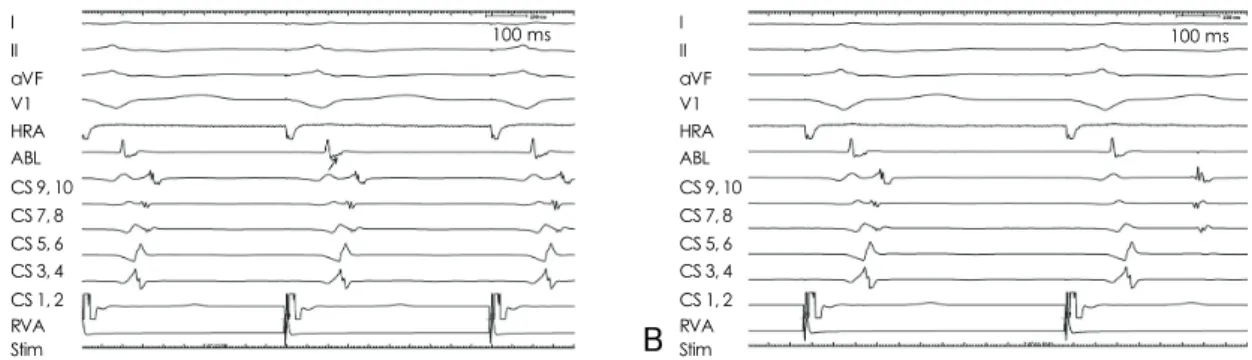
CS venography was performed and it demonstrated a diverticulum inserting into the proximal CS with a nar- row neck (Fig. 2). The ablation catheter was reposition- ed in the neck of the diverticulum and recorded an early retrograde atrial signal representing CS activity (Fig. 3A).
Ablation in this area under temperature control with a maximum pre-set energy of 15W achieved successful ab- olition of ventriculoatrial conduction via an accessory pathway (Fig. 3B).
Discussion
Epicardial accessory pathways are most commonly found in the posteroseptal and left posterior regions.
These accessory pathways are sometimes associated with CS anomalies, such as diverticula and fusiform or bul- bous enlargement. The finding of a steep negative delta wave in lead II is known to be predictive of epicardial accessory pathways. It has been reported that the sensi- tivity of a negative delta wave in lead II in identifying a CS accessory pathway is >70%.
4-6)The ECG of the present case showed a positive delta wave in lead I, an isoelectric delta wave in V1, and negative delta waves in leads II, III, and aVF. In addition, the R wave was gre- ater in amplitude than the S wave in lead V1. The ECG algorithm
5)to identify the location of an accessory path- way did not fit exactly in this case. Based on the ECG findings of a steep negative delta wave in the current case, it was necessary to predict the presence of an epicardial accessory pathway at the beginning of the procedure.
In the largest series to report the incidence of CS diverticula, of 480 patients with a posteroseptal or left posterior accessory pathway, Sun et al.
4)demonstrated a CS diverticulum in 36 (7.5%). Based on this landmark
Fig. 1. Surface ECG showing pre-excitation. ECG: electrocardiography.
Fig. 2. Coronary sinus venography showing a diverticulum with a narrow neck (arrows) near the ostium.
Sung-Won Jang, et al.·
391
clinical study,
4)CS diverticula were shown to contain myocardial fibers which connect to both the ventricle and the CS myocardial coat. The connections between the CS myocardial coat and the ventricle could serve as an accessory pathway. In patients with a CS diverticulum, an accessory pathway potential is often recorded from the neck of the diverticulum. A characteristic activation pattern can be recorded from the mapping catheter placed in the coronary venous system during retrograde conduction over epicardial posteroseptal accessory path- ways. The first potential is recorded from the CS diver- ticulum and is generated by the CS myocardial extension.
The second potential is recorded along the floor of the CS and usually has leftward activation sequence because of the fiber orientation of the CS musculature. In the case described herein, we observed a very early potential at the neck of the CS and successfully ablated the ac- cessory pathway.
Because of the close proximity of the CS ostium and posterolateral branch of the right coronary artery, cau- tion should be exercised during application of RF ener- gy into the CS. When the optimal ablation site is near the passage of the right coronary artery, saline-irrigated RF ablation or cryoablation is recommended to avoid a complication of coronary artery stenosis.
In summary, it is now well-appreciated that ablation of a posteroseptal accessory pathway needs a CS veno- graphy and careful evaluation of CS recordings for myo- cardial coat potentials. We have reported a patient who
had WPW syndrome with a posteroseptal accessory pa- thway associated with a CS diverticulum. RF ablation in the neck of the CS effectively eliminates the accessory pathway conduction. The current case highlights the potential importance of contrast CS venography and identification of myocardial coat potentials in patients with a posteroseptal accessory pathway which is difficult to ablate by the endocardial approach.
REFERENCES
1) Jackman WM, Wang XZ, Friday KJ, et al. Catheter ablation of accessory atrioventricular pathways (Wolff-Parkinson-White syn- drome) by radiofrequency current. N Engl J Med 1991;324:1605- 11.
2) Chen SA, Tsang WP, Hsia CP, et al. Radiofrequency catheter ab- lation for treatment of Wolff-Parkinson-White syndrome: short- and long-term follow-up. Int J Cardiol 1992;37:199-207.
3) Lee MH, Ahn S, Ku BK, et al. Catheter ablation of the postero- septal accessory pathways. Korean Circ J 1997;27:407-16.
4) Sun Y, Arruda M, Otomo K, et al. Coronary sinus-ventricular ac- cessory connections producing posteroseptal and left posterior accessory pathways: incidence and electrophysiological identifi- cation. Circulation 2002;106:1362-7.
5) Arruda MS, McClelland JH, Wang X, et al. Development and vali- dation of an ECG algorithm for identifying accessory pathway ablation site in Wolff-Parkinson-White syndrome. J Cardiovasc Electrophysiol 1998;9:2-12.
6) Takahashi A, Shah DC, Jais P, Hocini M, Clementy J, Haissague- rre M. Specific electrocardiographic features of manifest coronary vein posteroseptal accessory pathways. J Cardiovasc Electrophy- siol 1998;9:1015-25.
Fig. 3. Surface ECG and electrogram recordings during ventricular pacing. A: early retrograde sharp atrial potential representing coro- nary sinus activity (arrow). B: electrograms during ablation showing disconnection of the accessory pathway. ECG: electrocardio- graphy, HRA: high right atrium, ABL: ablation, CS: coronary sinus, RVA: right ventricular apex, Stim: stimulation.
I II aVF V1 HRA ABL CS 9, 10 CS 7, 8 CS 5, 6 CS 3, 4 CS 1, 2 RVA Stim
100 ms I 100 ms
II aVF V1 HRA ABL CS 9, 10 CS 7, 8 CS 5, 6 CS 3, 4 CS 1, 2 RVA Stim