Supraventricular tachyarrhythmias in patients with a persistent left superior vena cava
Jongmin Hwang 1† , Hyoung-Seob Park 2† , Jun Kim 3 *, Jeong Su Kim 1 , Jong Sung Park 4 , Ki-Hun Kim 5 , Myung Hwan Bae 6 , Sang-Hee Lee 7 , Young Soo Lee 8 , Seongwook Han 2 , Dae-Kyeong Kim 9 , Tae-Joon Cha 10 , Dong Gu Shin 7 , Byung Chun Jung 11 , and
Yoon-Nyun Kim 2
1Division of Cardiology, Department of Internal Medicine, Pusan National University Yangsan Hospital, 20, Geumo-ro, Mulgeum-eup, Yangsan-si, Gyeongsangnam-do 50612, Korea;2Division of Cardiology, Department of Internal Medicine, Keimyung University Dongsan Medical Center, 56, Dalseong-ro, Jung-gu, Daegu 41931, Korea;3Division of Cardiology, Department of Internal Medicine, Asan Medical Center, University of Ulsan College of Medicine, 88, Olympic-ro 43-gil, Songpa-gu, Seoul 05505, Korea;4Division of Cardiology, Department of Internal Medicine, Dong-A University Medical Center, 26, Daesingongwon-ro, Seo-gu, Busan 49201, Korea;5Division of Cardiology, Department of Internal Medicine, Inje University Haeundae Paik Hospital, Inje University College of Medicine, 875, Haeun-daero, Haeundae-gu, Busan 48108, Korea;6Division of Cardiology, Department of Internal Medicine, Kyungpook National University Hospital, 130, Dongdeok-ro, Jung-gu, Daegu 41944, Korea;7Division of Cardiology, Department of Internal Medicine, Yeungnam University Hospital, 170, Hyeonchung-ro, Nam-gu, Daegu 42415, Korea;8Division of Cardiology, Department of Internal Medicine, Daegu Catholic University Medical Center, 33, Duryugongwon-ro 17-gil, Nam-gu, Daegu 42472, Korea;9Division of Cardiology, Department of Internal Medicine, Inje University Busan Paik Hostpital, 75, Bokji-ro, Busanjin-gu, Busan 47392, Korea;10Division of Cardiology, Department of Internal Medicine, Kosin University Gospel Hospital, 262, Gamcheon-ro, Seo-gu, Busan 49267, Korea; and11Division of Cardiology, Department of Medicine, Daegu Fatima Hospital, 99, Ayang-ro, Dong-gu, Daegu 41199, Korea
Received 25 October 2016; editorial decision 17 April 2017; accepted 24 April 2017; online publish-ahead-of-print 20 June 2017
Aims A persistent left superior vena cava (PLSVC) is the most common thoracic venous anomaly. This venous anomaly can impact the evaluation and treatment of supraventricular tachyarrhythmia (SVA). The aim of this study was to assess the proportion and characteristics of PLSVC in adult SVA patients.
...
Methods and results
From July 2002 to July 2012, clinical and procedural data from databases of 10 cardiac electrophysiology laboratories in the Yeungnam region of the Republic of Korea were reviewed. Of 6662 adult SVA patients who underwent an EP study or cath- eter ablation of SVA during the 10-year study period, 18 patients had PLSVC (mean age 47.6 ± 14.8 years, 10 men). The pro- portion of PLSVC in adult SVA patients was 0.27% (18/6662). SVA type and procedural outcomes of radiofrequency (RF) catheter ablation in these patients were investigated and the results were as follows: successful slow pathway modification in six of seven patients with atrioventricular nodal reentrant tachycardia (AVNRT), successful ablation of accessory pathway in three of four patients with atrioventricular reentrant tachycardia, and successful ablation of atrial tachycardia (cavotricuspid isthmus-dependent in two, septal macroreentry in one, focal from the PLSVC in one) in three of four patients. In one patient with junctional tachycardia, catheter ablation failed. In two patients with atrial fibrillation, catheter ablation was successful.
...
Conclusion Among adult SVA patients who underwent an EP study or RF catheter ablation during the 10-year study period, 0.27% had PLSVC. The most common type of SVA was AVNRT. The success rate of catheter ablation was 82% in SVA patients with PLSVC. There were no procedure-related complications.
䊏 䊏 䊏 䊏 䊏 䊏 䊏 䊏 䊏 䊏 䊏 䊏 䊏 䊏 䊏 䊏 䊏 䊏 䊏 䊏 䊏 䊏 䊏 䊏 䊏 䊏 䊏 䊏 䊏 䊏 䊏 䊏 䊏 䊏 䊏 䊏 䊏 䊏 䊏 䊏 䊏 䊏 䊏 䊏 䊏 䊏 䊏 䊏 䊏 䊏 䊏 䊏 䊏 䊏 䊏 䊏 䊏 䊏 䊏 䊏 䊏 䊏 䊏 䊏 䊏 䊏 䊏 䊏 䊏 䊏 䊏 䊏 䊏 䊏 䊏 䊏 䊏 䊏 䊏 䊏 䊏 䊏 䊏 䊏 䊏 䊏 䊏 䊏 䊏 䊏 䊏 䊏 䊏 䊏 䊏 䊏 䊏 䊏 䊏 䊏 䊏 䊏 䊏 䊏 䊏 䊏 䊏 䊏 䊏 䊏 䊏 䊏 䊏 䊏 䊏 䊏 䊏 䊏 䊏 䊏 䊏 䊏 䊏 䊏 䊏 䊏 䊏 䊏 䊏 䊏 䊏 䊏 䊏 䊏 䊏 䊏 䊏 䊏 䊏 䊏 䊏 䊏 䊏 䊏 䊏 䊏 䊏 䊏 䊏 䊏 䊏 䊏 䊏 䊏 䊏 䊏 䊏 䊏 䊏 䊏 䊏 䊏 䊏 䊏 䊏 䊏 䊏 䊏 䊏 䊏 䊏 䊏 䊏 䊏 䊏 䊏 䊏 䊏 䊏 䊏 䊏 䊏 䊏 䊏 䊏 䊏 䊏 䊏 䊏 䊏 䊏 䊏 䊏 䊏 䊏 䊏 䊏 䊏 䊏 䊏 䊏 䊏 䊏 䊏 䊏 䊏 䊏 䊏 䊏 䊏 䊏 䊏
Keywords Superior vena cava • Supraventricular tachycardia • Catheter ablation
Introduction
A persistent left superior vena cava (PLSVC) is the remnant of the left cardinal vein, an embryological vessel, that is present during the
early developmental period.
1As the most common thoracic vein anomaly, the incidence of PLSVC is between 0.3 and 2% in individuals with a normal heart and 4.5–9% in those with a concomitant congeni- tal heart disease.
1–3PLSVC typically drains into the right atrium (RA)
* Corresponding author. Tel:þ82 2 3010 3164; fax: þ82 2 486-5918. E-mail address: [email protected]
†The first two authors contributed equally to the study.
Published on behalf of the European Society of Cardiology. All rights reserved.VCThe Author 2017. For permissions, please email: [email protected].
Downloaded from https://academic.oup.com/europace/article-abstract/20/7/1168/3876186 by KEIMYUNG UNIV MEDICAL LIBRARY user on 30 July 2019
via the coronary sinus (CS), which can become enlarged due to vol- ume overload.
4This enlarged CS can complicate mapping and abla- tion procedures of supraventricular tachyarrhythmias (SVA) arising from the CS ostia and the triangle of Koch.
5Also, excessive motion of a CS mapping catheter has been shown to preclude accurate elec- trogram localization.
6Furthermore, PSLVC itself can be a source of ectopy that initiates atrial fibrillation (AF), but catheter ablation tar- geting PLSVC has a risk of major complications.
7,8Thus, the presence of PLSVC can impact the evaluation and treatment of SVAs. Despite its clinical importance described above, only single case report or case-series studies have been previously reported and the research on the exact proportion of PLSVC in adult patients with SVA undergoing catheter ablation are scarce.
6–17Therefore, the aims of this study were (i) to evaluate the propor- tion of PLSVC in SVA patients and (ii) to investigate the clinical and electrophysiological (EP) characteristics as well as the procedural outcomes of radiofrequency (RF) catheter ablation in this group of patients.
Methods
Study population
This retrospective multicentre study included all consecutive adult SVA patients who underwent an EP study and/or catheter ablation between July 2002 and July 2012 in the Yeungnam region of the Republic of Korea.
The Yeungnam region includes the southeastern metropolitan cities of Busan, Ulsan, and Daegu as well as the Gyeongsangnam-do and Gyeongsangbuk-do provinces of Korea (see Supplementary material on- line, Figure S1). The area is 32 266.92 km
2and had a total population of 13 153 705 in July 2002 and 13 199 622 in July 2012, as indicated by resident registration-based population statistics, thereby accounting for approximately 24% of the total population of Korea.
18There are 13 EP centres located in this region, 10 of which participated in this study. A total of 6662 patients were reviewed, and from this population, 18 pa- tients with PLSVC were selected. Clinical characteristics, EP characteris- tics, outcomes of catheter ablations, and follow-up data for these patients were collected and retrospectively analysed.
Electrophysiological study and catheter ablation procedure
Before the procedure, careful evaluations were performed, including pa- tient history and physical examination, as well as a detailed 12-lead sur- face electrocardiographic (ECG) examination and two-dimensional transthoracic echocardiogram (TTE) to assess left ventricular (LV) ejec- tion fraction (EF) and to rule out any structural and/or valvular disease.
All patients signed informed consent for the procedure. The procedures were performed under a fasting state with light sedation after withdrawal
from antiarrhythmic drugs for a period of time equal to five-times the half-lives of the drugs. Multipolar electrode catheters were inserted per- cutaneously into the femoral vein, and, using single-plane fluoroscopy, were deployed into the CS, His bundle, right ventricular apex, respect- ively. The EP study included: (i) measurement of the conducting proper- ties of the atrium, AV node, ventricle, and accessory pathways (if present); (ii) initiation of SVAs; and (iii) determination of the mechanism of tachycardia. If tachycardia could not be induced in the baseline state, isoproterenol or atropine was used to facilitate induction. Induced SVAs were classified as atrioventricular nodal reentrant tachycardia (AVNRT), atrioventricular reentrant tachycardia (AVRT), typical atrial flutter (AFL), atrial tachycardia (AT), junctional tachycardia (JT), and AF. A three-di- mensional electroanatomic mapping system (CARTO, Biosense Webster Inc., Diamond Bar, CA, USA or NavX Ensite Velocity, St. Jude Medical Inc., Milwaukee, WI, USA) was used in selected patients. RF catheter abla- tion was tried if indicated. Catheter ablation was performed using a 4 mm tip non-irrigated ablation catheter. External irrigation catheter (3.5 or 4 mm tip) was used in AF patients and in some AT or AFL patients.
Endpoints of catheter ablation were: (i) slow pathway modification in AVNRT, (ii) bidirectional absence of accessory pathway conduction in AVRT, (iii) bidirectional conduction block across the critical isthmus in macro-reentrant AT, (iv) successful ablation of ectopic foci in focal AT or JT, (v) entrance and exit block of the pulmonary veins (PV) and non-PV foci ablation in AF, and (vi) elimination or dissociation of PLSVC poten- tials if it causes clinically significant SVA.
The presence of PLSVC was suspected if (i) CS dilatation was observed on preprocedural TTE or (ii) catheter for recording for CS electrogram was positioned in an unconventional manner (directed to the left main bronchus rather than the anterior basal left ventricle).
Subsequently, contrast venography using a long sheath or pigtail catheter was performed to confirm and delineate the PLSVC.
Statistics
Continuous variables are expressed as the mean value ± standard devi- ation. Categorical variables are expressed as numbers and percentages.
Only descriptive statistics are provided.
Ethics statement
The study protocol was reviewed and approved by the institutional re- view board of Pusan National University Yangsan Hospital (IRB No.
05-2015-141), and performed in accordance with the Declaration of Helsinki.
Results
Baseline characteristics of the study subjects
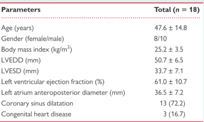
Table 1 summarizes the characteristics of SVA patients with PLSVC.
The mean age was 47.6 ± 14.8 years and 10 patients (55.5%) were male. Three patients (16.7%) had prior operations for congenital heart disease: one patient had atrial septal defect (ASD) closure, one patient had ASD with Cor triatriatum correction operation, and one patient had an operation for double outlet right ventricle, subaortic ventricular septal defect, pulmonic stenosis, and ASD with redo clos- ure of ASD later. Surgical patch closure was performed in all ASD.
There were no residual shunts or stenoses observed after surgical repair in these patients.
What’s new?
• Persistent left superior vena cava (PLSVC) was observed in 18 of 6662 patients (0.27%) with supraventricular tachyarrhythmia (SVA) during a 10-year study period.
• Ectopy from the PLSVC was found in 5 of 18 patients, and decremental property of PLSVC potential was found.
Downloaded from https://academic.oup.com/europace/article-abstract/20/7/1168/3876186 by KEIMYUNG UNIV MEDICAL LIBRARY user on 30 July 2019
The mean LV EF was 61.0 ± 10.7%. The mean LA anteroposterior diameter measured by echocardiography was 36.5 ± 7.2 mm. Dilated CS was found in 13 patients (72.2%) by two-dimensional TTE.
ECG examination during clinical tachycardia revealed narrow QRS tachycardia in 10 patients, wide QRS tachycardia in 1 patient, ven- tricular pre-excitation in 2 patients, AFL in 2 patients, and AF in 2 pa- tients. One patient had paroxysmal palpitations without documented SVA.
Types of supraventricular
tachyarrhythmia and proportion of persistent left superior vena cava
SVA diagnosis was confirmed by an EP study. Among the 18 SVA pa- tients with PLSVC, 7 had AVNRT, 4 had AVRT, 2 had typical AFL, 1 had AT from septal macroreentry, 1 had AT from the PLSVC, 1 had JT, and 2 had AF. The proportion of PLSVC in SVA patients was 0.27% (18/6662) during the 10-year study period. Table 2 shows the detailed proportion of PLSVC in patients with SVA.
Catheter ablation and follow-up results
Catheter ablation was tried in all but one patient. The patient number (No.), types of SVA, and procedures performed in individual patients are listed in Table 3.
Atrioventricular nodal reentrant tachycardia
Six patients (Patient Nos. 1–6) exhibited narrow QRS tachycardia on ECG, and EP studies revealed typical AVNRT. The mean cycle length was 405.7 ± 69.2 ms. Successful slow pathway modification was per- formed in these six patients. The mean procedure time was 92.5 ± 30.8 min and the mean fluoroscopy time was 21.4 ± 14 min.
Patient No. 18, who had an operation for congenital heart disease (double outlet right ventricle, subaortic ventricular septal defect, pul- monary stenosis, and ASD), presented with a wide QRS tachycardia on ECG, and an EP study revealed typical AVNRT with bundle branch block. The ablation of AVNRT in this patient was unsuccessful due to difficulty in mapping the triangle of Koch. After 4 months, the patient visited the emergency room due to typical AFL. Ablation of AFL was successful.
Atrioventricular reentrant tachycardia
The EP studies of two patients with narrow QRS tachycardia (Patient Nos. 7 and 8) and two patients with ventricular pre-excitation (Patient Nos. 9 and 10) revealed AVRT. The mean cycle length was 310.0 ± 99.6 ms. Successful ablation of the left lateral accessory path- way was performed in three patients. Patient No. 7 exhibited a left posterolateral bypass tract and underwent a failed catheter ablation.
In this patient, the irrigation catheter was not used by the operator at the time of the procedure and it was the main reason for failure. The mean procedure time was 83 ± 28.7 min and the mean fluoroscopy time was 17.9 ± 13.1 min.
Typical atrial flutter
Two patients exhibited a typical AFL and underwent successful abla- tion at the cavotricuspid isthmus (CTI) (Patient Nos. 11 and 12). The tachycardia cycle length was 300 and 220 ms. Patient No. 11 had undergone a previous ASD patch closure operation. The procedure time was 180 and 100 min, respectively, and the fluoroscopy time was 35 and 18.4 min, respectively.
Atrial tachycardia
Patient No. 13, who had a corrective operation for ASD with cor tri- atriatum, exhibited AT. The EP study revealed that the mechanism was septal macroreentry around the septal patch. The tachycardia cycle length was 285 ms. Linear ablation at the RA posterior septum was performed using an irrigation catheter. The procedure time was 210 min and the fluoroscopy time was 69.3 min. Patient No. 14 received an EP study due to paroxysmal palpitations without docu- mented arrhythmia. In the EP study, the existence of the PLSVC was confirmed and an AT from the PLSVC was noted. The cycle length was 365 ms. We did not ablate the PLSVC because there was an un- clear correlation between the induced arrhythmia and palpitations.
...
Table 1 Baseline clinical and echocardiographic characteristics
Parameters Total (n 5 18)
Age (years) 47.6 ± 14.8
Gender (female/male) 8/10
Body mass index (kg/m
2) 25.2 ± 3.5
LVEDD (mm) 50.7 ± 6.5
LVESD (mm) 33.7 ± 7.1
Left ventricular ejection fraction (%) 61.0 ± 10.7 Left atrium anteroposterior diameter (mm) 36.5 ± 7.2
Coronary sinus dilatation 13 (72.2)
Congenital heart disease 3 (16.7)
Data are expressed as the mean ± standard deviation or as a number (%).
LVEDD, left ventricular end-diastolic dimension; LVESD, left ventricular end-sys- tolic dimension.
...
Table 2 Proportion of PLSVC in patients with SVA Type of
arrhythmia
Number of patients
Number of PLSVC patients
Proportion of PLSVC in SVA patients (%)
AVNRT 2380 7 0.29
AVRT 2724 4 0.15
Typical atrial flutter 499 2 0.40
Atrial tachycardia 239 2 0.84
Junctional tachycardia 23 1 4.35 Atrial fibrillation 734 2 0.27
Total 6662 18 0.27
PLSVC, persistent left superior vena cava; SVA, supraventricular tachyarrhythmia;
AVNRT, atrioventricular nodal reentrant tachyarrhythmia; AVRT, atrioventricular reentrant tachycardia.
Downloaded from https://academic.oup.com/europace/article-abstract/20/7/1168/3876186 by KEIMYUNG UNIV MEDICAL LIBRARY user on 30 July 2019
Junctional tachycardia
Patient No. 15 exhibited JT from an anterior high septal site. The cycle length was 545 ms. RF catheter ablation of this patient was unsuccessful.
Atrial fibrillation
There were two patients with AF. Patient No. 16 successfully under- went pulmonary vein isolation with CTI ablation. The procedure time was 445 min and the fluoroscopy time was 116 min. Patient No.
17 was a 59-year-old man who exhibited paroxysmal AF. During an EP study after pulmonary vein isolation, complex fractionated elec- trograms were observed at the PLSVC, and a three-dimensional elec- troanatomical map demonstrated that the PLSVC acted as an arrhythmogenic substrate for AF. After focal ablation at the site at which fragmented potentials were observed, AF changed to a typical AFL. Additional ablation of the AFL was successfully executed. The procedure time in this patient was 328 min, and fluoroscopy time, 72 min. However, after 3 months, AT had occurred and another EP study exhibited reentrant AFL at the mitral isthmus. At this time, PLSVC was not directly involved in the pathogenesis of reentrant
AFL, and was activated from the entrance site at a proximal part that faced the LA. Thus, in this patient, the PLSVC presented as an arrhythmogenic substrate of paroxysmal AF and a bystander of AT.
Ectopic potentials from the persistent left superior vena cava
Five of 18 patients exhibited ectopic potentials and one patient among them showed arrhythmogenicity at the PLSVC. Patient No. 5, who had an AVNRT, showed atrial premature complex arising from the PLSVC. In Patient No. 10, who had an AVRT, AT was induced from the PLSVC after isoproterenol infusion. Fractionated potentials with decremental property under atrial pacing were seen at the PLSVC (Figure 1). Nevertheless, we did not treat the AT considering the risk of complication. During the follow-up period of 36 months, the patient had no recurrence of SVA. Patient No. 14 showed AT from the PLSVC in the EP study. We did not ablate the AT from PLSVC as described above. The patient did not complain of further palpitations following treatment with a low dosage of a beta-blocker.
Patient No. 15, who had JT, showed AT from the PLSVC, but ablation at the PLSVC was not attempted. In Patient No. 17, who exhibited ...
Table 3 Electrophysiological characteristics and procedural outcomes of individual patients Patient no. Sex Age Structural
heart disease
Tachycardia mechanism
Tachycardia cycle length
Arrhythmo- genicityof PLSVC
Procedure Success Recurrence Follow-up duration (months)
1 F 52 AVNRT, typical 360 No SP modification Yes No 5
2 M 46 AVNRT, typical 380 No SP modification Yes No 24
3 M 75 AVNRT, typical 295 No SP modification Yes No 12
4 F 66 AVNRT, typical 385 No SP modification Yes No 1
5 F 47 AVNRT, typical 465 Yes SP modification Yes No 29
6 F 23 AVNRT, typical 430 No SP modification Yes No 18
7 M 65 AVRT 305 No AP ablation No No 7
8 F 42 AVRT 450 No AP ablation Yes No 6
9 M 50 AVRT 265 No AP ablation Yes No 36
10 M 23 AVRT 220 Yes AP ablation Yes No 36
11 M 26 ASD closure op. AFL, typical 300 No CTI ablation Yes No 60
12 M 56 AFL, typical 220 No CTI ablation Yes No 4
13 F 66 ASD & Cor
triatriatum correction op.
Septal macroreentry
285 No RA septum ablation Yes No 12
14 F 40 AT from PLSVC 365 Yes EPS only No 12
15 F 37 JT 545 Yes Focus ablation No Yes 56
16 M 47 AF No PV isolationCTI
ablation
Yes No 36
17 M 59 AF Yes PV isolationCFAE in
CS ablation
Yes Yes (AFL) 3
18 M 37 Correction op. for DORV, subaortic VSD, PS, ASD. Redo ASD closure
AVNRT, typical 505 No SP modification No Yes (AFL) 8
AVNRT, atrioventricular nodal reentrant tachycardia; SP, slow pathway; AVRT, atrioventricular reentrant tachycardia; AP, accessory pathway; AFL, atrial flutter; CTI, cavotricus- pid isthmus; ASD, atrial septal defect; RA, right atrium; AT, atrial tachycardia; PLSVC, persistent left superior vena cava; EPS, electrophysiological study; JT, junctional tachycardia;
AF, atrial fibrillation; PV, pulmonary veins; CFAE, complex fractionated atrial electrogram; CS, coronary sinus; DORV, double outlet right ventricle; VSD, ventricular septal de- fect; PS, pulmonic stenosis.
Downloaded from https://academic.oup.com/europace/article-abstract/20/7/1168/3876186 by KEIMYUNG UNIV MEDICAL LIBRARY user on 30 July 2019
Figure 1 Cardiac tracings from a 23-year-old man with WPW syndrome. (A) AT was induced from PLSVC after isoproterenol infusion. Cardiac tracings are displayed from top to bottom: surface electograms (I, II, III, and V1), HRA, proximal to distal His bundle electrograms (His 7–8, 5–6, 3–4, 1–2), unipolar electrograms of the proximal to distal CS (designated as CS 9–10, 7–8, 5–6, 3–4, 1–2), and electrograms from the RVA. (B) Response of PLSVC potentials (arrows) upon atrial extrastimulation pacing. With an extrastimulus coupling interval decremented from 380 to 200 ms, decre- mental property of the PLSVC could be demonstrated. Cardiac tracings are displayed from top to bottom: surface electograms (I, II, III, and V1), HRA, unipolar electrograms of the PLSVC, unipolar electrograms of the proximal to distal CS and electrograms from the RVA. WPW, Wolff–
Parkinson–White; AT, atrial tachycardia. PLSVC, persistent left superior vena cava; HRA, high right atrial electrogram; CS, coronary sinus; RVA, right ventricular apex.
Downloaded from https://academic.oup.com/europace/article-abstract/20/7/1168/3876186 by KEIMYUNG UNIV MEDICAL LIBRARY user on 30 July 2019
AF, the PLSVC showed complex fragmented potential. Ablation at these foci resulted in organization of AF into AFL.
Procedural outcomes
A total of 17 RF catheter ablations were performed and 14 proced- ures were successful (Patient No. 14 only received an EP study). An attempt to perform catheter ablation on three patients failed to ameliorate the AVNRT of Patient No. 18, AVRT of Patient No. 7, and JT of Patient No. 15. The procedural success rate was 82% (14/
17). There were no procedure-related complications.
In patients with successful ablation of SVA, there was no recur- rence of atrial arrhythmias during the median follow-up duration of 18 months (1–60 months).
Discussion
Major findings
The major findings of this study are the following: First, PLSVC was observed in 0.27% of adult SVA patients who underwent EP studies or RF catheter ablations during the 10-year study period. This inci- dence rate is similar to that seen in the general population. Second, repaired congenital heart disease was present in a relatively small portion (3 of 18, 16.7%) of patients. Third, the most common type of SVA was AVNRT, which is consistent with results of previous re- ports. Fourth, ectopy from the PLSVC was found in 5 of 18 patients.
Proportion of persistent left superior vena cava in supraventricular
tachyarrhythmia patients
In our report, 6662 adult SVA patients underwent an EP or RF cath- eter ablation procedure in the Yeungnam region of Korea during the 10-year study period. In this period, PLSVC was observed in 18 SVA patients. Therefore, the prevalence of PLSVC in our SVA population is 0.27%. This is similar to the prevalence of PLSVC in the general population: between 0.3 and 2% in individuals with a normal heart and 4.5–9% in those with congenital heart defects.
1,3,19Furthermore, this trend was maintained in each subtype of SVA as the rate of PLSVC was 0.29% in AVNRT, 0.15%, in AVRT, 0.84% in AT, 0.4% in AFL, and 0.27% in AF. Unlike other arrhythmias, PLSVC was observed in 4.35% of JT patients. However, this may be due to the small total number of JT patients.
Recently, Ouchi et al.
20reported that the incidence of PLSVC was 0.27% (19 out of 6936 patients) in adult patients who underwent car- diac CT images for cardiac diseases or preprocedural planning. This reported incidence rate exactly matches with that of our study results.
Embryologic/anatomic considerations of persistent left superior vena cava
Embryologically, PLSVC results from the abnormal development of the sinus venosus during foetal life. Usually, the proximal portion of the left sinus horn persists as the CS, whereas the distal left horn and the left cardinal vein obliterate, thereby forming the ligament of Marshall in the adult. When the left cardinal vein persists, it becomes the PLSVC and generally drains into the RA through an enlarged
CS.
2–4A PLSVC is usually suspected when an unusually large CS on two-dimensional TTE views is observed (appears as dilated, echo- free space posteriorly in the AV groove between the LA and left ven- tricle).
21In our study, 13 of the 18 PLSVC patients (72%) exhibited CS dilatation on preprocedural TTE. Thus, echocardiography can be an appropriate method for screening PLSVC patients in catheter ab- lation candidates. In addition, PLSVC can be suspected when the catheter for recording for CS electrogram is positioned in an uncon- ventional manner (directed to the left main bronchus rather than an- terior basal left ventricle). Therefore, if the conditions described above are met, it is recommended to look for the presence of PSLVC before or during the procedure.
Clinical impact of persistent left superior vena cava in supraventricular
tachyarrhythmias
Distorted CS anatomy due to PLSVC can be an obstacle to treating AVNRT and AVRT. Okishige et al.
12reported the difficulty of RF abla- tion for AVNRT associated with PLSVC. The authors described that the patients with PLSVC have significantly enlarged CS ostia, and therefore, the course of the slow pathway may be displaced. The His bundle may also be displaced, increasing the risk of damaging the AV node during an ablation of the slow pathway. Moreover, the contact of the ablation catheter may have been insufficient.
15In AVRT, a CS electrode catheter cannot be used for guidance during ablation of the left-sided accessory pathway and may not allow for a stable electrode position at an endocardial target site.
10,14Despite these difficulties, successful catheter ablation of AVNRT and AVRT was performed in previous case reports.
5,13,14,22–24Our study also showed an acceptable ablation success rate (82%, 9/11).
Our two failed patients had additional reasons for failure: the surgery of congenital heart disease contributed to the failure in the AVNRT patient, and inappropriate catheter selection was responsible in the failed AVRT patient. Therefore, catheter ablation can be a reasonable method in the treatment of AVNRT and AVRT patients with PLSVC.
In our study, five patients showed ectopic potentials from the PLSVC, and one patient among them who demonstrated PLSVC acted as an arrhythmogenic substrate for AF. It has previously been well described that the major thoracic veins have an established role in the genesis and maintenance of AF.
25,26These include the vein and ligament of Marshall, which are the developmental remnants of the embryonic left superior vena cava.
27Furthermore, PLSVC has been demonstrated to be an arrhythmogenic source of paroxysmal or per- sistent AF in patients.
16In our study, one patient exhibited arrhyth- mogenic potentials from PLSVC, which induced AF, and this patient successfully received RF catheter ablation at the PLSVC. Four pa- tients showed ectopy from the PLSVC. Ablation was not performed due to the uncertain role of these electric potentials. During the fol- low-up period, SVA recurrence due to the PLSVC did not occur in these patients.
Study limitations
There are several limitations to our study. First, this is a retrospective study, which has inherent limitations. Second, because of the rela- tively small number of study subjects, we could not perform a sub- group analysis for each SVA. Third, a pharmacologic provocation test
Downloaded from https://academic.oup.com/europace/article-abstract/20/7/1168/3876186 by KEIMYUNG UNIV MEDICAL LIBRARY user on 30 July 2019
using multiple drugs or a high dose infusion of isoproterenol to evalu- ate arrhythmogenic property was not conducted in all patients. If those maneuvers were performed, arrhythmogenic property of the PLSVC could have been determined in more patients. Fourth, since venography was not routinely performed in all patients, the incidence of PLSVC in SVA patients who underwent EP studies or RF catheter ablations might not be true incidences. Also, the classification of the subtype of PLSVC could not be assessed.
Conclusions
In our study, the proportion of adult SVA patients with a PLSVC who underwent an EP study and RF catheter ablation procedure was 0.27% during the 10-year study period. The most common type of SVA was AVNRT. The success rate of catheter ablation in SVA pa- tients with PLSVC was 82%. There were no complications related to the procedure. In patients with successful ablation, the recurrence of SVA was not observed during the median follow-up period of 18 months. In some patients, electrical potentials of the PLSVC were demonstrated.
Supplementary material
Supplementary material is available at Europace online.
Conflict of interest: none declared.
References
1. Steinberg I, Dubilier W Jr, Lukas DS. Persistence of left superior vena cava. Dis Chest 1953;24:479–88.
2. Buirski G, Jordan SC, Joffe HS, Wilde P. Superior vena caval abnormalities: their oc- currence rate, associated cardiac abnormalities and angiographic classification in a paediatric population with congenital heart disease. Clin Radiol 1986;37:131–8.
3. Fraser RS, Dvorkin J, Rossall RE, Eidem R. Left superior vena cava: a review of associated congenital heart lesions, catheterization data and roentgenologic find- ings. Am J Med 1961;31:711–6.
4. Nsah EN, Moore GW, Hutchins GM. Pathogenesis of persistent left superior vena cava with a coronary sinus connection. Pediatr Pathol 1991;11:261–9.
5. Siliste C, Margulescu AD, Vinereanu D. Successful slow pathway ablation in a pa- tient with persistent left superior vena cava. Europace 2010;12:141–2.
6. Ma CS, Hu D, Fang Q, Shang LH, Wang LF, Belz MK et al. Catheter ablation of left- sided accessory pathway with left superior vena cava. Am Heart J 1995;130:613–5.
7. Liu H, Lim KT, Murray C, Weerasooriya R. Electrogram-guided isolation of the left superior vena cava for treatment of atrial fibrillation. Europace 2007;9:775–80.
8. Wissner E, Tilz R, Konstantinidou M, Metzner A, Schmidt B, Chun KR et al.
Catheter ablation of atrial fibrillation in patients with persistent left superior vena cava is associated with major intraprocedural complications. Heart Rhythm 2010;7:1755–60.
9. Davis D, Pritchett EL, Klein GJ, Benson DW, Gallagher JJ. Persistent left superior vena cava in patients with congenital atrioventricular preexcitation conduction abnormalities. Am Heart J 1981;101:677–9.
10. Chiou CW, Chen SA, Chiang CE, Wu TJ, Tai CT, Lee SH et al. Radiofrequency catheter ablation of paroxysmal supraventricular tachycardia in patients with congenital heart disease. Int J Cardiol 1995;50:143–51.
11. Chiang CE, Chen SA, Yang CR, Cheng CC, Wu TR, Tsai DS et al. Major coron- ary sinus abnormalities: identification of occurrence and significance in radiofrequency ablation of supraventricular tachycardia. Am Heart J 1994;127:1279–89.
12. Okishige K, Fisher JD, Goseki Y, Azegami K, Satoh T, Ohira H et al.
Radiofrequency catheter ablation for AV nodal reentrant tachycardia associated with persistent left superior vena cava. Pacing Clin Electrophysiol 1997;20:2213–8.
13. Katsivas A, Koutouzis M, Nikolidakis S, Lazaris E, Arealis G, Kyriakides ZS.
Persistent left superior vena cava associated with common type AV nodal reen- trant tachycardia and AV reentrant tachycardia due to concealed left lateral ac- cessory pathway. Int J Cardiol 2006;113:E124–5.
14. Horlitz M, Schley P, Thiel A, Shin DI, Muller M, Klein RM et al. Wolff-Parkinson- White syndrome associated with persistent left superior vena cava. Clin Res Cardiol 2006;95:133–5.
15. Nakamura T, Hachiya H, Suzuki M, Sugiyama K, Yagishita A, Tanaka Y et al.
Three-dimensional electroanatomical mapping for atrioventricular nodal reen- trant tachycardia associated with persistent left superior vena cava. J Arrhythm 2013;29:228–31.
16. Hsu LF, Jais P, Keane D, Wharton JM, Deisenhofer I, Hocini M et al. Atrial fibrilla- tion originating from persistent left superior vena cava. Circulation 2004;109:828–32.
17. Elayi CS, Fahmy TS, Wazni OM, Patel D, Saliba W, Natale A. Left superior vena cava isolation in patients undergoing pulmonary vein antrum isolation: impact on atrial fibrillation recurrence. Heart Rhythm 2006;3:1019–23.
18. Statistics Korea. Korean Statistical Information Service: Population Statistics Based on Resident Registration. http://kosis.kr/eng/statisticsList/statisticsList_01List.jsp?vwcd
=MT_ETITLE&parentId=A#SubCont (1 August 2016, date last accessed).
19. Campbell M, Deuchar DC. The left-sided superior vena cava. Br Heart J 1954;16:423–39.
20. Ouchi K, Sakuma T, Kawai M, Fukuda K. Incidence and appearances of coronary sinus anomalies in adults on cardiac CT. Jpn J Radiol 2016;34:684–90.
21. Gonzalez-Juanatey C, Testa A, Vidan J, Izquierdo R, Garcia-Castelo A, Daniel C et al. Persistent left superior vena cava draining into the coronary sinus: report of 10 cases and literature review. Clin Cardiol 2004;27:515–8.
22. Ernst S, Ouyang F, Linder C, Hertting K, Stahl F, Chun J et al. Modulation of the slow pathway in the presence of a persistent left superior caval vein using the novel magnetic navigation system Niobe. Europace 2004;6:10–4.
23. Sakabe K, Fukuda N, Wakayama K, Nada T, Shinohara H, Tamura Y.
Radiofrequency catheter ablation for atrioventricular nodal reentrant tachycardia in a patient with persistent left superior vena cava. Int J Cardiol 2004;95:355–7.
24. Bortone A, Pasquie JL, Messaoudene SA, Lacroix D. Ablation of a concealed ac- cessory pathway within a persistent left superior vena cava: role of the LocaLisa navigation system. Europace 2006;8:1038–40.
25. Chen PS, Wu TJ, Hwang C, Zhou S, Okuyama Y, Hamabe A et al. Thoracic veins and the mechanisms of non-paroxysmal atrial fibrillation. Cardiovasc Res 2002;54:295–301.
26. Zipes DP, Knope RF. Electrical properties of the thoracic veins. Am J Cardiol 1972;29:372–6.
27. Hwang C, Wu TJ, Doshi RN, Peter CT, Chen PS. Vein of marshall cannulation for the analysis of electrical activity in patients with focal atrial fibrillation.
Circulation 2000;101:1503–5.