Yonsei Med J 48(6):1048 - 1051, 2007 DOI 10.3349/ymj.2007.48.6.1048
Yonsei Med J Vol. 48, No. 6, 2007
Catheter ablation of the left free-wall accessory pathways (APs) is normally performed by the retrograde transaortic approach via a femoral artery or the transseptal approach.
Here we report a case of an overt left free-wall AP, which was successfully ablated with a retrograde transaortic ap- proach via the radial artery without any vascular compli- cations. The patient has remained free of any symptoms or pre-excitation observed on the ECG during a 10-month post- ablation follow-up.
Key Words: Catheter ablation, pre-excitation, radial artery
INTRODUCTION
Radiofrequency (RF) catheter ablation of left free-wall accessory pathways (APs) is performed with the retrograde transaortic or transseptal approach depending on the operator's preference with an equal success rate.
1Transradial coronary angiography and intervention are preferred because of the early ambulation and avoidance of complications related to the femoral arterial punc- ture.
2-6In contrast to the widespread use of the radial artery in coronary intervention, there are few reports on its use in RF catheter ablation.
Here we report a case of an overt left free- wall bypass tract that was successfully ablated via a radial artery.
CASE REPORT
A 37-year-old man with Wolff-Parkinson-White syndrome was referred for an electrophysiologic study (EPS) and ablation. The patient had been suffering from recurrent episodes of palpitations for 2 years. The 12-lead ECG during sinus rhythm exhibited delta waves with a negative polarity in leads I and aVL and positive polarity in lead V1, indicating a left free-wall AP (Fig. 1A). The physi- cal examination, chest X-ray, 2-D and Doppler echocardiography were normal. He had neither claudications nor evidence of peripheral arterial disease upon physical examination. After a writ- ten informed consent was obtained from the patient, an EPS was performed with the patient in a fasting state and sedation with midazolam and fentanyl. Three 6-French multipolar electrode catheters were introduced into the right femoral vein and positioned in the high right atrium (HRA), His-bundle recording region (HIS) and the right ventricular apex (RVA). A 6-French decapolar electrode catheter was introduced into the right internal jugular vein and advanced into the coronary sinus (CS). Programmed electrical stimu- lation was performed with a stimulus of 0.2 ms duration at twice the diastolic threshold. The surface and intracardiac ECGs were simultane- ously recorded by way of a dedicated computer- ized system (CardioLab
TM, Purucka Engineering Inc., Houston, TX, USA). The local intracardiac electrogram was filtered between 50-500 Hz. The basic intervals were as follows: sinus cycle length
= 995 ms, PR interval = 120 ms, QRS duration
=140 ms, QT interval = 400 ms, PA interval = 55 ms, AH interval = 90 ms and HV interval = -14 ms.
Catheter Ablation of a Left Free-Wall Accessory Pathway via the Radial Artery Approach
Dong Won Lee, Jun Kim, Han-Cheol Lee, June Hong Kim, Kook Jin Chun, Taek Jong Hong, and Yung Woo Shin
Division of Cardiology, Department of Internal Medicine, Pusan National University College of Medicine, Busan, Korea.
Received June 7, 2006 Accepted August 27, 2006
Reprint address: requests to Dr. Jun Kim, Department of Inter- nal Medicine, Pusan National University Hospital, Amidong 1-10 Seo-Gu, Busan 602-739, Korea. Tel: 82-51-240-7794~5, Fax: 82- 51-240-7796, E-mail: [email protected]
Catheter Ablation via Radial Artery
Yonsei Med J Vol. 48, No. 6, 2007
The earliest anterograde ventricular and retrograde atrial electrograms were recorded at CS 3, 4 (Fig.
2A and 2B). The effective refractory period (ERP) and block cycle length (BCL) of the AP were as follows: anterograde ERP of the AP = 390 ms at a basic pacing CL (S1S1) of 600 ms, anterograde AP BCL = 450 ms, retrograde ERP of the AP = 280 ms at a basic pacing CL of 600 ms and retrograde AP BCL < 320 ms. Maximal preexcitation on 12-lead ECG was noted during rapid atrial pacing (Fig.
1B). A regular, narrow QRS tachycardia with a cycle length of 330 ms could be induced by extra-
stimulus pacing or burst pacing from the HRA or RV during the baseline state (Fig. 1C). The earliest activation site during the tachycardia was re- corded in the distal CS (Fig. 2C). Prolongation of the tachycardia cycle length upon development of a left bundle branch block without any change in the atrial activation sequence supported the par- ticipation of the bypass tract in the reentrant circuit of the tachycardia. The patient consented to the use of the radial artery as the access site after a discussion with the operator regarding the potential benefits and risks of its use. After con- firmation of a normal result of the Allen's test, cannulation of the left radial artery was per- formed and a 7-French ablation catheter with a 4 mm distal tip was advanced through the artery to map the mitral annulus. The earliest ventricular activation preceding the onset of the local V by 14 ms was recorded during the sinus rhythm, with the catheter positioned in the 2 o'clock region of the mitral annulus. The delta wave disappeared 3 s after the application of the RF energy to that site with a temperature of 55 . The RF energy was delivered for 60 s. There was neither anterograde nor retrograde conduction through the AP there- after. The patient remained free from any palpita-
Fig. 1. 12-lead ECGs during sinus rhythm (A), rapid atrial pacing (B), and an induced supraventricular tachycardia (C).
Fig. 2. Surface lead electrocardiograms and intracardiac electrograms during the baseline (A), ventricular pacing at a cycle length of 320 ms (B) and a supraventricular tachycardia (C). ECG leads I, aVF, and V1 are displayed together with the electrograms from the high right atrium (HRA), proximal to distal His bundle (His), proximal to distal coronary sinus (CS), and right ventricular apex (RVA) catheters.
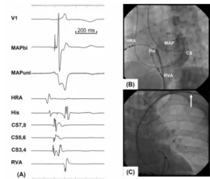
Fig. 3.Intracardiac electrograms (A) and 40 left anterior oblique fluoroscopic view from the ablation site (B). (A) The local electrogram recorded from the mapping catheter (MAP) exhibited a tri-phasic fused atrio-ventricular elec- trogram with a possible accessory pathway potential, and the unipolar electrogram recorded from the MAP (MAPuni) revealed a QS pattern. (B), (C) The ablation catheter was passed over the left subclavian artery (arrow) and positioned on the mitral annulus.
Dong Won Lee, et al.
Yonsei Med J Vol. 48, No. 6, 2007
tions or pre-excitation on the 12-lead ECG during a 10-month post-ablation follow-up.
DISCUSSION
This is the first report on the use of the radial artery as an access site for catheter ablation. Some electrophysiologists prefer transseptal catheteriza- tion in patients with left AV bypass tracts on the premise it avoids potential injuries to the aorta and aortic valve and makes mapping of the mitral annulus easier than the retrograde transaortic approach.
7,8However, it can cause potentially dangerous complications such as an aortic punc- ture or cardiac tamponade and requires special training.
9To avoid the use of additional instru- ments and potentially catastrophic side-effects, other electrophysiologists use the retrograde transaortic approach as an initial approach.
10,11However, access site-related complications such as hematomas, pseudoaneurysms, or arteriovenous fistulas, can occur with the retrograde transaortic approach.
12,13Although there are several types of percutaneous vascular closure devices to over- come these complications, a meta-analysis demon- strated the ineffectiveness of these devices in preventing vascular complications.
14To avoid complications related to the femoral artery access and to promote early ambulation, the radial artery is widely used in percutaneous coronary angiog- raphy and intervention.
2-5Because of the relatively small size of the radial artery and its predisposition to spasms, it has not been considered an adequate access site for inter- ventional electrophysiology. With the increase in geriatric population with tachyarrhythmias, the number of patients with co-morbid conditions such as severe peripheral artery disease, aortic atherosclerosis, or tortuous aortas, will also in- crease, and the chance of encountering non-acces- sible femoral arteries will grow. In those situa- tions, if electrophysiologists are not confident with a transseptal catheterization, the radial artery might be one of the last options to map the mitral annulus or left ventricle.
There are several methods to decrease the risks of radial arterial spasms. Generous use of anti- spastic agents, including the calcium-channel
blocker, nicorandil, nitrate and the use of a small diameter mapping catheter, will lessen the development of spasms.
15,16As clockwise or coun- terclockwise rotation of the catheter will promote radial artery spasms, rapid mapping of the optimal site with minimal rotation of the catheter will reduce the risk of spasms.
In summary, we report a case of a manifest left free-wall AP, which was successfully ablated by a retrograde transaortic approach via a radial artery. The use of the radial artery in the catheter ablation should be considered when patients have severe peripheral artery disease or an aortic pathology precluding a femoral arterial access.
ACKNOWLEDGMENTS
The authors thank Mr. John Martin for his linguistic assistance with this manuscript.
REFERENCES
1. Lesh MD, Van Hare GF, Scheinman MM, Ports TA, Epstein LA. Comparison of the retrograde and trans- septal methods for ablation of left free wall accessory pathways. J Am Coll Cardiol 1993;22:542-9.
2. Park EH, Kim MH, Park TH, Ahn SJ, Jung DS, Paik JH, et al. Feasibility of transradial coronary angiography using a single Judkins left catheter. Korean Circ J 2005;
35:253-7.
3. Slagboom T, Kiemeneij F, Laarman GJ, van der Wieken R. Outpatient coronary angioplasty: feasible and safe.
Catheter Cardiovasc Interv 2005;64:421-7.
4. Han KR, Park WJ, Oh DJ, Park DG, Jung WC, Jung KJ, et al. Feasibility and problems in transradial coronary angiography and intervention. Korean Circ J 2000;30:
1083-91.
5. Kim JY, Yoon J, Jung HS, Ko JY, Yoo BS, Hwang SO, et al. Feasibility of the radial artery as a vascular access route in performing primary percutaneous coronary intervention. Yonsei Med J 2005;46:503-10.
6. Choussat R, Black A, Bossi I, Fajadet J, Marco J.
Vascular complications and clinical outcome after coro- nary angioplasty with platelet IIb/IIIa receptor block- ade. Comparison of transradial vs transfemoral arterial access. Eur Heart J 2000;21:662-7.
7. Liu TJ, Lai HC, Lee WL, Wang KY, Wu TJ, Huang JL, et al. Immediate and late outcomes of patients undergoing transseptal left-sided heart catheterization for symptomatic valvular and arrhythmic diseases. Am Heart J 2006;151:235-41.
Catheter Ablation via Radial Artery
Yonsei Med J Vol. 48, No. 6, 2007 8. Swartz JF, Tracy CM, Fletcher RD. Radiofrequency
endocardial catheter ablation of accessory atrioventri- cular pathway atrial insertion sites. Circulation 1993;87:
487-99.
9. Roelke M, Smith AJ, Palacios IF. The technique and safety of transseptal left catheterization: the Massa- chusetts General Hospital experience with 1,279 pro- cedures. Cathet Cardiovasc Diagn 1994;32:332-9.
10. Lesh MD, Van Hare GF, Schamp DJ, Chien W, Lee MA, Griffin JC, et al. Curative percutaneous catheter abla- tion using radiofrequency energy for accessory path- ways in all locations: results in 100 consecutive patients.
J Am Coll Cardiol 1992;19:1303-9.
11. Calkins H, Langberg J, Sousa J, el-Atassi R, Leon A, Kou W, et al. Radiofrequency catheter ablation of accessory atrioventricular connections in 250 pateints.
Abbreviated therapeutic approach to Wolff-Parkinson- White syndrome. Circulation 1992;85:1337-46.
12. Louvard Y, Benamer H, Garot P, Hildick-Smith D, Loubeyre C, Rigattieri S, et al. Comparison of transradial and transfemoral approaches for coronary angiography
and angioplasty in octogenarians (the OCTOPLUS study). Am J Cardiol 2004;94:1177-80.
13. Juergens CP, Hallani H, Leung DY, Crozier JA, Robinson JT, Lo S, et al. Comparison of 6 and 7 French guiding catheters for percutaneous coronary interven- tion: results of a randomised trial with a vascular ultrasound endpoint. Catheter Cardiovasc Interv 2005;
66:528-34.
14. Nikolsky E, Mehran R, Halkin A, Aymong ED, Mintz GS, Lasic Z, et al. Vascular complications associated with arteriotomy closure devices in patients under- going percutaneous coronary procedure: a meta-an- alysis. J Am Coll Cardiol 2004;44:1200-9.
15. Kim SH, Kim EJ, Kim MK, Yun IS, Park WJ, Han SJ, et al. Comparison of the effects of nicorandil and cocktail solution to prevent radial artery spasm during coronary angiography. Korean Circ J 2006;36:133-9.
16. Yoo BS, Lee SH, Ko JY, Lee BK, Kim SN, Lee MO, et al. Procedural outcomes of repeated transradial coro- nary procedure. Catheter Cardiovasc Interv 2003;58:
301-4.