CASE REPORT pISSN 1225-7737/eISSN 2234-8042 http://dx.doi.org/10.12701/yujm.2014.31.2.113
Yeungnam Univ J Med 2014;31(2):113-116YUJM VOLUME 31, NUMBER 2, DECEMBER 2014 113
완전방실차단을 동반한 감염성 심내막염 환자에서 판막치환술 후 관정맥동을 통해 좌심실을 조율하는 심박조율기 시술
조관훈, 김인호, 안서희, 오용석
가톨릭대학교 의과대학 서울성모병원 내과학교실, 순환기내과
Implantation of a permanent pacemaker through the coronary sinus in a patient who underwent mechanical valve replacement for infective endocarditis
with a complete atrioventricular block
Kwan Hoon Jo, Inho Kim, Soe Hee Ann, Yong Seog Oh
Division of Cardiology, Department of Internal Medicine, The Catholic University of Korea College of Medicine, Seoul, Korea
A 52-year-old man was referred to our hospital due to fever and myalgia that occurred 2 weeks earlier.He showed a complete atrioventricular block on his electrocardiogram, and his vital signs were unstable.
On his transthoracic echocardiograph, the 1.5 cm vegetation in the aortic valve with severe aortic regur- gitation suggested infective endocarditis. His transesophageal enchocardiograph showed abscess in his mi- tral-aortic intervalvular fibrosa and vegetation was suspected on his anterior mitral valve leaflet. The patient underwent an emergent operation for valve replacement with temporary epicardial pacing. Intraoperatively, the septal leaflet of his tricuspid valve was injured during the debridement of the abscess pocket that was extended to the membranous septum. The aortic, mitral, and tricuspid mechanical valves were replaced with annular reconstruction without complications. After 14 days of intravenous antibiotics, we successfully changed the epicardial pacemaker into a transvenous DDD-type permanent pacemaker by placing a left ventricular lead via the coronary sinus and an atrial lead in the right atrium appendage. The patient was discharged in a tolerable state and was examined uneventfully in our hospital’s outpatient clinic for 8 months.
Keywords: Infective endocarditis; Heart block; Artificial cardiac pacemaker; Coronary sinus;
Cardiac valve prosthesis
Received: August 29, 2013, Revised: September 23, 2013, Accepted: September 30, 2013
Corresponding Author: Yong Seog Oh, Division of Cardiology, Seoul St. Mary’s Hospital, 222 Banpo-daero, Seocho-gu, Seoul 137-701, Korea
Tel: +82-2-2258-1141, Fax: +82-2-2258-1142 E-mail: [email protected]
서 론
대동맥 판막의 감염성 심내막염은 판막의 파괴 및 천공 (perforation), 폐쇄 부전 외에도 판막 주위 조직으로 염증이 파급되어 약 42%의 환자에서 대동맥 판막하 합병증이 발생한 다고 알려져 있다[1]. 이는 농양(abscess), 거짓동맥류(pseduo-
aneurysm) 및 천공의 임상 양상으로 mitral-aortic intervalvular fibrosa (MAIVF) 및 승모판 전엽, 건삭 드물게 심실 중격을 침 범한다[1]. 본 증례는 경흉부 및 경식도 심초음파 검사를 통 해 대동맥 판막 및 승모판 전엽의 증식(vegetation)과 MAIVF 의 농양을 확인하여 감염성 심내막염을 진단하고, 완전방실 차단과 중증의 대동맥판막 폐쇄부전으로 환자가 혈역학적으 로 불안정한 상태를 감안하여 응급으로 심외막 심박조율과 대동맥 및 승모판의 인공판막치환술을 시행하였다. 또한 중격 의 막성 부위(membranous septum) 농양 제거 시, 삼첨판륜 및 삼첨판막이 손상되어 판막륜의 재건술 및 삼첨판의 인공 판막치환술을 함께 시행하였다. 수술 후 완전방실차단이 지속 되어, 관정맥동을 통해 영구 박동조율기를 삽입하였으며, 별
Kwan Hoon Jo et al.
114 YUJM VOLUME 31, NUMBER 2, DECEMBER 2014
Fig. 1. (A) Electrocardiograpy upon admission that shows com- plete atrioventricular block. (B) Chest X-ray, anteroposterior view, that shows acute pulmonary edema in the perihilar area.
Fig. 2. Transthoracic echocardiography that shows (A) a 1.5 cm echogenic mass in the aortic valve from the parasternal long-axis view of the end-diastole, (B) combined with moderate aortic regur- gitation (color doppler image).
Fig. 3. Transesophageal echocardiography at 53 degrees at the mid-esophageal level that shows multiple irregularly margined vegetations in the (A) LCC and (B) RCC (arrow). At 130 degrees at the mid-esophageal level, (C) fluctuating motion of the vegeta- tion was noted and abscess formation in the mitral-aortic inter- valvular fibrosa (arrow) was suspected with the vegetation on the AML. LA, left atrium; LCC, left coronary cusp; RA, right atrium;
NCC, noncoronary cusp; RCC, right coronary cusp; RV, right ventricular; AML, anterior mitral leaflet.
다른 합병증 없이 퇴원하였기에 보고하는 바이다.
증 례
내원 2주전부터 발생한 발열, 오한 및 근육통을 주소로 52세 남자 환자가 입원하였다. 키 167.7 cm, 몸무게 67.9 kg 이었고, 기저질환으로 고혈압 외에 특이 병력은 없었으나 만성 알코올 중독 상태였다. 입원 후, 갑작스런 호흡곤란 및 허탈 증상을 보여 시행한 이학적 검사에서 혈압 90/40 mmHg로 저혈압 및 맥압의 증가 소견을 보였고, 체온은 36.
4℃였다. 청진상 우측 빗장뼈 부근에서 3/6 강도의 이완기 심잡음 및 양측 폐에서 수포음이 청진되었다. 혈액 검사에서 백혈구 16,650/mm3 (중성구 77.6%), 혈소판 283,000/mm3 였으며, 적혈구침강속도 120 mm/hour, C-반응단백질 16.68 mg/dL로 증가되어 있었다. Creatine kinase-MB 5.80 ng/mL, troponin-I 1.921 ng/mL로 심근효소 수치가 증가되어 있었 고, 아스파르테이트아미노전이효소/알라닌아미노전이효소 는 각각 21/15 (U/L)였다. 총빌리루빈은 0.5 mg/dL로 간수치 는 정상 소견을 보였고, 임시로 보고된 혈액배양 결과에서 그람 양성 구균이 동정되었다. 흉부 방사선 촬영에서 심비대 및 폐부종 소견 및 심전도 검사에서 박동수 50회의 완전방실
차단을 보여 응급 심초음파 검사를 시행하였다(Fig. 1). 경흉부 심초음파 검사에서는 좌심실 구혈률 67%, 약 1.5 cm 크기의 증식이 대동맥판막에 부착되어 좌심실로 오르내리는 모습이 관찰되었으며, 중증도의 대동맥판막 폐쇄부전이 동반되어 있었다(Fig. 2). 경식도 심초음파 검사에서 MAIVF의 농양 및 승모판 전엽의 증식이 의심되어(Fig. 3) modified Duke criteria [2]에 따라 감염성 심내막염으로 진단하였다. 환자가 혈역학적으로 불안정하고, 증식으로 인한 대동맥판막 폐쇄 부전과 3도 완전방실차단이 있어 응급 수술을 계획하였으며, 이는 American College of Cardiology/American Heart Associ- ation 가이드라인의 수술 적응증[3]에 따른 것이다. 수술 중 직경 1.5 cm의 우종이 우 관상동맥엽(right coronary cusp)에 서 관찰되었고, 여러 개의 작은 증식이 승모판 전엽에서 확인 되었다. 또한 2 cm 크기의 농양이 중격의 막성 부위에서 있 어, 증식과 농양을 제거하였다. 중격 막성부위의 농양을 제거 하는 과정에서 삼첨판의 중격부 판막이 손상되어 판막륜을 재건하면서 대동맥, 승모판 및 삼첨판의 인공판막치환술을 시행하였고, 심외막 심박조율기를 삽입하여 수술을 마쳤다.
수술 후 판막 배양 검사 결과
Streptococcus viridians
(⍺- hemolyticus)가 동정되었고, 이는 처음 내원 시 시행한 혈액 배양 검사결과와 동일하였다. 이에 항생제(3세대 세팔로스Implantation of pacemaker via coronary sinus after valve replacement
YUJM VOLUME 31, NUMBER 2, DECEMBER 2014 115
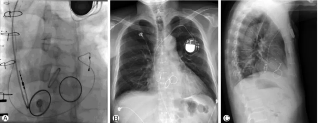
Fig. 4. Fluoroscopy after the permanent pacemaker implantation that shows the location of the ventricular pacemaker lead in the posterior cardiac vein via the coronary sinus (A) and a chest X-ray (B, C).
포린과 젠타마이신)를 2주간 투여하였고, 재검한 혈액배양 검사에서 균 음전 소견으로 보고되어 영구형 심박동기 삽입 을 계획하였다. 삼첨판막 치환술을 받은 환자로 혈관 내 접근 을 통한 심박동기를 삽입하였을 경우 발생할 수 있는 삼첨판 의 손상을 고려하여 박동조율기의 심실 전극은 심장 정맥굴을 통해 위치하기로 하였고[4], 심방 전극은 우심방에 삽입하기 로 하였다. 영구 박동조율기는 DDD형으로(Adapta ADD01- Medtronic), 시술은 환자의 좌상대정맥으로 접근하여 유도 철사를 심장 정맥굴에 위치시키고 조영제를 이용하여 유도 도관의 위치를 확인하는 방법으로, 심실 전극을 후방 심정맥 에, 심방 전극은 우심방에 삽입하였다(Fig. 4). 심박수는 분당 최소 60회, 최대 130회로 설정하였으며, 전압 2.5 V, 교류 저항(impedance)은 심방 432 ohms, 심실 1,171 ohms, 문턱 값(threshold)은 심방 2.0-2.8 Mv, 심실 15.68 Mv로 모두 적정 수준이었다. 시술 후 단순 흉부 방사선 촬영 및 심전도, 심장 초음파 검사에서 별다른 이상 소견 없었고, 이후 전신 상태가 호전되어 와파린 3 mg의 항응고 요법을 유지하기로 하고 퇴 원하였다. 시술 10개월 후 교류 저항은 심방 534 ohms, 심실 1,221 ohms, 문턱값은 심방 5.6 Mv, 심실 15.68 Mv로 적정 수준을 유지하고 있었다.
고 찰
삼첨판 인공판막치환술을 받은 환자들에서 인공 심박동기 를 삽입하게 되면 발생할 수 있는 문제에 대해 그 동안 많은 논의가 있었다. 삼첨판 치환술을 받은 환자에게 심실 전극을 삼첨판을 경유하여 삽입할 경우 성공적으로 심박 조율을 했 다는 보고도 있다. 하지만, 삼첨판 파열 등을 포함한 삼첨판 기능부전의 위험이 증가하고, 심박조율기의 전극이 조기 파
열될 가능성이 있어, 삼첨판 치환술을 시행한 환자에게 있어 서 심내막 조율은 적절치 않은 것으로 되어 있다[5-7]. 이전에 삼첨판 인공판막 치환술을 받은 환자에서 전신마취가 필요한 개흉술에 의한 심외막 심박조율기 삽입이 시행되었으나 이 방법은 전신 마취가 필요하고 감염, 좌심실 파열 등의 합병증 발생 위험성 및 수술 후 인공 심박조율기의 문턱값 상승 가능 성이 있는 것으로 알려져 있다[8].
최근 심부전의 치료로 양심실조율기가 널리 사용되면서, 전극선이 개선되어 관정맥동을 통한 심박조율기의 삽입이 가능해졌다. 이에 Bai 등[9]이 대심정맥을 통한 영구 박동조 율기 이식의 성공을 보고하였고, Curnis 등[10]은 대동맥판 막, 승모판 및 삼첨판을 기계식 인공판막으로 대치한 환자의 완전방실차단 치료법으로 심장 정맥굴을 통한 박동조율기의 성공적인 이식을 보고하였다. 심장 정맥굴을 통한 좌심실 조 율의 경우 조율역치(pacing threshold)의 상승, 항응고제 사용 의 필요성, 횡경막 자극, 심장 정맥굴 박리 및 전극선 이탈 등이 보고된 바가 있다[11]. 본 증례에서 사용한 전극의 경우 심실 전극은 Medtronic 4196을 사용하였으며, 심실 길이는 78.88 cm, 전극 사이의 길이는 21 mm이고, bipolar이다. 특징 적으로 전극 끝부분에 steroid로 코팅되어 있어 염증반응으 로 인한 조율역치의 상승의 가능성을 줄였으며, 횡경막 자극 의 경우 전극선이 왼쪽 가로막 신경이나 횡격막을 직접 자극 하여 발생할 수 있어 주의를 요하나, 본 증례에서는 보고되지 않았다.
최근 들어 심장 정맥굴을 통한 좌심실 조율을 위해 고안된 새로운 전극선들의 이용이 가능해짐에 따라 Hansky 등[12]은 99%에 이르는 심장 정맥굴을 통한 좌심실 조율의 성공률을 보였다. 그리고, Winter 등[13]은 삼첨판 치환술을 시행 받은 환자에게 심장 정맥굴을 통해 인공 심박동기 전극선을 삽입
Kwan Hoon Jo et al.
116 YUJM VOLUME 31, NUMBER 2, DECEMBER 2014
하여, 4주 후 재 시술 후에 약 29개월 동안 전극선 이탈 등의 부작용 없이 추적 관찰되어 심장 정맥굴을 통한 영구 박동조 율기의 이식이 비교적 안전한 대안으로 제안되고 있다. 저자 들은 감염성 심내막염으로 인한 대동맥 판막 하 농양과 완전 방실차단이 동시에 발생한 환자에게 대동맥판막치환술, 승 모판막치환술, 삼첨판막치환술 및 경피적 심박조율기를 삽 입한 뒤, 2주간의 항생제 치료 후, 성공적으로 심장 정맥굴을 통한 영구 박동조율기로 대체 삽입하였다. 향후 삼첨판의 기 계식 판맥 대치술을 받은 환자 등에 영구 박동조율기의 삽입 이 필요한 경우, 치환된 삼첨판을 경유하여 전극선을 삽입하 거나 전신마취가 필요한 경피적 심박조율기를 삽입하기보다 심장 정맥굴을 통한 좌심실 조율이 우선 고려될 수 있는 안전 한 방법으로 생각되어 보고하는 바이다.