443 REVIEW
DOI 10.4070 / kcj.2009.39.11.443
Print ISSN 1738-5520 / On-line ISSN 1738-5555 Copyright ⓒ 2009 The Korean Society of Cardiology
Open Access
Cardioembolic Stroke in Atrial Fibrillation-Rationale for Preventive Closure of the Left Atrial Appendage
Boris Leithäuser, MD and Jai-Wun Park, MD
Asklepios General Hospital Harburg, 1st Medical Department, Cardiology, Intensive Care Medicine, Hamburg, Germany ABSTRACT
Atrial fibrillation is the most common cardiac arrhythmias, and a major cause of morbidity and mortality due to cardioembolic stroke. The left atrial appendage is the major site of thrombus formation in non-valvular atrial fi- brillation. Loss of atrial systole in atrial fibrillation and increased relative risk of associated stroke point strongly toward a role for stasis of blood in left atrial thrombosis, although thrombus formation is multifactorial, and much more than blood flow irregularities are implicated. Oral anticoagulation with vitamin-K-antagonists is currently the most effective prophylaxis for stroke in atrial fibrillation. Unfortunately, this treatment is often contraindi- cated, particularly in the elderly, in whom risk of stroke is high. Moreover, given the risk of major bleeding, there is reason to be skeptical of the net benefit when warfarin is used in those patients. This work reviews the patho- physiology of cardioembolic stroke and critically spotlights the current status of preventive anticoagulation the- rapy. Various techniques to exclude the left atrial appendage from circulation were discussed as a considerable alternative for stroke prophylaxis. (Korean Circ J 2009;39:443-458)
KEY WORDS: Atrial appendage; Atrial fibrillation; Thromboembolism; Stroke; Prostheses and implants; Prognosis.
Introduction
Atrial fibrillation (AF) is the most common cardiac rhythm disturbance observed in clinical practice, and has numerous potential complications; of these, stroke is the most serious and life threatening. Prevalence of AF increases with age, up to 15% in octogenerians, and continues to grow rapidly due to the increasing pro- portion of aging in the population. Moreover, the el- derly population of today has a higher prevalence of predisposing conditions for AF, such as diabetes, heart failure, hypertension, and coronary heart disease.1-6) Lifetime risk for development of AF is 1 in 4 for men and women 40 years of age and older.7)
Absence of a regular contraction of the fibrillating atria leads to an increase of atrial pressure and dilata-
tion, which, together with hemoconcentration,8)9) endo- thelial dysfunction, and a prothrombotic state, is pre- requisite for thrombus formation.10) Echocardiography and autopsy studies have shown that more than 90% of all thrombi in patients with AF originating in the left atrium form in the left atrial appendage (LAA).11-15) Consequently, LA thrombi are responsible for at least one fourth of ischemic stroke, which is more frequently associated with persistent and severe disability, compar- ed to ischemic events attributable to vascular disease.16-19) Associated mortality is 27% in 12 months, and a five year recurrence rate of up to 34%.6) Socio-economical dimensions of this disease, therefore, are relevant.
The main risk of an embolic event in patients with atrial fibrillation is lack of adequate oral anticoagulation (OAC). Vitamin-K-antagonists (VKA) are highly effective for stroke prevention in patients with AF, but a sub- stantial number of patients are not eligible for chronic therapy with coumadines due to their narrow therapeu- tic range and bleeding complications that are potentially fatal. Even eligible patients may have reservations about long-term OAC with VKA, due to the need for contin- uous laboratory monitoring and possible interactions with food, drugs, and individual lifestyle. Patients at high risk of embolic stroke, but with contraindications
Correspondence: Jai-Wun Park, MD,Asklepios General Hospital Harburg, 1st Medical Department, Cardiology, Intensive Care Medicine, Eissendorfer Pferdeweg 52, 21075 Hamburg, Germany
Tel: 49-40-1818-86-2215, Fax: 49-40-1818-86-2431 E-mail: [email protected]
○cc This is an Open Access article distributed under the terms of the Creative Commons Attribution Non-Commercial License (http://creativecommons.
org/licenses/by-nc/3.0) which permits unrestricted non-commercial use, distribution, and reproduction in any medium, provided the original work is properly cited.
444·Prevention of Stroke With LA Appendage Closure
for OAC are in a need of an alternative approach that is not associated with long-term risk of hemorrhage or other attendant circumstances. This is particularly true for those who have survived intracranial hemorrhage but remain at high risk for cardiogenic embolism. A rea- sonable alternative may be exclusion of the LAA cavity from circulation, using either surgical or percutaneous catheter-based procedures. Currently, excision of the LAA at the time of mitral valve surgery is recommended for reduction of future stroke risk.20) Efficacy of LAA ex- clusion in patients undergoing elective coronary artery bypass graft surgery was shown in the LAA Occlusion Study (LAAOS).21)
In this review, we discuss pathophysiology of throm- bogenesis in the LAA, and its consequential role in AF- related embolic stroke. Further emphasis is put on te- chnical and clinical data from different percutaneous transcatheter devices for occlusion of the LAA for pro- tection of cardiogenic embolism.
Anatomy and Function of the Left Atrial Appendage
The LAA is a long, tubular, often multilobed and tra- beculated stucture, a remnant of the embryonic left atrium (LA), whereas the smooth LA cavity is derived from an outgrowth of the pulmonary veins.22) The ori- fice of the LAA is oval in shape, located between the left ventricle and the left upper pulmonary vein. The body of the appendage extends over the atrioventricular groove and the surface of the left ventricle, towards the left cir- cumflex artery and the great cardiac vein in an anterior direction.23-25) LAA structure was initially characterized from 1,842 autopsy hearts.26) Anatomy of the LAA is complex and varies considerably in terms of volume (0.7-19.2 mL), length (16-51 mm), and size of the orifice (5-40 mm).25)27)28) The structure of the cardiac muscle cells of the LAA appears similar to that of the surround- ing myocardium.29) The muscular mass of the LAA con- tains the majority of cardiac atrial natriuretic factor (ANF).30) ANF increases sodium excretion, diuresis, and natriuresis, and, thus, opposes the sodium-conserving actions of the renin-angiotensin-aldosterone system.31) Tabata et al.32) found that the most important factor in- creasing plasma concentration of ANF in patients with left-sided cardiac dysfunction is distension of the LAA wall. Moreover, stretch sensitive receptors within the LAA are involved in regulation of heart rate.33)
After primary ex vivo characterization of LAA anatomy, use of transoesophageal echocardiography (TOE) has made clear in vivo imaging of the LAA possible, so that its size, shape, flow patterns, and content can be assess- ed in health and disease.34-37) However, due to its varia- ble and complex anatomy, standard definitions of to- mographic imaging planes are nearly impossible. On the
other hand, assessment of LAA function by Doppler echocardiography and detection of LAA spontaneous echocardiographic contrast (SEC) is of considerable cli- nical relevance. New insights into LAA anatomy, in terms of angulation and motility, have recently been provided by computed tomography, which also confirmed the echocardiographic finding of a broad interindividual variation in LAA morphology.38-40) Also, using magnetic resonance imaging, three dimensional analysis of the LAA is possible and will be of considerable interest in the future, due to avoidance of radiation for the patient.41)
In sinus rhythm, the LAA shortens to a greater extent than the rest of the left atrium and has a distinct pat- tern of contraction,34) although this seems to be of minor importance for overall cardiac performance.42) Consider- ing emptying and filling waves, TOE Doppler flow wi- thin the LAA was described as quadriphasic in the ma- jority of healthy subjects with sinus rhythm.43)44) In AF without thrombus, the LAA appears to empty passively and fill with multiple small fibrillatory contractions that do not contribute to LV filling. LA pressure is the essen- tial determinant of LAA flow. Hence, the magnitude of LAA filling and emptying is influenced primarily by both left ventricular function and heart rate.43)44) The- refore, in a heart disease state with both increased atrial and left ventricular end diastolic pressure, the LAA may compensate for consequent volume overload due to its distensibility.45)46)
Thrombogenesis in Atrial Fibrillation
The LAA is the site most commonly associated with thrombus formation, particularly in patients with non- valvular AF.11-15)47)48) A multivariable analysis of cohorts followed prospectively in clinical trials and other care settings revealed that thrombi have been identified using TOE in 15-20% of patients with AF who have clinical risk factors for ischemic stroke.12)48) Pathogenesis of LAA thrombus formation has not been fully elucidated, but the precondition is likely to result from a hypercoagula- ble state explained by Virchow’s triad of thrombogenesis- i. e., abnormal changes of the vessel wall, blood flow, and blood constituents.49)50) Nowadays, this is translated as follows: “Abnormal blood flow” refers to reduced flow up to stasis due to lack of contraction in combination with increased volume and size of the LAA; “abnormal blood constituents” are represented by activated coagu- lation factors and platelets, and “abnormal vessel wall”
in this case refers to structural and functional changes of endothelial or endocardial cells.
Abnormal blood flow
Volume and size of the LAA increases in atrial fibril- lation in what is termed as atrial remodeling.34)38)48)51)
Larger LA and LAA sizes are associated with lower LAA
Boris Leithäuser, et al.·445
flow velocity47) and risk of ischemic stroke.52)53) Dimi- nished contractility of the appendage understandably leads to reduction of blood flow as well,34)42) which is associated with increased thrombogenicity within this trabeculated blind spot.50)54)55) Notably, risk of ischemic stroke due to diminished LAA flow appears not to be related to the underlying cardiac rhythm which led to the consideration of LAA velocity as a surrogate para- meter for risk stratification.56) Under conditions of low LAA blood flow, SEC may occur on TOE47)48)50)56-59) in strong association with LAA thrombus formation and systemic embolism.15)52)60-64) SEC is thought to be related to an intensified interaction between fibrinogen and erythrocytes,65) but a low level of hemoglobin is not as- sociated with lower prevalence of SEC when controlled for clinical and echocardiographic variables.66) Density of SEC increases and LAA velocities show significant and progressive decline together with accumulation of clinical risk factors for stroke, as evaluated by the CH- ADS2 score (see below).67) Furthermore, there is a high likelyhood of cerebral embolism and death, despite an- ticoagulant therapy, in patients with low LAA emptying velocity and dense SEC.68)69) It is important to note that anticoagulation does not influence the presence of SEC because it does not change underlying hemodynamic abnormalities. In chronic congestive heart failure (CHF), there is a negative correlation between LAA emptying velocity, LV ejection fraction, and LV end-diastolic pres- sure, possibly explaining the increased incidence of st- roke in patients with atrial fibrillation and CHF.70-74) Furthermore, independent predictors for the presence of thrombus and dense SEC included left ventricular ejection fraction <40% and left atrial dimension >50 mm.75)
Abnormal blood constituents
Presence of a LA thrombus is a result of a dynamic process of coagulation activation and fibrinolysis. The- refore, a high fibrin turnover is a cause rather than a consequence of cardiogenic embolism in patients with AF, in whom laboratory markers of both activated coa- gulation and impaired fibrinolysis can be found.49) Pro- thrombin fragments 1+250)76) and thrombin-antithrom- bin-complexes (TAT),50)57) as a measure of thrombin ge- neration, as well as markers for platelet activation,50) are increased in AF. Elevated D-dimer plasma levels indicat- ing increased fibrin formation and degradation50)57)76-79) are independently predictive of the presence of LAA thrombi on TOE,80) and, most importantly, are associat- ed with future thromboembolic events.72)81) Initiation of OAC reduces D-dimer values;79)82) however, in patients with thromboembolism, these values remain at a higher level compared to those without events.72) This led to the assumption that, comparable to the diagnostic algori- thm in deep vein thrombosis/pulmonary embolism, D-
dimers could be helpful in predicting the absence of LAA thrombi in patients with AF.80)83)84) Activation of coagulation is directly related to diminished LAA func- tion, in that an inverse correlation was shown between both LAA flow velocities and LAA diameter, and TAT as well as D-dimer.57)85)86) Tissue factor expression in- duced by local inflammation is involved in pathogenesis of thrombosis in patients with nonvalvular atrial fibril- lation.87) Levels of coagulation activation markers also rise, together with the number of risk factors for stroke.88-90) SEC visible on TOE shows a significant correlation to prothrombin fragments 1+2, D-dimer, and TAT.91)
Studies on the fibrinolytic system in AF are few, and conflicting results regarding clinical relevance have been reported, which may be due to the natural course of the fibrinolytic response to coagulation activation. Hyperfi- brinolysis as a consequence of a strong coagulation stim- ulus is more likely in early inflammatory states than in later or chronic states, where predominance of inhibitors leads to hypofibrinolysis.92) Tissue type plasminogen ac- tivator (tPA) plays a crucial role in inititation of fibri- nolysis and can be found in elevated levels in AF.93) Plas- min is the most important component of the fibrinoly- tic system generated in response to thrombin formation, but it is rapidly inactivated by α2-antiplasmin. The acti- vator-inhibitor-complex (plasmin-α2-antiplasmin; PAP) has a short half-life in plasma, and therefore indicates recent fibrinolytic activity.94) In a subgroup of patients enrolled in the Stroke Prevention in Atrial Fibrillation (SPAF) III study, PAP levels in patients with AF were associated with clinical characteristics predictive of th- romboembolism, including older age and reduced LV function.95) PAP levels are also elevated in acute stroke96) and myocardial infarction,97) and may therefore indicate nonspecific cell damage, inflammation, and endothelial dysfunction. The same is true for both tissue plasmino- gen activator (t-PA) and its inhibitor (PAI-1),72)98) which can be correlated with severity of inflammatory disor- ders.99)
The significance of platelet activation in atrial fibrilla- tion is uncertain and may also be seen in the light of a nonspecific inflammatory reaction. Procoagulant mem- brane vesicles derived from activated platelets, known as microparticles, are elevated in patients with conditions that are associated with atrial fibrillation (e. g., hyperten- sion, coronary artery disease, diabetes, stroke).100) Solu- ble sP-selectin is well known as a marker of platelet acti- vation and is elevated in the plasma of patients with AF.50)74)101-105) Recently, Choudhury et al.100) found that both AF patients and disease control subjects had signi- ficantly higher levels of platelet derived microparticles and sP-selectin compared to healthy control subjects;
however, there was no difference between AF patients and disease control subjects. β-thromboglobulin is ano- ther platelet-specific protein that indicates activation and
446·Prevention of Stroke With LA Appendage Closure
another examplary representative of flow-dependent he- mostatic activation, as it was found to be highest in pa- tients with the lowest LAA flow velocities and the grea- test atrial dilatation.85) Plasma levels of β-thromboglo- bulin are dependent on duration of AF, and showed a significant increase 12 hours after onset of paroxysmal AF.106) It is worthy of mention that results concerning the effect of antithrombotic treatment on markers of platelet activation in AF have been uncertain. In studies by Kamath et al.107)108) treatment with warfarin or aspirin either failed to demonstrate any significant benefit on platelet activation (β-thromboglobulin, sP-selectin), or showed an effect in favor of aspirin on the absolute amount of P-selectin per platelet in patients with AF.108) The clinical significance of this finding remains to be elucidated. Likewise, the prognostic relevance of platelet activation in terms of future thromboembolic events is ambivalent. A substudy of the SPAF III trial revealed no association,103) whereas the Rotterdam study found plas- ma levels of sP-selectin to be predictive of clinical adverse outcomes in AF.104) On the whole, evidence of platelet activation in AF patients seems most likely due to under- lying cardiovascular disease, rather than arrhythmia per se.
Abnormal endothelium and endocardium
Further insight into the hypercoagulable state in atrial fibrillation is provided by studies of von Willebrand factor (vWF), a hemostatic mediator involved in platelet aggregation and clot stabilization, derived from endo- thelial cells and thrombocytes.109) Endothelial activation or disturbance is indexed by elevated plasma levels of vWF.110) Increased plasma vWF can be found in patients with AF,50)76)78)79)86)105)111) and tends to increase further with concomitant heart failure.74) Most important, the level of vWF is intimately related to recognized inde- pendent risk factors for stroke (heart failure, age, dia- betes, previous stroke) and might itself be predictive of future stroke.102)112)113) As expected, increased plasma levels of vWF were found to be associated with presence of LAA thrombus, visible by TOE,50) whereas Fukuchi et al.114) found a significant correlation between degree of endocardial expression of vWF and extent of platelet adhesion/thrombus formation in the LAA. Endocar- dial overexpression of vWF may occur during the process of atrial structural remodeling in chronic AF.115)116) These structural changes also affect the extracellular matrix117)118) and, therefore, the system of matrix metalloproteinases (MMP). There are hints to a link between the MMP sys- tem and a prothrombic state.119)
Increased levels of circulating endothelial cells (CEC) have been demonstrated in conditions associated with endothelial damage.120) Freestone et al.111) found that CEC levels in patients with AF and an acute cardiovas- cular or cerebrovascular event were significantly elevated compared to patients with stable, chronic AF. Therefore,
in the interim balance, it can be stated that AF itself is the major contributory factor to thrombogenesis, which implies activation of the coagulation cascade, rather than platelets, and is the key to excess thromboembolic risk in AF.
Stroke-Risk Asessment in Atrial Fibrillation
Risk of stroke varies considerably among patients with AF. On the one hand, individual risk for stroke must be considered prior to prescription of VKA for anticoagu- lation; on the other hand, risk of stroke must be outwei- ghted against the risk of bleeding and burden on the patient due to the need for continuous laboratory INR monitoring or possible interactions associated with food, drugs, and individual lifestyle or preferences.121) Several stroke risk stratification schemes are available to help clinicians with this decision.68)122-125) Two useful resources stand out in clinical practice: The Framingham risk score, derived by Wang et al.,68) and the CHADS2 score pu- blished by Gage et al.123) Both use a five step calculation to predict the risk for stroke in patients with AF. The former considers age, gender, systolic blood pressure, dia- betes, and prior stroke or TIA. Each category is assigned different grading, and predicts 5-year stroke risk in the absence of anticoagulation. Concerning the latter, the C stands for recent congestive heart failure, the H for hypertension, the A for age 75 or older, the D for dia- betes, and the S for prior stroke or TIA. Each category is assigned one point, except for stroke or TIA, which re- ceives two points due to high association with subsequent stroke. A high score on this index correlates with raised annual stroke rate. The CHADS2 score may be easier to use, but is less precise.126)
The Effect of Cardioversion
One of the theoretic benefits of cardioversion in pa- tients with AF is the assumption that restoring normal atrial electromechanical activity may diminish the risk of cardiogenic thromboembolism and, therefore, spare the need for anticoagulation. However, there is a body of evidence to demonstrate that a temporary worsening of LAA function (“stunning”) after cardioversion is re- sponsible for development of new clots.127)128) The hall- mark of LAA stunning is reduction of post cardiover- sion LAA flow velocity in sinus rhythm compared to those in AF, regardless of iatrogenic (electrical or ph- armocological) or spontaneous cardioversion.129)130) Even after succesful restoration of sinus rhythm, SEC can be seen in up to 37% of patients after 3 months,131) indi- cating the persistence of a hypercoagulable state after cardioversion.78)132) The strategy of rhythm control has been directly compared with simple rate control in se-
Boris Leithäuser, et al.·447
veral randomized clinical trials. In a pooled analysis, fre- quency of ischemic stroke in the group of patients as- signed to rate control was comparable to that of patients assigned to rhythm control.133) It is amazing, therefore, that Cox et al.134) reported on 306 patients who un- derwent the maze procedure for treatment of medically refractory atrial fibrillation. Only 2 perioperative strokes occurred, and in 265 patients followed up to 11.5 years after the maze procedure, there was only one late minor stroke, which has since been completely resolved. On the one hand, this good result may be explained by pre- dominance of sinus rhythm, and, on the other hand, by the absence of the LAA and restoration of left atrial me- chanical function.135)
Hypothetically, restoring mechanical activity to the LAA with cardioversion may also result in systemic em- bolism due to wash out of pre-existing LAA thrombi, which may explain the occurence of stroke shortly after treatment.136)137) The Assessment of Cardioversion Using Transesophageal Echocardiography (ACUTE) trial138) compared a TOE-guided strategy combined with short- term anticoagulation using a conventional 3-week oral anticoagulation pre cardioversion strategy. Although there was significant difference in the composite end point of major and minor bleeding and a shorter time to cardioversion, there was no difference in the compo- site end point of stroke, transient ischemic attack, and peripheral embolism. Therefore, current guidelines for cardioversion in patients with atrial fibrillation lasting longer than 48 hours recommend coumadin treatment for at least three weeks prior to cardioversion, and for a minimum of four weeks afterward. Alternatively, perfor- mance of a cardioversion without anticoagulation is jus- tifiable if direct previous TOE shows no thrombi present in the LAA.121)
Cardiac Imaging for Thrombus Detection
During the past thirty years, TOE has become a valu- able tool for diagnosis of thrombus within the appenda- ge by allowing semi-invasive, highly accurate imaging of the LAA.139) It is, therefore, an essential part of the gui- delines for management of AF,121) and the modality of choice for detecting LA or LAA thrombi with a sensiti- vity and specificity of approximately 95% to 100%.127) Nevertheless, detection of LAA thrombus is prone to misdiagnosis because clots may remain hidden due to the three-dimensional complexity of the LAA, and a false-positive diagnosis of thrombus may stem from false interpretation of a prominent pectinate muscle. The aforementioned sensitivity and specificity regarding thrombus detection by TOE was found in comparison with intraoperative observations;13) however, no compa- rison has been made using direct left atrial angiography.
Sensitivity for thrombus detection during the left ven- tricular phase of pulmonary angiogram was found to be 100% compared to later surgical inspection.140) In our own experience with transcatheter LAA occlusion and by means of direct angiography, we detected LAA thrombi not seen with simultaneously performed TOE in ap- proximately 10% of patients (Fig. 1).141) Furthermore, we revealed additional LAA lobes in some patients during repeated LAA angiography. The “collapsed” lobes seem to be “blown up” by contrast injections into the LAA.
Consequently, in these cases, the procedure was halted and postponed, causing inconvenience and additional risk for patients, as well as increased overall treatment costs. Furthermore, this finding strengthens the hypo- thesis of cardiac embolism due to wash out of pre-exist- ing LAA thrombi after successful cardioversion. Hence, an imaging modality with higher accuracy of preproce- dural LAA thrombus detection would be very helpful for planning of LAA occlusion procedures. Further te- chnical development of echocardiography seems pro- mising. Recent introduction of an RT-m3D TOE trans- ducer permits virtually instantaneous 3D imaging while preserving spatial and temporal resolution, thereby sig- nificantly enhancing visualization of complex 3D struc- tures such as the LAA.142) Further improvement of B- mode image quality of 3D-TOE will be available in the near future. The combination of tissue doppler imaging and use of contrast agents may help to better characte- rize thrombogenic structures within the LAA.58) Also, with respect to LAA imaging, few studies using MRI
Fig. 1. Angiographic contrast filling defect in the contast shadow of the LAA (white arrows) indicating a spherical thrombus, which was not diagnosed by TOE. Note the TOE probe at the left mar- gin and the loop of the pigtail catheter (striped arrow), indicating the position of the aortic valve. LAA: left atrial appendage, TOE:
transoesophageal echocardiography.
448·Prevention of Stroke With LA Appendage Closure
and spiral CT scan have been reported on detection of thrombus and SEC.38)39)143-146) Regarding LAA occlusion procedures, the advantages of cardiac CT/MRA include the following: 1) preprocedural imaging of the anatomi- cal characteristics of the LAA and neighbouring atrial structures; 2) assessment of the anatomical relationship of the LA, esophagus, and adjacent vascular structures;
3) postprocedural detection of structural and functional changes of the LA and LAA.147)
Oral Anticoagulation: Therapeutic Gold Standard in Atrial Fibrillation
Of the VKAs, warfarin is the most widely used and investigated coumarin derivative, exerting its anticoag- ulant effect through modulation of c-carboxylation of glutamic acid (GLA) residues of the vitamin K-depen- dent coagulation factors II, VII, IX, and X, resulting in production of coagulation factors with reduced coagulant capacity.148)149) Dose adjusted (INR 2.0-3.0) OAC is well established in patients with nonvalvular AF, and has been associated with a decreased risk of cardioembolic events of greater than 60%.150)151) Effectiveness of OAC depends on intensity of treatment,77) in that insufficient doses of anticoagulants reduce the therapeutic benefit, while excessive anticoagulation increases the risk of bleed- ing.152) The benefit of dose adjusted OAC is confirmed by a significant decrease of markers of coagulation acti- vation,72)79) whereas fixed low-dose warfarin or aspirin- warfarin combination treatment did not substantially reduce markers of thrombogenesis.153) Despite the pro- ven benefit, OAC with warfarin or other VKA remains underused in clinical practice,154) although underutiliza- tion implies that a decision not to use warfarin is rea- ched after assessment of the balance between benefit (prevention of thromboembolic stroke) and risk (bleed- ing), which suggests a net benefit. With regard to anti- thrombotic therapy, there are two groups of AF patients:
Those with no contraindications for therapy who never received warfarin, and those who are on therapy, but with an INR below or above the range of 2.0-3.0, having in- creased risk for either stroke or bleeding. Overall, 55%
of warfarin-eligible patients actually use the drug, with the lowest rates seen in the oldest patients-those at high- est risk of stroke.155)156) In the United States, AF patients on warfarin spend only about one-half of the time within therapeutic INR range,157-159) and of those patients ad- mitted to hospital with a stroke while receiving warfarin therapy, most have subtherapeutic international nor- malized ratios.160)161) Leckey et al.162) found that only 13%
of patients with ischemic stroke and known AF before stroke were taking warfarin. Considering the worst case scenario, that half of patients are untreated and the other half are out of range 50 percent of the time, only one fourth receive optimal treatment.
Review of the literature has identified several barriers to prescription of VKA, which are related to the patient, the physician, and the health care system.163) One of the strongest patient related predictors of warfarin withhold- ing is age. Warfarin use increases with a history of ische- mic stroke, and decreases with age >80 years. The most important physician related reasons not to anticoagulate include 1) the perception of benefit vs. risk of therapy, insofar as the risk for embolism, relative to hemorrhage, is judged to be lower, and 2) the relative contraindication to therapy due to lack of patient reliability or patient noncompliance as a reason for difficulties in monitor- ing the prothrombin ratio.163)
Acetylsalicylic acid (ASA, Aspirin) as an anticoagulant has some advantages over warfarin. These include sub- stantially less potential for drug-drug or drug-diet inter- actions, a wider therapeutic index, and no need for INR- monitoring. For AF patients with increased risk of bleed- ing, it is often prescribed instead of warfarin on the pre- sumption that it is safer. A meta-analysis of six random- ized controlled trials suggests that ASA does reduce the risk for ischemic stroke in AF (22% relative risk reduc- tion for ASA compared with placebo).12)164) It is notewor- thy that only one of these trials {the Stroke Prevention in Atrial Fibrillation (SPAF) study165)} reported a statis- tically significant difference. Recently, the Birmingham Atrial Fibrillation Treatment of the Aged Study, BAF- TA,166) reaffirmed that warfarin is superior to ASA in stroke prevention. On the other hand, the risk of major hemorrhage, including hemorrhagic stroke, was similar in both aspirin (2.0% per year) and warfarin (1.9% per year) treated patients. The authors recommended use of anticoagulation (warfarin) for all people over 75 years who have atrial fibrillation, unless there are contraindi- cations, or the patient decides that the size of the benefit is not worth the inconvenience of the treatment.165)
Bleeding Risk Under Oral Anticoagulation
The emphasis on avoidance of hemorrhagic stroke or traumatic intracranial hemorrhage and other iatrogenic events may cause physicians and patients to choose the- rapy that minimizes side effects, rather than therapy that maximizes benefit. In association with anticoagulation, bleeding is the complication of greatest concern; there- fore, selection of appropriate therapy should weigh ad- vantages and disadvantages carefully.167) In anticoagulat- ed patients, annual risk of major bleeding, meaning a hemorrhage requiring >2 units of blood or requiring hospitalization, ranges between 1.1-1.7% and 0.3-0.6%
for intracranial hemorrhage.124)168) However, this low level of risk has rarely been replicated in contemporary stu- dies, and then, perhaps, only in patients who are stable on long-term anticoagulation. Higher rates of hemor-
Boris Leithäuser, et al.·449
rhage were found in the Stroke Prevention in Atrial Fi- brillation II Study, which consisted of 2 parallel trials of patients aged >75 years and ≤75 years.169) Annual rates of major bleeding were 4.2% and 1.7%, respectively.
Hylek and coworkers170) studied a cohort of 472 patients of whom one third were ≥80 years of age, compared with a total of 20 patients >75 years in the pooled an- alysis of 5 randomized trials that proved efficacy of anticoagulation,150) and 91% had ≥1 stroke risk factor.
Cumulative incidence of major hemorrhage for patients
≥80 years of age was 13.1 per 100 person-years and 4.7 for those <80 years of age (p=0.009). Thus, the increas- ing risk of bleeding with increasing age is clearly dem- onstrated in this study. Moreover, during the first 90 days of warfarin treatment, age ≥80 years and interna- tional normalized ratio (INR) ≥4.0 were associated with increased risk, despite trial-level anticoagulation control.
Within the first year, 26% of patients who were ≥80 years of age stopped taking warfarin. Perceived safety issues accounted for 81% of them. The most obvious results to be emphasized from this study are that the risk of major hemorrhage increases by a factor of 10-fold between CHADS2 scores of 0 and ≥4, and also that warfarin termination was highest among patients with CHADS2 scores ≥3.170) The finding that bleeding risk is highest within the first year of therapy was confirmed by results from the ACTIVE-W study: annual risk of major hemorrhage over the duration of the study is quoted as 2.6% and 2.0% for warfarin-naive and warfa- rin-experienced patients, respectively. In contrast, the risks are more like 6% to 7% and 4%, respectively, in the first year.171) Moreover, an ancillary analysis from the ACTIVE-W trial reports that risk of major bleeding dur- ing OAC was lower among patients with a CHADS2
score of 1 (1.36% per year) compared with CHADS2 >1 (2.75% per year).172)
The following patient characteristics are considered risk factors for anticoagulation-related bleeding compli- cations: Advanced age, uncontrolled hypertension, his- tory of myocardial infarction or ischemic heart disease, cerebrovascular disease, anemia or a history of bleeding, and concomittant use of other drugs, such as antiplatelet agents.173) Moreover, primary clinical and health-related problems associated with typical geriatric syndrome in older adults, and which make the decision regarding use VKA more complex, include functional decline, frailty and falls, polypharmacy, nutritional deficiencies, and cognitive dysfunction.167) All of these conditions are of- ten cited as reasons to preclude the elderly from antico- agulation.174) A prospective observational study of 207 AF patients in an acute-care setting hospital, assessing frail and non-frail patients, demonstrated that frail pa- tients were less likely to receive warfarin, both on hos- pital admission and on discharge. Notably, such patients at the same time were at greater risk of experiencing em-
bolic stroke (12.3% vs. 3.9% in frail and non-frail pa- tients). Aronow et al.175) described 312 AF patients with an average age of 84 years residing in a chronic care fa- cility. Rates of stroke over three years in those not anti- coagulated were 56% in those with no prior history of thromboembolism and 81% in those with prior history of thromboembolism. These frail patients also had a tendency to sustain a greater risk of major/severe he- morrhage, as well as greater mortality.176)
Comparable to existing stroke risk stratification sche- mes, there are bleeding risk models:177-182) Beyth and coworkers178) identified four independent risk factors for bleeding: Age ≥65 years, history of GI bleeding, his- tory of stroke, and one or more of four specific comorbid conditions. They found a cumulative incidence of major bleeding at 48 months of 53% in high-risk paitients (three or four risk factors), 12% in middle-risk patients (one or two risk factors), and 3% in low-risk patients (no risk factors). Kuijer and colleagues179) developed another prediction model based on age, gender, and presence of malignancy. In patients classified at high, middle, and low risk, frequency of major bleeding was 7%, 4%, and 1%, respectively, after 3 months of therapy.
Comparable bleeding rates for comparable risk classes were found by Shireman et al.181) (5.4%, 2.0%, and 0.9%, respectively, after 90 days of treatment) using the fol- lowing criteria: Age >70 years, gender, remote bleeding, recent bleeding, alcohol/drug abuse, diabetes, anemia, and antiplatelet use. Gage et al.182) gave 2 points for a prior bleed and 1 point for each of 10 further stroke risk factors, and claimed the highest accuracy of all other bleed prediction schemes. Nevertheless, none of these risk prediction schemes has been fully validated in large prospective AF patient cohorts.
The outlined clinical complexity of pathophysiology and prevention of thromboembolism in AF patients is most impressively illustrated by the fact that those at highest risk of stroke, and, therefore with the greatest need for antithrombotic therapy, also experience the most bleeding. Thus, an alternative that combines high efficacy in stroke prevention with low risk of bleeding is warranted.
Left Atrial Appendage Occlusion for Stroke Risk Reduction
The frequency of thrombus formation in the LAA of patients with AF and its suspected role as a source of embolism led to the hypothesis that resection or obli- teration of the LAA might reduce the risk of stroke.
Hellerstein et al.183) was the first to show the feasibility of LAA resection in dogs. The first resection of the LAA for prophylaxis of recurrent arterial embolism in men was performed in 1949 by Madden,184) followed by Beal et al.185) in 1950. Johnson and coworkers performed atrial appendectomies in 437 patients during cardiac
450·Prevention of Stroke With LA Appendage Closure
surgery. No strokes were attributed to AF, and no pa- tients were found to have atrial clots on TOE during follow-up.4) Odell and coworkers186) demonstrated in dogs and human cadavers that thoracoscopic exclusion of the LAA using either a stapler or an endoloop is also feasible and effective; Blackshear and colleagues187) eval- uated left atrial appendage obliteration in 14 high-risk patients with atrial fibrillation who had clinical risk factors for stroke and an absolute contraindication to or failure of prior thrombosis prevention with warfarin.
One fatal stroke occurred 55 months after surgery, and one non-disabling stroke occurred three months after surgery. For the LAA Occlusion Study,21)188) 77 patients with risk factors for stroke were randomized to either LAA occlusion or control (52 patients for LAA occlu- sion, 25 patients in the control group) at the time of cor- onary artery bypass graft (CABG). Two patients (2.6%), both randomized to the LAA occlusion group, had pe- rioperative thromboembolic events: One had an intrao- perative ischemic stroke, and the other a TIA occurring on the third postoperative day. The former patient was in AF and had echocardiographic evidence of a patent foramen ovale and bilateral carotid stenoses. After a mean follow-up of 13±7 months, no further strokes or TIAs occured in the LAA occlusion group.21) Nonethe- less, surgical or thoracoscopic LAA closure, other than as an adjunctive procedure, as recommended by ACC guidelines20) in patients undergoing mitral valve surgery, has not been enthusiastically accepted due to its invasive nature. Thus, based on surgical experience, development of a less invasive percutaneous approach to close the LAA by implantation of a mechanical device was a lo- gical consequence.189)190)
The Percutaneous Left Atrial Appendage Trans- catheter Occlusion system
The Percutaneous Left Atrial Appendage Transcathe- ter Occlusion (PLAATO) (EV3, Inc., Plymouth, MN, USA) device was the first to be successfully deployed for use in humans,191) but was withdrawn from the market by the manufacturer in 2006. The PLAATO system con- sists of an implant and a delivery catheter. The implant is a self-expanding nitinol cage covered with an occlu- sive expanded polytetrafluoroethylene membrane. The expanded membrane has intimate contact with the inner wall of the appendage, so that complete closure of the
ostium can be achieved. The diameter of the nitinol cage ranges from 15 to 32 mm. Small hooklets along the struts and passing through the membrane assist with device anchoring. The device was delivered through a custom 12 Fr transseptal sheath curved to point at the left atrial appendage.
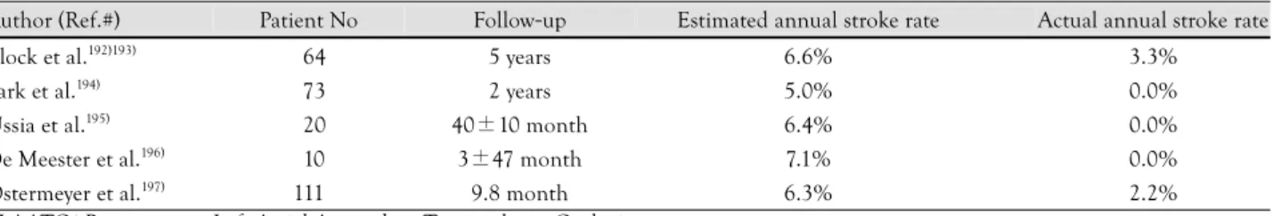
Several reports demonstrate efficiacy in stroke pre- vention using PLAATO (Table 1).
However, related serious adverse events occurred in every patient cohort procedure; these included vessel perforation during vascular access and cardiac tampo- nade after transseptal puncture. Some patients experi- enced pericardial effusions, which were mostly uneven- tful, but also lead to pericardiocentesis and a prolonged hospital stay. The worst case in our series was a peripro- cedural death due to device embolization, resulting in acute occlusion of the left ventricular outflow tract. Im- plant anchoring in another patient appeared unstable in LA angiography, so that the device was explanted, and the LAA of the patient was occluded by open heart sur- gery on the catheter table.194) Our LAA occlusion pro- gram was halted following these disastrous cases, and extensive revision was undertaken for all 73 prior im- plantations. As a result, we discovered a rotation of the device at the moment of release from the delivery sys- tem in a significant number of cases which resulted in loss of contact between parts of the anchor rows and the LAA wall (unpublished observation). One further theoretical concern was de novo formation of thrombi on the atrial surface of the implant which, to the best of our knowledge, has never been reported on the basis of clinical data. Nevertheless, there are conflicting results concerning formation of neo-endothelium on the de- vice, which is the prerequisite to absence of thrombo- genicity. Post mortem analysis by Omran et al.198) dem- onstrated a PLAATO device completely covered by ne- oendothelium on the atrial surface one year after implan- tation, whereas we found no endothelialization of the luminal side 2.5 years after LAA occlusion.199)
The Watchman Left Atrial Appendage system The second device specifically designed for percuta- neous transcatheter LAA exclusion is the Watchman Left Atrial Appendage System (Atritech Inc., Plymouth, MN, USA). This three-part system consists of a trans- septal sheath, a delivery catheter, and an implantable
Table 1. Currently published data for stroke prevention using the PLAATO system
Author (Ref.#) Patient No Follow-up Estimated annual stroke rate Actual annual stroke rate
Block et al.192)193) 064 5 years 6.6% 3.3%
Park et al.194) 073 2 years 5.0% 0.0%
Ussia et al.195) 020 40±10 month 6.4% 0.0%
De Meester et al.196) 010 3±47 month 7.1% 0.0%
Ostermeyer et al.197) 111 9.8 month 6.3% 2.2%
PLAATO: Percutaneous Left Atrial Appendage Transcatheter Occlusio
Boris Leithäuser, et al.·451
device. The implant is a selfexpanding nitinol frame structure with fixation barbs and a permeable polyester fabric that covers the atrial side, and is available in dia- meters ranging from 21-33 mm. The device has been implanted since 2002 in Europe and since 2003 in the United States.200) Two patients experienced device em- bolization; both implants were successfully retrieved per- cutaneously. Five pericardial effusions (two of them need- ing pericardiocentesis) and one major air embolism oc- curred without long-term sequelae. The WATCHMAN Left Atrial Appendage System for Embolic PROTEC- Tion in Patients With Atrial Fibrillation (PROTECT AF) study was designed to demonstrate safety, efficacy, and non-inferiority of the WATCHMAN device against chronic warfarin therapy in patients with nonvalvular atrial fibrillation who are eligible for long-term OAC.201)202) Of 707 patients enrolled, 463 were randomly assigned to LAA closure and 244 to warfarin therapy. The device was successfully implanted in 91% of the patients in whom it was attempted. Patients were followed for an aggregate of 1,065 patient-years. After 6 months, 355 (92%) of patients with an implanted device were able to
discontinue warfarin therapy. For the control group, plasma warfarin concentration was in the therapeutic INR range (between 2.0 and 3.0) 66% of the time. Rate of ischemic stroke was higher in the intervention group than in the control group. Five patients had periproce- dural events, mainly air embolism. After the periproce- dural timeframe, ischemic stroke occurred in nine pa- tients in the intervention group, compared with six pa- tients in the control group. In both groups, all ischemic strokes that had INR measurements available at the time of the event occurred at a subtherapeutic INR level. He- morrhagic strokes were less frequent in the intervention group than in the control group. Five of the six hemorr- hagic strokes in the control group were fatal, and all oc- curred in patients with therapeutic INR levels. Device embolisation occurred in three patients; one was noted during the procedure and two were discovered by TEE on day 45. One device embolisation was removed per- cutaneously by use of a vascular snare; the other two pa- tients underwent surgery, one of whom had concomi- tant aortic valve replacement. Endpoint data of adverse events are shown in Table 2.202)
Nevertheless, this initial study shows substantial draw- backs: 12.3% of patients had serious procedural com- plications and 2.2% of attempted implantations result- ed in cardiovascular surgical intervention due to device- related complications. Therefore, a substantial learning curve must be considered in association with device im- plantation. The primary efficiacy estimate of the PROT- ECT-AF-study is less precise, due to the small number of participants, and nearly 30% of patients receiving de- vices had a CHADS2 score of 1, and were therefore can- didates for aspirin therapy without warfarin, even with- out LAA occlusion.203)
The AMPLATZER Cardiac Plug system
The AMPLATZER Cardiac Plug (ACP) is a transca-
Table 2. Results of the PROTECT-AF-Study202): adverse events Intervention
(n=463) (%)
Control (n=244) (%) Serious pericardial effusion* 22 (4.8) 0
Major bleeding† 16 (3.5) 10 (4.1)
Procedure related ischemic stroke 05 (1.1) 0
Device embolisation 03 (0.6) 0
Hemorrhagic stroke‡ 01 (0·2) 06 (2·5)
Other§ 02 (0·4) 0
*Defined as the need for percutaneous or surgical drainage. †Major bleeding is defined as a bleeding event that required at least 2 units of packed red blood cells or surgery to correct. ‡Of the seven he- morhagic strokes, six resulted in death (intervention group, n=1;
control group, n=5). §An oesophageal tear and a procedurerelated arrhythmia
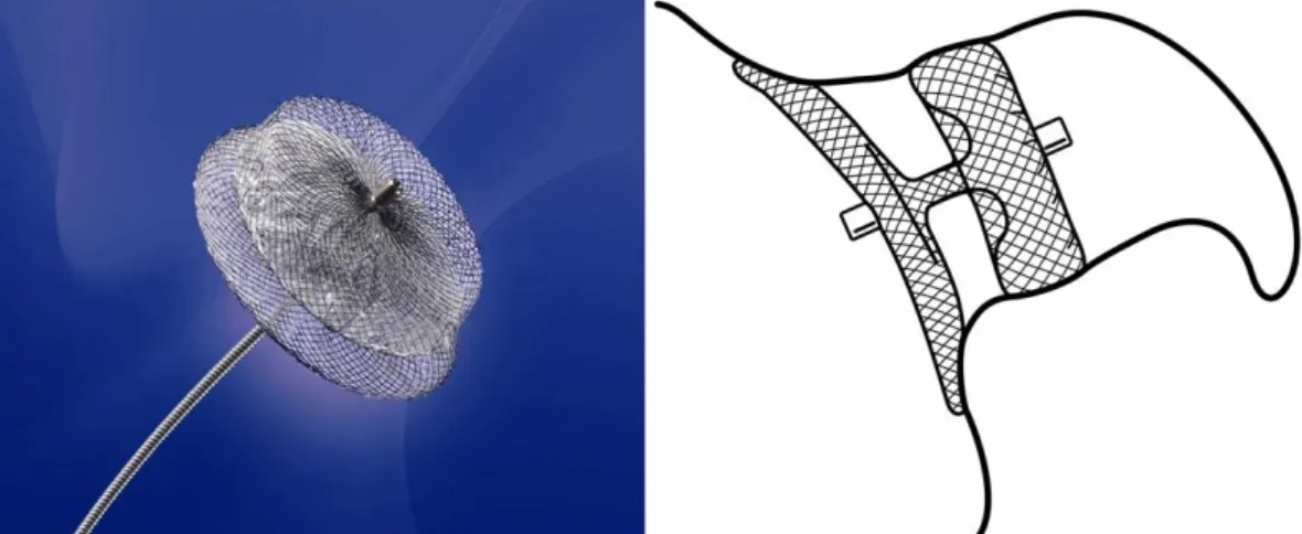
Fig. 2. The AMPLATZER Cardiac Plug (ACP). On the right, the ideal position within the LAA is sketched. The lobe of the device is anchored in the “landing zone” 1-2 cm distal of the LAA orifice, while the disc fully covers the outer shape and enables endothelialization from the surrounding atrial wall. These images were provided by, and are property of AGA, Inc., Minneapolis, MN, USA. LAA: left atrial appendage.
452·Prevention of Stroke With LA Appendage Closure
theter, self-expanding device constructed from a nitinol mesh and polyester patch (Fig. 2). The ACP consists of a lobe and a disc connected by a central waist. It is availa- ble in eight diameter sizes (16, 18, 20, 22, 24, 26, 28, and 30 mm). The lobe has stabilizing wires to improve device placement and retention. The device has thread- ed screw attachments at each end for connection to the delivery and loading cable. Radio-opaque markers at each end and at the stabilizing wires assist with fluo- roscopic positioning. The ACP is a further development based on the AMPLATZER double-disk septal occluder, which was designed for closure of atrial septal defects and patent foramen ovale. In principle, this device can also be used for occlusion of the LAA; however, results of a feasibility trial were disappointing, as an emboliza- tion occured in one of 16 patients.204) The currently un- published initial experience with ACP is encouraging.
Its implantation is rather demanding technically, how- ever, the disadvantages and risks associated with use of the PLAATO system are apparently eliminated (Fig. 3).
A multicenter prospective registry trial to evaluate tech- nical and short term success is approaching.
Concerns Over Left Atrial Appendage Closure
Controversy persists in regard to the risks and benefits of LAA occlusion for prevention of embolic stroke.
Adverse hemodynamic and physiological effects may result from LAA obliteration.205) Fluid retention is a potential late complication of LAA occlusion. Human atrial appendages contain 30% of total cardiac ANF.30) Experimental data have shown that bilateral appenda- gectomy in dogs eliminates ANF release and blunts re- nal excretion of sodium and water during acute volume load.206) Clinically evident postoperative fluid retention after the maze procedure with bilateral appendagectomy has been reported.207) Another study of the maze proce- dure reported on diminished ANF secretion accompani-
ed by increased need for postoperative diuretics and do- pamine.208) However, this effect was abolished when the right atrial appendage was preserved.209) There are clues, however, that in the natural course of permanent atrial fibrillation, atrial degeneration may at least lead to de- creased ANF secretion.210) However, to date, many cases of surgical LAA exclusion with long term follow-up have not shown deleterious results. Pathophysiological conse- quences of implanting a foreign body into the LAA re- main to be fully elucidated. Small iatrogenic atrial septal defects can be created after transseptal puncture. They usually disappear within 6 months of the procedure.
Furthermore, every implanted foreign material bears the risk of infection.211)
Conclusion
AF is known to confer a risk of stroke; however, this risk is not homogeneous. Chronic oral anticoagulation as the prophylactic measure of choice has a number of major limitations associated with its narrow therapeutic range. A great deal of overlap evidently exists in throm- boembolic stroke risk and risk of bleeding. Such over- lapping risk creates a difficult management problem.
Given the demonstrated risk of major bleeding, there is reason to be skeptical about net benefit when warfarin is used in some elderly patients with AF. Whether or not the needs of high-risk patients can be met by newer pharmacological and nonpharmacological antithrom- botic/antiembolic therapies remains to be determined.
It would seem that risk stratification schemes must be refined to incorporate data available from imaging stu- dies that enhance predictive value for ischemic events and risk factors for bleeding. Occlusion of the LAA ori- fice, therefore, offers a theoretically appealing way to re- duce incidence of stroke in patients who cannot be an- ticoagulated, or who developed stroke despite being on OAC. Nevertheless, there are limitations to this approach in that it cannot be easily applied prophylactically to
Fig. 3. Images of the AMPLATZER Cardiac Plug (ACP) in situ. A: TOE. B: fluoroscopy after implantation.
B A
Boris Leithäuser, et al.·453
large numbers of patients. Concerns about procedural safety and need for long-term follow up should be ad- dressed before this potentially important technology is widely deployed.
REFERENCES
1) The Stroke Prevention in Atrial Fibrillation Investigators. Pre- dictors of thromboembolism in atrial fibrillation: I. clinical fea- tures of patients at risk. Ann Intern Med 1992;116:1-5.
2) Falk RH. Atrial fibrillation. N Engl J Med 2001;344:1067-78.
3) Go AS, Hylek EM, Phillips KA, et al. Prevalence of diagnosed atrial fibrillation in adults: national implications for rhythm ma- nagement and stroke prevention: the Anticoagulation and Risk Factors in Atrial Fibrillation (ATRIA) Study. JAMA 2001;285:
2370-5.
4) Johnson WD, Ganjoo AK, Stone CD, Srivyas RC, Howard M.
The left atrial appendage: our most lethal human attachment!:
surgical implications. Eur J Cardiothorac Surg 2000;17:718-22.
5) Kannel WB, Benjamin EJ. Status of the epidemiology of atrial fibrillation. Med Clin North Am 2008;92:17-40, ix.
6) Lloyd-Jones D, Adams R, Carnethon M, et al. Heart disease and stroke statistics - 2009 update: a report from the American Heart Association Statistics Committee and Stroke Statistics Subcom- mittee. Circulation 2009;119:e21-181.
7) Lloyd-Jones DM, Wang TJ, Leip EP, et al. Lifetime risk for de- velopment of atrial fibrillation: the Framingham Heart Study.
Circulation 2004;110:1042-6.
8) Yamada H, Sugiyama T, Ashida T, Fujii J. Sustained hemocon- centration in patients with chronic atrial fibrillation, a potential risk for stroke and thromboembolic complications: a retrospec- tive study. Jpn Heart J 1998;39:715-20.
9) Kamath S, Blann AD, Chin BS, Lip GY. Platelet activation, hae- morheology and thrombogenesis in acute atrial fibrillation: a comparison with permanent atrial fibrillation. Heart 2003;89:
1093-5.
10) Al-Saady NM, Obel OA, Camm AJ. Left atrial appendage: struc- ture, function, and role in thromboembolism. Heart 1999;82:
547-54.
11) Aberg H. Atrial fibrillation: I. a study of atrial thrombosis and systemic embolism in a necropsy material. Acta Med Scand 1969;185:373-9.
12) Blackshear JL, Odell JA. Appendage obliteration to reduce stroke in cardiac surgical patients with atrial fibrillation. Ann Thorac Surg 1996;61:755-9.
13) Manning WJ, Weintraub RM, Waksmonski CA, et al. Accuracy of transesophageal echocardiography for identifying left atrial thrombi: a prospective, intraoperative study. Ann Intern Med 1995;123:817-22.
14) Tsai LM, Chen JH, Lin LJ, Yang YJ. Role of transesophageal echocardiography in detecting left atrial thrombus and sponta- neous echo contrast in patients with mitral valve disease or non- rheumatic atrial fibrillation. J Formos Med Assoc 1990;89:270-4.
15) Leung DY, Black IW, Cranney GB, Hopkins AP, Walsh WF.
Prognostic implications of left atrial spontaneous echo contrast in nonvalvular atrial fibrillation. J Am Coll Cardiol 1994;24:
755-62.
16) Lin HJ, Wolf PA, Kelly-Hayes M, et al. Stroke severity in atrial fibrillation: the Framingham Study. Stroke 1996;27:1760-4.
17) Hylek EM, Go AS, Chang Y, et al. Effect of intensity of oral an- ticoagulation on stroke severity and mortality in atrial fibrilla- tion. N Engl J Med 2003;349:1019-26.
18) Anderson DC, Kappelle LJ, Eliasziw M, Babikian VL, Pearce LA, Barnett HJ. Occurrence of hemispheric and retinal ischemia
in atrial fibrillation compared with carotid stenosis. Stroke 2002;
33:1963-7.
19) Spratt N, Wang Y, Levi C, Ng K, Evans M, Fisher J. A pro- spective study of predictors of prolonged hospital stay and disa- bility after stroke. J Clin Neurosci 2003;10:665-9.
20) Bonow RO, Carabello BA, Kanu C, et al. ACC/AHA 2006 gui- delines for the management of patients with valvular heart di- sease: a report of the American College of Cardiology/American Heart Association Task Force on Practice Guidelines (writing committee to revise the 1998 Guidelines for the Management of Patients With Valvular Heart Disease): developed in collabora- tion with the Society of Cardiovascular Anesthesiologists: en- dorsed by the Society for Cardiovascular Angiography and In- terventions and the Society of Thoracic Surgeons. Circulation 2006;114:e84-231.
21) Healey JS, Crystal E, Lamy A, et al. Left Atrial Appendage Occlusion Study (LAAOS): results of a randomized controlled pilot study of left atrial appendage occlusion during coronary bypass surgery in patients at risk for stroke. Am Heart J 2005;
150:288-93.
22) Brown NA, Anderson RH. Symmetry and laterality in the human heart: developmental implications. In: Harvey RP, Rosenthal N editors. Heart Development. San Diego: Academic Press;1999.
p.447-61.
23) Veinot JP, Harrity PJ, Gentile F, et al. Anatomy of the normal left atrial appendage: a quantitative study of age-related changes in 500 autopsy hearts: implications for echocardiographic exami- nation. Circulation 1997;96:3112-5.
24) Kerut EK. Anatomy of the left atrial appendage. Echocardiogra- phy 2008;25:669-73.
25) Su P, McCarthy KP, Ho SY. Occluding the left atrial appendage:
anatomical considerations. Heart 2008;94:1166-70.
26) Sharma S, Devine W, Anderson RH, Zuberbuhler JR. The deter- mination of atrial arrangement by examination of appendage mor- phology in 1842 heart specimens. Br Heart J 1988;60:227-31.
27) Ernst G, Stollberger C, Abzieher F, et al. Morphology of the left atrial appendage. Anat Rec 1995;242:553-61.
28) Stollberger C, Ernst G, Bonner E, Finsterer J, Slany J. Left atrial appendage morphology: comparison of transesophageal images and postmortem casts. Z Kardiol 2003;92:303-8.
29) Lannigan RA, Zaki SA. Ultrastructure of the myocardium of the atrial appendage. Br Heart J 1966;28:796-807.
30) Chapeau C, Gutkowska J, Schiller PW, et al. Localization of im- munoreactive synthetic atrial natriuretic factor (ANF) in the heart of various animal species. J Histochem Cytochem 1985;
33:541-50.
31) Peterson TV, Benjamin BA. The heart and control of renal ex- cretion: neural and endocrine mechanisms. FASEB J 1992;6:
2923-32.
32) Tabata T, Oki T, Yamada H, Abe M, Onose Y, Thomas JD. Rela- tionship between left atrial appendage function and plasma con- centration of atrial natriuretic peptide. Eur J Echocardiogr 2000;
1:130-7.
33) Kappagoda CT, Linden RJ, Mary DA. Gradation of the reflex response from atrial receptors. J Physiol 1975;251:561-7.
34) Pollick C, Taylor D. Assessment of left atrial appendage function by transesophageal echocardiography: implications for the de- velopment of thrombus. Circulation 1991;84:223-31.
35) Li YH, Hwang JJ, Ko YL, et al. Left atrial spontaneous echo contrast in patients with rheumatic mitral valve disease in sinus rhythm: implication of an altered left atrial appendage function in its formation. Chest 1995;108:99-103.
36) Valocik G, Kamp O, Mihciokur M, et al. Assessment of the left atrial appendage mechanical function by three-dimensional echo- cardiography. Eur J Echocardiogr 2002;3:207-13.