INTRODUCTION
Atrial fibrillation (AF) is the most common arrhythmia and is related with cardiovascular disorders, including heart failure and stroke, and doubles the associated mortality rate.1 Several studies have indicated diastolic dysfunction to be an indepen- dent predictor of AF.2 The presence and severity of diastolic
dysfunction might be associated with the left atrium (LA) sub- strate of AF and progressive atrial mechanical remodeling due to increased LA pressure.3 Increased left ventricular (LV) fill- ing pressure has been linked with all-cause mortality, an in- creased frequency of LA appendage thrombus, and stroke in non-valvular AF patients.4
We hypothesized that the AF recurrence is associated with the degree of diastolic dysfunction in patients with persistent AF after cardioversion. Several studies have demonstrated that increased LV filling pressure may affect AF recurrence af- ter cardioversion.2,4 Therefore, this study set out to determine whether increased LV filling pressure is associated with an in- creased risk of AF recurrence after cardioversion in patients with persistent AF. In contrast to previous studies, to limit the effect of LA structural remodeling which is in the pathogene- sis of AF, we enrolled patients without extreme LA enlarge- ment. A limited number of studies have investigated how dia- stolic parameters may predict AF recurrence after cardioversion
Left Ventricular Filling Pressure as Assessed by the E/e’
Ratio Is a Determinant of Atrial Fibrillation Recurrence after Cardioversion
Hyemoon Chung, Byoung Kwon Lee, Pil-Ki Min, Eui-Young Choi, Young Won Yoon, Bum-Kee Hong, Se-Joong Rim, Hyuck Moon Kwon, and Jong-Youn Kim
Cardiology Division, Department of Internal Medicine, Gangnam Severance Hospital, Yonsei University College of Medicine, Seoul, Korea.
Purpose: Left ventricular (LV) filling pressure affects atrial fibrillation (AF) recurrence. We investigated the relationship between diastolic dysfunction and AF recurrence after cardioversion, and whether LV filling pressure was predictive of AF recurrence.
Materials and Methods: Sixty-six patients (mean 58±12 years) with newly diagnosed persistent AF were retrospectively enrolled.
We excluded patients with left atrial (LA) diameters larger than 50 mm, thereby isolating the effect of LV filling pressure. We eval- uated the differences between the patients with (group 1) and without AF recurrence (group 2).
Results: Group 1 showed increased LA volume index (LAVI) and E/e’ compared to group 2 (p<0.05). During a mean follow-up period of 25±19 months, AF recurrence after cardioversion was 60.6% (40/66). The area under the receiver operating characteris- tics curve of E/e’ for AF recurrence was 0.780 [95% confidence interval (CI): 0.657–0.903], and the optimal cut-off value of the E/e’
was 9.15 with 75.0% of sensitivity and 73.1% of specificity. A Kaplan-Meier survival curve showed that the cumulative recurrence- free survival rate was significantly lower in patients with higher LV filling pressure (E/e’>9.15) compared with patients with lower LV filling pressure (E/e’≤9.15) (log rank p=0.008). Cox regression analysis revealed that E/e’ [hazards ratio (HR): 1.100, 95% CI:
1.017–1.190] and LAVI (HR: 1.042, 95% CI: 1.002–1.084) were independent predictors for AF recurrence after cardioversion.
Conclusion: LV filling pressure predicts the risk of AF recurrence in persistent AF patients after cardioversion.
Key Words: Diastolic dysfunction, left ventricular filling pressure, atrial fibrillation, cardioversion Yonsei Med J 2016 Jan;57(1):64-71
http://dx.doi.org/10.3349/ymj.2016.57.1.64 pISSN: 0513-5796 · eISSN: 1976-2437
Received: December 17, 2014 Revised: May 13, 2015 Accepted: May 29, 2015
Corresponding author: Dr. Jong-Youn Kim, Cardiology Division, Department of Internal Medicine, Gangnam Severance Hospital, Yonsei University College of Medi- cine, 211 Eonju-ro, Gangnam-gu, Seoul 06273, Korea.
Tel: 82-2-2019-3310, Fax: 82-2-3463-3882, E-mail: [email protected]
•The authors have no financial conflicts of interest.
© Copyright: Yonsei University College of Medicine 2016
This is an Open Access article distributed under the terms of the Creative Com- mons Attribution Non-Commercial License (http://creativecommons.org/ licenses/
by-nc/3.0) which permits unrestricted non-commercial use, distribution, and repro- duction in any medium, provided the original work is properly cited.
by using echocardiography. Here, we investigated the predic- tors of AF recurrence in patients undergoing continued anti- arrhythmic drug therapy after cardioversion.
MATERIALS AND METHODS
Study population
The study retrospectively enrolled 66 patients (57 males, mean 58±12 years) with newly diagnosed non-valvular, lone, and persistent AF from January 2009 to December 2012 at the Gangnam Severance Hospital, Yonsei University College of Medicine. Persistent AF was defined as continuous AF sus- tained greater than 7 days, according to the expert consensus statement.5 For patients with newly diagnosed AF, heart rate was controlled below 110 beats/minute and maintained by using beta blockers or calcium channel blockers. After heart rate control, pre-treatment with flecainide for 2 weeks was performed in all patients before electrical cardioversion.
Those patients who were converted to sinus rhythm by pre- treatment with flecainide were labeled as the chemical car- dioversion group. Electrical cardioversion was performed in the remaining patients who were not converted to sinus rhythm by flecainide treatment. These patients were labeled as the electrical cardioversion group. All 66 patients were con- verted to sinus rhythm by either flecainide treatment or elec- trical cardioversion. After cardioversion, flecainide was ad- ministered continuously in order to maintain sinus rhythm.
All patients who underwent electrical cardioversion had taken oral anticoagulation with vitamin K antagonists such as war- farin for 4 weeks and had maintained optimal prothrombin time (PT) international normalized ratio (INR) range of 2.0 to 3.0 before the cardioversion. Exclusion criteria were: 1) LV ejection fraction (EF) <50%, 2) LA anterior-posterior (AP) di- mension >50 mm, and 3) known coronary artery disease (CAD) or suspected CAD. The patients were divided into two groups according to AF recurrence: group 1, with AF recurrence (n=40); and group 2, without AF recurrence (n=26). Electronic medical records were reviewed, and pertinent data points were recorded. All patients provided written, informed consent.
Echocardiographic study
After rate control, two-dimensional transthoracic echocar- diography (TTE) was performed. All the echocardiographic studies were performed using an iE33 (Philips Ultrasound, Bothell, WA, USA) with an S3 probe. Comprehensive echo- Doppler and M-mode evaluation were assessed in all patients before cardioversion. Left ventricle wall thicknesses was mea- sured during end-diastole phases. LA AP dimension was mea- sured at end-systole from the parasternal long axis view. All measurements were done according to current American So- ciety of Echocardiography guidelines.6 The modified Simp- son’s rule was used to calculate LV volumes and EF from api-
cal 2- and 4-chamber views. The prolate ellipse method was used to calculate LA volume from apical 4-chamber and para- sternal long-axis views at ventricular end-systole, then LA vol- umes were indexed to body surface area.
Peak early (E) and late (A) diastolic mitral inflow velocities were measured in apical 4-chamber view. Tissue Doppler in- terrogation was done in septal mitral annulus in apical 4-cham- ber view, and then peak systolic mitral annulus velocity and early diastolic mitral annulus peak velocity (e’) were measured, and the ratio of E/e’ was calculated. Pulsed Doppler and pulsed tissue Doppler parameters were measured as the aver- age of three cardiac cycles, and the R-R intervals were relatively regular except during lasting atrial fibrillation.
Follow-up
After the diagnosis of AF, patients were treated by flecainide for two weeks. If sinus rhythm was not achieved after chemi- cal cardioversion, electrical cardioversion was performed. All patients were scheduled for regular follow-up visits after car- dioversion, including clinical examination, and 12-lead elec- trocardiography (ECG) every 3 months and Holter monitoring every 6 months. In the case of symptom recurrence between follow-up visits, patients were evaluated by additional clinical examination, ECG, and Holter monitoring. AF recurrence af- ter cardioversion was defined as any documented supraven- tricular tachyarrhythmia, such as AF, atrial flutter, or atrial tachycardia episode, lasting >30 seconds.
Statistical analysis
Continuous variables that are normally distributed are report- ed as mean±SD or 95% confidence interval (CI). Student t-test was used to compare the means of continuous variables that were approximately normally distributed between the two groups. Continuous variables that were not normally distrib- uted are reported as median (25–75 percentile range) and are compared using the Kruskal-Wallis test. Normality was deter- mined using the Kolmogorov-Smirnov goodness-of-fit test.
Categorical variables are reported as count (percentage) and are compared using Fisher’s exact test.
Independent predictors of AF recurrence after cardiover- sion were assessed using univariate Cox proportional hazards regression models, and multivariable models were assessed based on the results of univariate models. Multivariate analy- sis (stepwise forward and enter method) was done with vari- able with p<0.05 in univariate analysis. Differences in cumu- lative event-free survival rate between patients with and without elevated LV filling pressure were explored using the Kaplan-Meier method followed by log-rank test. Receiver op- erating characteristic (ROC) analysis was performed to evalu- ate the values for predicting AF recurrence and identify the optimal cut-off values for E/e’ level. A p-value of ≤0.05 was considered statistically significant.
The SPSS statistical package (IBM, Markham, Canada) was
used to perform all statistical evaluations.
RESULTS
Patient characteristics
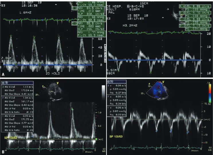
The baseline characteristics of the patients are presented in Table 1. There were no differences in the age, gender, hyper- tension, diabetes, dyslipidemia, stroke, smoking and CHA2DS2- Vasc score between the groups. There were no differences in the LA AP dimension, LV mass index, or LV EF between the groups. Group 1 had an increased LA volume index (LAVI;
30.30±8.32 mL/m2 vs. 25.47±7.75 mL/m2, p=0.021) and E/e’
(10.9±3.7 vs. 8.3±2.3, p=0.002) compared with group 2. The difference of E/e’ between two groups is described with rep- resentative cases in Fig. 1, and depicted by scatter plots in Fig.
2. There were no significant difference in the prescribed med- ications such as beta blocker, calcium channel blocker, angio- tensin-converting enzyme inhibitor and angiotensin II recep- tor blocker.
Diastolic function and recurrence rate of AF after cardioversion
The conversion to sinus rhythm during pre-treatment with
flecainide was achieved in 39/66 patients (59.1%). Electrical cardioversion was carried out in the remaining patients, 27/66 (40.9%). All patients were converted to sinus rhythm with ei- ther chemical or electrical cardioversion. During a mean fol- low-up period of 25±19 months, the recurrence rate of AF af- ter cardioversion was 60.61% (40/66; 48.7% vs. 77.8% in chemical cardioversion vs. electrical cardioversion). The chemical cardioversion group had a decreased LA volume in- dex (47.66 mL/m2 vs. 65.44 mL/m2, p<0.001) compared to the electrical cardioversion group. There was no difference in the value of E/e’ (9.7±3.3 vs. 10.6±4.1, p=0.351) between these two groups.
The area under the ROC curve of E/e’ for AF recurrence was 0.780 (95% CI: 0.657–0.903). ROC analysis showed the optimal cut-off value for the E/e’ for predicting AF recurrence level to be 9.15 with 75.0% of sensitivity and 73.1% of specificity (Fig. 3).
Univariate Cox proportional hazards regression analysis showed that LAVI [hazards ratio (HR): 1.045, 95% CI: 1.007–
1.085, p=0.020], LV mass index (HR: 1.022, 95% CI: 1.001–
1.042, p=0.036) and the ratio of E/e’ (HR: 1.113, 95% CI: 1.031–
1.202, p=0.006) were significant predictors of AF recurrence after cardioversion. Multivariate Cox proportional hazards re- gression analysis with stepwise forward method revealed that E/e’ (HR: 1.100, 95% CI: 1.017–1.190, p=0.017) and LAVI (HR:
Table 1. Baseline Characteristics
Group 1 (n=40) Group 2 (n=26) p value
Age, yrs 58±13 58±11 0.988
Male, n (%) 35 (86.4) 22 (85.0) 1.000
Underlying diseases, n (%)
Hypertension 20 (48.8) 14 (56.0) 0.619
Diabetes 9 (22.0) 3 (12.0) 0.512
Stroke 6 (14.6) 1 (4.0) 0.239
Dyslipidemia 8 (19.5) 2 (8.0) 0.297
Medication, n (%)
BB 9 (22.5) 3 (11.5) 0.126
CCB 32 (80.0) 22 (84.6) 0.252
ACEi 2 (5.0) 3 (11.5) 0.243
ARB 17 (42.5) 8 (30.8) 0.108
Aspirin 22 (55.0) 17 (65.4) 0.176
Warfarin 16 (40.0) 9 (34.0) 0.168
CHADS2 score 1.1±1.3 0.7±0.7 0.080
CHA2DS2-Vasc score 1.6±1.5 1.3±1.1 0.379
LA diameter, mm 39.63±5.43 37.69±5.37 0.161
LA volume index, mL/m2 30.30±8.32 25.47±7.75 0.021
LV mass index, g/m2 91.56±17.19 84.98±15.97 0.123
LV EF, % 65±7 66±9 0.780
E, cm/s 0.80±0.26 0.70±0.19 0.110
e’, cm/s 0.08±0.03 0.09±0.03 0.325
E/e’ 10.9±3.7 8.3±2.3 0.002
DT, ms 219±72 192±48 0.370
LA, left atrium; LV, left ventricle; EF, ejection fraction; E/e’, the ratio of mitral peak velocity of early filling (E) to early diastolic mitral annular velocity (e’); DT, de- celeration time; BB, beta blocker; CCB, calcium channel blocker; ACEi, angiotensin-converting enzyme inhibitor; ARB, angiotensin ll receptor blocker.
1.042, 95% CI: 1.002–1.084, p=0.042) were independent pre- dictors for AF recurrence after cardioversion (Table 2 and 3).
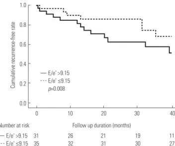
Fig. 4 shows the Kaplan-Meier survival curves for AF recur-
rence with or without elevated LV filling pressure. The cumu- lative recurrence-free survival rate was significantly lower in patients with elevated LV filling pressure (E/e’>9.15) than pa- tients without elevated LV filling pressure (E/e’≤9.15) (log rank p=0.008).
DISCUSSION
This study investigated the relationship between LV filling pressure and AF recurrence after cardioversion. In line with previous reports, overall AF recurrence rate in our study pop- ulation was 60.6% (40/66).7,8 Diastolic dysfunction is linked with poorer cardiovascular outcomes, development of AF, more severe AF symptoms, as well known.9 The main finding of this study was that elevated LV filling pressure, as indicated by the increased E/e’ ratio, was correlated significantly with AF recurrence after cardioversion. Our present study enrolled only patients who were newly diagnosed with AF in the ab- sence of extremely enlarged LA. In this way, we were able to more effectively isolate the effect of LV filling pressure on AF A
B
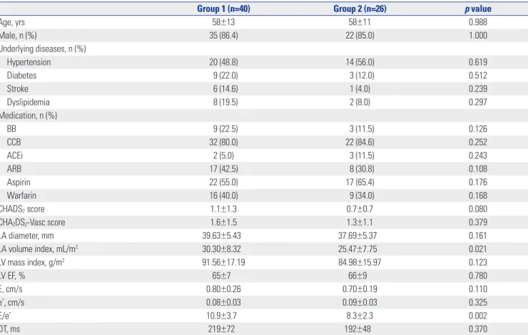
Fig. 1. The representative cases which reveal the difference of E/e’ between two groups. (A) In patients without recurrence, early (E) diastolic mitral in- flow peak velocity and early diastolic mitral annulus peak velocity (e’) were measured, and the ratio of E/e’ was calculated. (B) In patients with recur- rence, the ratio of E/e’ was calculated by the same way.
Fig. 2. The scatter plots of E/e’ with or without AF recurrence. AF, atrial fi- brillation; E/e’, the ratio of mitral peak velocity of early filling (E) to early di- astolic mitral annular velocity (e’).
25
20
15
10
5
0
AF recur (-) AF recur (+)
E/e’
recurrence. The results suggested that elevated LV filling pres- sure may predict the outcome after cardioversion in patients with persistent AF.
Evaluation of diastolic function in AF patients and echocardiographic parameters
Evaluation of diastolic function involves integration of multi- ple echocardiographic parameters. Diastolic dysfunction is characterized by a progressive decrease in LV compliance with corresponding impairment in myocardial relaxation, re- sulting in elevated LV end diastolic pressure despite normal end diastolic volume.2 Clinically useful parameters, which re- flect the pressure gradient between the LA and LV, include mitral inflow Doppler patterns of early filling peak velocity (E), atrial peak velocity (A), the E/A ratio, and deceleration time (DT). These transmitral flow parameters are affected by load- ing status, and DT varies as it is dependent on the cardiac cy- cle length. Therefore, they need to be considered in combina- tion with tissue Doppler imaging, which is less dependent on preload. Since, atrial systolic waves (A wave, atrial peak veloc- ity) are lost in AF patients, it is difficult to evaluate diastolic function based on transmitral flow velocity.
Mitral annular velocity (e’), determined by pulsed wave tis- sue Doppler, is a relatively preload-independent variable, and has been demonstrated to be an excellent marker of LV relax- ation.10 Therefore, E/e’ reflects the LV filling pressure and is useful for evaluating diastolic function. Indeed, several stud- ies have reported that E/e’ is associated with LV filling pres- sure, even in AF patients, with sensitivities >70% and specifici- ties >90%.11 Furthermore, Okura, et al.9 reported that E/e’ was a strong predictor for heart failure in AF patients. In our study, 1.0
0.8
0.6
0.4
0.2
0.00.0 0.2 0.4
1-specificity ROC curve
0.6 0.8 1.0
Sensitivity
Fig. 3. Receiver operating characteristic (ROC) curves for E/e’ for AF re- currence after cardioversion. Area under the ROC curve for E/e’ was 0.780 (95% confidence interval: 0.657–0.903, p<0.001). AF, atrial fibrillation;
E/e’, the ratio of mitral peak velocity of early filling (E) to early diastolic mi- tral annular velocity (e’).
Table 2. Univariate Cox Proportional Hazards Regression Analysis for Predicting AF Recurrence after Cardioversion
Variables HR 95% CI p value
Age, per 1 yr increase 1.003 0.975–1.031 0.851
Male gender 0.847 0.546–1.313 0.457
Hypertension 0.925 0.489–1.750 0.811
Diabetes mellitus 0.820 0.376–1.791 0.619
Dyslipidemia 1.176 0.490–2.821 0.717
CHADS2 score 1.088 0.838–1.411 0.527
CHA2DS2-VASc score 1.004 0.803–1.257 0.969
LV EF, per 1% increase 0.985 0.949–1.023 0.440
LA diameter, per 1 mm increase 1.033 0.971–1.098 0.305
LA volume index, per 1 mm3/m2 1.045 1.007–1.085 0.020
LV mass index, per 1 g/m2 increase 1.022 1.001–1.042 0.036
E/e’, per 1 increase 1.113 1.031–1.202 0.006
AF, atrial fibrillation; HR, hazards ratios; CI, confidence interval; LV, left ventricle; EF, ejection fraction; LA, left atrium; E/e’, the ratio of mitral peak velocity of ear- ly filling (E) to early diastolic mitral annular velocity (e’).
Table 3. Multivariate Cox Proportional Hazards Regression Analysis for Predicting AF Recurrence after Cardioversion
Variables Stepwise forward method Enter method
HR 95% CI p value HR 95% CI p value
LA volume index, per 1 mm3/m2 1.042 1.002–1.084 0.042 1.040 1.000–1.082 0.050
E/e’, per 1 increase 1.100 1.017–1.190 0.017 1.079 0.992–1.174 0.076
LV mass index, per 1 g/m2 increase 1.013 0.991–1.036 0.256
AF, atrial fibrillation; HR, hazards ratios; CI, confidence interval; LA, left atrium; LV, left ventricle; E/e’, the ratio of mitral peak velocity of early filling (E) to early diastolic mitral annular velocity (e’).
the ratio of E/e’ was a significant independent predictor of AF recurrence after cardioversion, indicating that LV filling pres- sure may be associated with AF recurrence. Since estimating E/e’ during AF is less accurate than the same measure taken during sinus rhythm, we measured these parameters while the R-R intervals were relatively regular after rate control.
Recurrence of AF after cardioversion according to LV filling pressure
The association between the recurrence of AF after cardiover- sion and echocardiographic parameters reflecting diastolic dysfunction remains inadequately assessed. In the present study, we found that LV filling pressure was significantly cor- related with AF recurrence after cardioversion.
When LV compliance is reduced, pressure essentially backs up, causing an increase in LA pressure. This atrial pressure overload leads to atrial electrical and structural remodeling including atrial stretching, dilatation and fibrosis. These changes provide a vulnerable substrate for AF.12 The mecha- nism of AF initiation and recurrence in patients with diastolic dysfunction stems from this progressive remodeling.3,13 Long- term volume overload could be predicted by both the LAVI and E/e’, while long-term pressure overload of the LA leads to progressive LA enlargement and electrical instability, which reflect the severity of diastolic dysfunction. Therefore, AF re- currence after cardioversion is associated with elevated LV filling pressure and LA remodeling. Some studies have dem- onstrated LAVI to be an independent predictor of AF recur- rence.14 The duration of AF reflects the degree of atrial remod- eling and has also been demonstrated to be an important predictor of the success of cardioversion.14
Some studies have demonstrated that diastolic dysfunction might predict AF recurrence after cardioversion therapies.2,4
The LA structural remodeling reflects the chronicity of expo- sure to abnormal filling pressures as consequence of diastolic dysfunction.14 Caputo, et al.4 identified an enlarged LA vol- ume and an elevated E/e’ as predictors of AF recurrence after electrical cardioversion. However, they enrolled AF patients with no regard to LA size. This resulted in their study popula- tion to include patients with extremely enlarged atria. It is highly likely that elevated LV filling pressure, resulted from di- astolic dysfunction, might cause the intrinsic pathologic physiologic change of LA before LA enlargement. LA dysfunc- tion had been observed before LA enlargement in patients with paroxysmal AF, and this might be due to the reduction of booster pump function of LA, such as intrinsic active relax- ation and contractility rather than conduit function.15,16 Our study enrolled only patients who were newly diagnosed with AF in the absence of extremely enlarged LA. In these patients, the atria have not been given ample time to structurally re- model, thus resulting in an unchanged LA size.
Importantly, several studies have reported an LA diameter above 50 mm predicts the AF recurrence.17 By design, our study excluded patients with LA diameters larger than 50 mm in order to avoid the associated effects on AF recurrence. In this way, we could more effectively isolate the effect of LV fill- ing pressure on AF recurrence, and found that E/e’ was an in- dependent predictor for AF recurrence after cardioversion, and an elevated E/e’ was associated with poorer outcomes af- ter cardioversion in patients with persistent AF patients.
Therefore, we conclude that LV filling pressure, estimated by E/e’, is predictive of AF recurrence after cardioversion even in the absence of extreme atrial enlargement.
Clinical implication
It is important to manage diastolic dysfunction with optimal medical therapy. We can possibly do more intensive observa- tion of symptoms and volume status of the patients with high- er E/e’ and without extreme LA enlargemt, and more aggres- sive medical treatment. The elevated LV filling pressure could be an early sign of left atrial pathological process before LA re- modeling, presented as LA enlargement. Based on the result of this study, we carefully suggest that early medical interven- tion could improve the clinical prognosis regarding AF recur- rences before the extreme LA enlargement observed by TTE.
Proper management can attenuate the structural and electri- cal remodeling of the atria, typically associated with diastolic dysfunction, thereby limiting AF recurrence after cardiover- sion. Therefore, medical therapy targeted at reducing LV fill- ing pressure is important before and after cardioversion. Re- nin-angiotensin system (RAS) inhibitors, such as angiotensin- converting enzyme inhibitor or angiotensin ll receptor blocker might reduce LV filling pressure. These medications may at- tenuate structural remodeling and electrical remodeling, and subsequently the recurrence of AF, by promoting the regres- sion of atrial fibrosis.18 Indeed, Fukuda, et al.19 demonstrated 1.0
0.8
0.6
0.4
0.2
0.0
0 10
E/e’ >9.15 E/e’ ≤9.15 p=0.008
E/e’ >9.15 31 26 21 19 11 E/e’ ≤9.15 35 32 31 30 27
20 Follow up duration (months) Number at risk
30 40
Cumulative recurrence-free rate
Fig. 4. The Kaplan-Meier survival curves for AF recurrence in patients af- ter cardioversion with or without increased LV filling pressure. AF, atrial fibrillation; E/e’, the ratio of mitral peak velocity of early filling (E) to early diastolic mitral annular velocity (e’); LV, left ventricle.
that E/e’ was higher in non-RAS inhibitor medication group than RAS inhibitor medication group. Although two prospec- tive studies reported that RAS blockers could not suppress AF recurrence,20,21 Ishikawa, et al.22 demonstrated that RAS inhib- itor significantly reduced the AF recurrence after pulmonary vein isolation. Generally, therefore, these previous studies support that these drugs might reduce AF recurrence by de- creasing LV filing pressure, the primary risk factor for AF re- currence assessed in this study. Based on the present results, further assessment of LV filling pressure as a valuable predic- tor of clinical outcomes associated with AF recurrence may help evaluate the effectiveness of these drugs. Moreover, a prospective study is needed to evaluate whether medical therapies aimed at lowering LV filling pressure could reduce AF recurrence after cardioversion and improve clinical out- comes in AF patients.
Limitations
The present study has several limitations. This study was per- formed at a tertiary referral hospital, and there might be some selection and referral bias. The relatively small number of en- rolled patients in this study is also a limitation. Additional echocardiographic parameters, such as left atrial appendage velocities using transesophageal echocardiography, were not considered in this study. Until now, there is no established method for evaluating LV diastolic function in AF patients, al- though several studies have reported clinical usefulness of E/
e’ to estimate LV filling pressure in this population.9 Therefore, we could not identify an association between diastolic dys- function and AF recurrence. Additionally, Doppler parame- ters measured during AF could be less accurate than in the si- nus rhythm, although we minimized this complication by measuring these parameters while the R-R intervals were rela- tively regular. Our study indicated that the echocardiographic parameters, indicating diastolic function such as E/e’ ratio, can be reasonably assessed in AF patients. We think that more prospective studies are needed to firmly confirm the relation- ship between the medical treatment of diastolic dysfunction and the improvement of clinical prognosis regarding AF re- currences.
Conclusions
Our study provides data that support the assessment of LV fill- ing pressure to predict clinical outcomes after cardioversion.
The presence of LV diastolic dysfunction is a crucial mediator of AF development and recurrence. After cardioversion, pa- tients with an elevated LV filling pressure, estimated by the E/
e’ ratio, had an increased AF recurrence rate without extreme LA enlargement. These results suggest that elevated LV filling pressure might predict the outcome after cardioversion of AF regardless of the degree of mechanical atrial remodeling.
Therefore, it is important to manage diastolic dysfunction, in- cluding reduction of LV filling pressure, in order to reduce the
AF recurrence rate and improve the outcome in patients with persistent AF after cardioversion. For early detected AF pa- tients without extreme LA enlargement, reduction of LV filling pressure after cardioversion might decrease the risk of AF re- currence.
ACKNOWLEDGEMENTS
We thank the study participants and supporting medical staffs for making this study possible.
REFERENCES
1. European Heart Rhythm Association; European Association for Cardio-Thoracic Surgery, Camm AJ, Kirchhof P, Lip GY, Schotten U, et al. Guidelines for the management of atrial fibrillation: the Task Force for the Management of Atrial Fibrillation of the Euro- pean Society of Cardiology (ESC). Eur Heart J 2010;31:2369-429.
2. Melduni RM, Cullen MW. Role of left ventricular diastolic dys- function in predicting atrial fibrillation recurrence after success- ful electrical cardioversion. J Atr Fibrillation 2012;5:87-94.
3. Huang JL, Tai CT, Lin YJ, Ting CT, Chen YT, Chang MS, et al. The mechanisms of an increased dominant frequency in the left atri- al posterior wall during atrial fibrillation in acute atrial dilatation.
J Cardiovasc Electrophysiol 2006;17:178-88.
4. Caputo M, Urselli R, Capati E, Navarri R, Sinesi L, Furiozzi F, et al.
Usefulness of left ventricular diastolic dysfunction assessed by pulsed tissue Doppler imaging as a predictor of atrial fibrillation recurrence after successful electrical cardioversion. Am J Cardiol 2011;108:698-704.
5. Fuster V, Rydén LE, Cannom DS, Crijns HJ, Curtis AB, Ellenbo- gen KA, et al. 2011 ACCF/AHA/HRS focused updates incorpo- rated into the ACC/AHA/ESC 2006 guidelines for the manage- ment of patients with atrial fibrillation: a report of the American College of Cardiology Foundation/American Heart Association Task Force on practice guidelines. Circulation 2011;123:e269-367.
6. Lang RM, Bierig M, Devereux RB, Flachskampf FA, Foster E, Pel- likka PA, et al. Recommendations for chamber quantification: a report from the American Society of Echocardiography’s Guide- lines and Standards Committee and the Chamber Quantification Writing Group, developed in conjunction with the European As- sociation of Echocardiography, a branch of the European Society of Cardiology. J Am Soc Echocardiogr 2005;18:1440-63.
7. Prystowsky EN, Benson DW Jr, Fuster V, Hart RG, Kay GN, Myer- burg RJ, et al. Management of patients with atrial fibrillation. A statement for healthcare professionals. From the subcommittee on electrocardiography and electrophysiology, American Heart Association. Circulation 1996;93:1262-77.
8. Kim H, Lee JP, Yoon HJ, Park HS, Cho YK, Nam CW, et al. Associ- ation between Doppler flow of atrial fibrillatory contraction and recurrence of atrial fibrillation after electrical cardioversion. J Am Soc Echocardiogr 2014;27:1107-12.
9. Okura H, Takada Y, Kubo T, Iwata K, Mizoguchi S, Taguchi H, et al. Tissue Doppler-derived index of left ventricular filling pres- sure, E/E’, predicts survival of patients with non-valvular atrial fi- brillation. Heart 2006;92:1248-52.
10. Bolognesi R, Tsialtas D, Barilli AL, Manca C, Zeppellini R, Javern- aro A, et al. Detection of early abnormalities of left ventricular function by hemodynamic, echo-tissue Doppler imaging, and mitral Doppler flow techniques in patients with coronary artery
disease and normal ejection fraction. J Am Soc Echocardiogr 2001;14:764-72.
11. Watanabe T, Iwai-Takano M, Oikawa M, Yamaki T, Yaoita H, Maruyama Y. Optimal noninvasive assessment of diastolic heart failure in patients with atrial fibrillation: comparison of tissue doppler echocardiography, left atrium size, and brain natriuretic peptide. J Am Soc Echocardiogr 2008;21:689-96.
12. Khan A, Moe GW, Nili N, Rezaei E, Eskandarian M, Butany J, et al. The cardiac atria are chambers of active remodeling and dy- namic collagen turnover during evolving heart failure. J Am Coll Cardiol 2004;43:68-76.
13. Chin JY, Youn HJ. The effect of ablation for paroxysmal atrial fi- brillation on left atrial volume and function: a one-year follow- up study. Yonsei Med J 2014;55:895-903.
14. Tsang TS, Barnes ME, Gersh BJ, Bailey KR, Seward JB. Left atrial volume as a morphophysiologic expression of left ventricular di- astolic dysfunction and relation to cardiovascular risk burden.
Am J Cardiol 2002;90:1284-9.
15. Kojima T, Kawasaki M, Tanaka R, Ono K, Hirose T, Iwama M, et al. Left atrial global and regional function in patients with parox- ysmal atrial fibrillation has already been impaired before en- largement of left atrium: velocity vector imaging echocardiogra- phy study. Eur Heart J Cardiovasc Imaging 2012;13:227-34.
16. Lancellotti P, Henri C. The left atrium: an old ‘barometer’ which can reveal great secrets. Eur J Heart Fail 2014;16:1047-8.
17. Olshansky B, Heller EN, Mitchell LB, Chandler M, Slater W, Green M, et al. Are transthoracic echocardiographic parameters associated with atrial fibrillation recurrence or stroke? Results from the atrial fibrillation follow-up investigation of rhythm management (AFFIRM) study. J Am Coll Cardiol 2005;45:2026- 33.
18. Schneider MP, Hua TA, Böhm M, Wachtell K, Kjeldsen SE, Schmieder RE. Prevention of atrial fibrillation by Renin-Angio- tensin system inhibition a meta-analysis. J Am Coll Cardiol 2010;55:2299-307.
19. Fukuda Y, Fukuda N, Morishita S, Tamura Y. Preventive effect of renin-angiotensin system inhibitor on left atrial remodelling in patients with chronic atrial fibrillation: long-term echocardio- graphic study. Eur J Echocardiogr 2011;12:278-82.
20. ACTIVE I Investigators, Yusuf S, Healey JS, Pogue J, Chrolavicius S, Flather M, et al. Irbesartan in patients with atrial fibrillation. N Engl J Med 2011;364:928-38.
21. GISSI-AF Investigators, Disertori M, Latini R, Barlera S, Franzosi MG, Staszewsky L, et al. Valsartan for prevention of recurrent atri- al fibrillation. N Engl J Med 2009;360:1606-17.
22. Ishikawa K, Yamada T, Yoshida Y, Takigawa M, Aoyama Y, Inoue N, et al. Renin-angiotensin system blocker use may be associated with suppression of atrial fibrillation recurrence after pulmonary vein isolation. Pacing Clin Electrophysiol 2011;34:296-303.