281 https://e-kcj.org
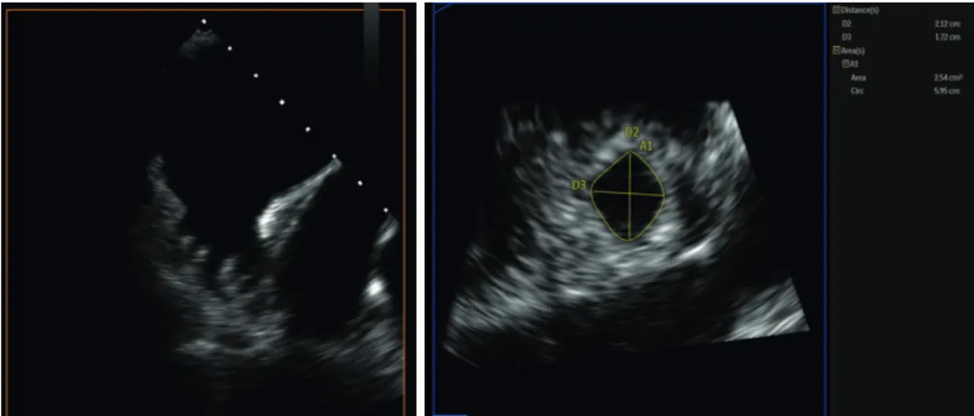
A 79-year-old woman in permanent atrial fibrillation with CHA
2DS
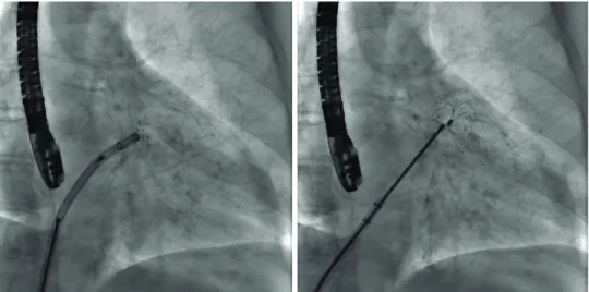
2-VASc score 6 and under acenocumarol therapy was referred to our department for percutaneous left atrial appendage (LAA) closure after suffering a hemorrhagic cerebrovascular accident. Transesophageal echocardiography (TEE) revealed a thrombus in LAA (Figure 1). We planned to implant LAmbre™ (Lifetech Scientific Corp., Shenzhen, China) with simultaneous use of a cerebral protection device, Sentinel™ (Claret Medical, Santa Rosa, CA, USA) (Figure 2). The procedure was guided by TEE without any contrast injection. A partial umbrella delivery of a LAmbre 24/30 mm was done in front of LAA ostium and the whole system was advanced up to the point immediately before thrombus in LAA superior lobe. At this point, the umbrella delivery was completed and afterward the cover part was immediately unsheathed (Figure 3).
TEE revealed a proper position of LAmbre, so the device was eventually released (Figure 4).
Sentinel™ did not contain any debris. The patient's postoperative course was uneventful.
Korean Circ J. 2020 Mar;50(3):281-283 https://doi.org/10.4070/kcj.2019.0305 pISSN 1738-5520·eISSN 1738-5555
Images in
Cardiovascular Medicine
Received: Sep 20, 2019 Accepted: Oct 23, 2019 Correspondence to Mohsen Mohandes, MD
Interventional Cardiology Unit, Cardiology Division, Joan XXIII University Hospital, IISPV Universitat Rovira Virgili, Calle Dr Mallafré Guasch 4, Tarragona 43007, Spain.
E-mail: [email protected] Copyright © 2020. The Korean Society of Cardiology
This is an Open Access article distributed under the terms of the Creative Commons Attribution Non-Commercial License (https://
creativecommons.org/licenses/by-nc/4.0) which permits unrestricted noncommercial use, distribution, and reproduction in any medium, provided the original work is properly cited.
ORCID iDs Mohsen Mohandes
https://orcid.org/0000-0003-1045-3639 Conflict of Interest
The authors have no financial conflicts of interest.
Author Contributions
Conceptualization: Mohandes M, Pernigotti A; Data curation: Morr CI; Writing - original draft: Mohandes M; Writing - review & editing:
Bardají A.
Mohsen Mohandes , MD 1 , Alberto Pernigotti, MD 1 , Carlos Igor Morr, MD 2 , and Alfredo Bardají, MD, PhD, FESC 3
1
Interventional Cardiology Unit, Cardiology Division, Joan XXIII University Hospital, IISPV, Rovira i Virgili University, Tarragona, Spain
2
Echocardiography and Image Department, Cardiology Division, Joan XXIII University Hospital, IISPV, Rovira i Virgili University, Tarragona, Spain
3