. . . .
. . . .
The electroanatomical remodelling of the left
atrium is related to CHADS
2
/CHA
2
DS
2
VASc
score and events of stroke in patients
with atrial fibrillation
Jae Hyung Park
1, Boyoung Joung
1, Nak-Hoon Son
2, Jae Min Shim
1,
Moon Hyung Lee
1, Chun Hwang
3, and Hui-Nam Pak
1*
1
Yonsei University Health System, Seoul, Republic of Korea;2
Department of Biostatistics, Yonsei University, Seoul, Republic of Korea; and3
Krannert Heart Institute, Indiana University, Indianapolis, IN, USA
Received 29 December 2010; accepted after revision 6 April 2011; online publish-ahead-of-print 15 May 2011
Background Although atrial fibrillation (AF) increases the risk of stroke, its relationship with atrial remodelling has not yet been
studied. We hypothesized that the degree of electroanatomical remodelling of the left atrium (LA) is related to
CHADS2/CHA2DS2VASc score and events of stroke.
Methods and results
We compared CHADS2/CHA2DS2VASc score (0, 1,≥2) or events of stroke with mean and regional LA volume
[by three-dimensional (3D) computed tomography images] or LA endocardial voltage (by 3D-electroanatomical map) in 348 patients who underwent catheter ablation of AF (78.4% male, 55.4+11.0 years old, paroxysmal
AF:per-sistent AF ¼ 215:133). We graded LA volume index as Grade 1 (,48.3 mL/m2; n ¼ 80), grade 2 (48.3 – 63.0 mL/m2,
n ¼ 82), grade 3 (63.0 – 99.0 mL/m2; n ¼ 94), and grade 4 (≥99.0 mL/m2; n ¼ 92). Results (i) The percentage volume
of anterior portion of LA enlarged at the early stage of LA remodelling (Grade 1 vs. grade 2, P ¼ 0.006) and the voltage of posterior venous LA was significantly reduced with the degree of LA remodelling (P ¼ 0.001). (ii) Mean
LA volume/body surface area (BSA), especially anterior portion of LA, was greater in patients with high CHADS2/
CHA2DS2VASc score (P ¼ 0.002). Mean LA voltage was significantly lower in patients with high CHA2DS2VASc
score than low score (P ¼ 0.007). (iii) In patients who experience stroke (n ¼ 22), LA volume/BSA, especially anterior LA, was greater (P ¼ 0.012), and LA endocardial voltage was lower (P ¼ 0.039) than those without stroke.
Conclusion Electroanatomical remodelling of LA, estimated by LA volume and endocardial voltage, has significant relationship
with the risk scores or events of stroke in patients with non-valvular AF.
-Keywords Atrial fibrillation † CHADS2score † Stroke † Left atrium † Voltage
Introduction
Atrial fibrillation (AF) is the most common arrhythmia disorder, affecting up to 9% of the population by the age of 80 years, and
is a significant risk factor for thromboembolic stroke.1,2CHADS2
scores have been utilized as an excellent predictor of stroke and a guideline for anti-thrombotic therapy in patients with
non-valvular AF.3–5Although the CHADS2score is an effective
predic-tor of ischaemic stroke in patients with AF, the pathophysiologic
mechanisms remain to be studied. Substrate or tissue factors
related to CHADS2scores have to be considered. Recurrent
fibril-latory activation of AF induces progressive electrical and tissue
structural remodelling,6–8and reduction of left atrial (LA)
endo-cardial voltage in the presence of fibrosis.9We previously reported
that AF-related electroanatomical remodelling changes both entire and regional LA volume, endocardial voltage, conduction velocity, and distribution of complex fractionated atrial
electro-gram.10–12 However, the relationship between the degree of
*Corresponding author: 250 Seungsanno, Seodaemun-gu, Seoul 120-752, Republic of Korea. Tel:+82 2 2228 58459; fax: +82 2 393 2041, Email: [email protected]
Published on behalf of the European Society of Cardiology. All rights reserved.&The Author 2011. For permissions please email: [email protected].
at YONSEI UNIVERSITY MEDICAL LIBRARY on November 10, 2013
http://europace.oxfordjournals.org/
electroanatomical remodelling and CHADS2score and the event
of stroke has not yet been studied. Therefore, we hypothesized that the degree of electroanatomical remodelling of LA is related
to CHADS2 scores and to the event of stroke. The purposes of
this study were to evaluate the regional volume change of LA depending on the degree of structural remodelling, and to compare the degree of mean and regional LA volume enlargement
or LA endocardial voltage depending on CHADS2score and the
events of stroke. We also compared the degree of electroanatomi-cal remodelling with newly suggested risk scores for stroke in
non-valvular AF—CHA2DS2VASc score.13
Methods
Patient selection
The study protocol was approved by the Institutional Review Board of our institute. All patients provided written informed consent. The study enrolled 348 patients with AF (male:female ¼ 273:75, mean age ¼ 55.4+11.0 years old) who underwent radiofrequency catheter ablation (RFCA). Among them, 215 patients had paroxysmal AF, and 133 had persistent AF (PeAF). The exclusion criteria were as follows: (i) permanent AF refractory to the electrical cardioversion; (ii) LA sizes .55 mm measured on echocardiogram; (iii) AF with rheu-matic valvular disease; (iv) associated structural heart disease other than left ventricular (LV) hypertrophy; (v) prior AF ablation; and
(vi) sinus rhythm not maintained for LA voltage mapping before RFCA. The patients with the presence of an LA thrombus were excluded by transoesophageal echocardiography. We imaged all patients with a three-dimensional (3D) spiral computed tomography (CT) (64 Channel, Light Speed Volume CT, Philips, Brilliance 63, Amsterdam, Netherlands) to visually define the anatomy of LA and pulmonary veins (PVs). Trans-thoracic echocardiography was per-formed in every patient and LV systolic and diastolic functions were measured by ejection fraction (EF) and mitral valve area tissue Doppler (E/E’), respectively.
Electrophysiological mapping procedure
Intracardiac electrograms were recorded using the Prucka Cardi-oLabTM Electrophysiology system (General Electric Medical Systems
Inc., Easton Turnpike, Fairfield, USA), and catheter ablation procedures were performed on all patients using 3D electroanatomical mapping (NavX, St Jude Medical Inc., Minnetonka, MN, USA) merged with 3D spiral CT. Before catheter ablation, we generated an LA 3D electroa-natomical map and a voltage map by obtaining contact bipolar electro-grams from 350 – 400 points of the LA endocardium, during atrial pacing with a pacing cycle length of 500 ms. Bipolar electrograms were filtered from 32 to 300 Hz. Colour-coded voltage maps were generated by recording bipolar electrograms and measuring peak-to-peak voltage. The acquisition of an LA voltage map was aban-doned if frequently re-initiating AF required electrical cardioversion more than three times. Only patients for whom the LA voltage map was available were included in the study.
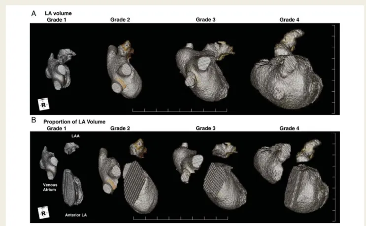
Figure 1 (A) Right lateral views of left atrium in the reconstructed three-dimensional spiral computed tomography image depending on the degree of left atrial remodelling. (B) left atrium images were divided into the venous atrium, the anterior left atrium, and the left atrial appen-dage. Volumetric measurement reveals significantly higher relative volume of the anterior left atrium in Stage 2 remodelling than in Stage 1.
at YONSEI UNIVERSITY MEDICAL LIBRARY on November 10, 2013
http://europace.oxfordjournals.org/
Volumetric and curvilinear analyses
of three-dimensionalspiral computed
tomography imaging
The 3D spiral CT images of LA were analysed on an imaging processing workstation (Aquarius, Terarecon Inc., San Mateo, CA, USA) as described before.11The curvilinear lengths on LA were measured at
the linear ablation sites: bilateral antral ablation line, roof line, posterior inferior line, left lateral isthmus line, anterolateral line, and anteroseptal line. Each LA image was divided into portions by embryological origin as follows: the venous LA (posterior LA including the antrum and posterior wall), LA appendage (LAA), and anterior LA (excluding the LAA and venous LA).14 Although both LAA and anterior LA are
embryologically of primordial atrial origin, they differ in geometry, myocardial fibre orientations, and distribution of autonomic inner-vations. Therefore, we divided LAA and anterior LA, and referenced them to the points of inflection on the 3D spiral CT image. The absol-ute and relative volumes of each portion were calculated and compared.
Off-line analyses of colour-coded
three-dimensional maps of left atriums
We analysed colour-coded voltage maps of both anterior – posterior (AP) and posterior – anterior views that had been converted to image files as previously reported.11PVs were not included in the analysis. The digital measurements of colour-coded voltage maps
were performed by a single student, using a consistent method, who was blinded to the clinical information of the maps. The percentage of colour-coded areas in each quadrant of the voltage maps was ana-lysed by customized software (Image Pro, Silver Spring, MD, USA), and referenced to the colour scale bars. NavX detected peak-to-peak voltage differences of each contact bipolar electrogram, and changed them to colour codes. Low-voltage areas, defined as LA voltage ≤0.2 mV, were coded grey; high-voltage areas (.5.0 mV) were coded purple. The mean LA voltage was calculated by summation of % area of each colour multiplied by representative voltage, and then divided by the total area of LA. The reference distance was measured by the inter-electrode distances of coronary sinus catheters (duodeca-polar catheter, St Jude Medical Inc.).
Data analyses
We classified patients according to CHADS2 scores (0, n ¼ 154; 1,
n ¼ 124; and ≥2, n ¼ 70), CHA2DS2VASc score (0, n ¼ 146; 1,
n ¼ 106; and≥2, n ¼ 96), the quartiles of 3D spiral CT measured LA volume index (Grade 1≤48.3 mL/m2
, n ¼ 87; Grade 2, 48.3–63.0 mL/m2, n ¼ 87; Grade 3, 63.0–99.0 mL/m2, n ¼ 87; and Grade 4≥ 99.0 mL/m2, n ¼ 87), and the patients with (n ¼ 22) or without (n ¼ 326) stroke. We compared them by absolute or relative volumes or curvilinear lengths adjusted by BSA, mean LA voltage, LV systolic and diastolic func-tion, and duration of AF. Data are expressed as the mean+standard deviation. The statistical significance of these comparisons was assessed
. . . . Table 1 Comparison ofleft atrial morphology and left ventricular function according to atrial fibrillation -related left atrial size
Grade 1 Grade 2 Grade 3 Grade 4 ANOVA ANOVA (n 5 87) (n 5 87) (n 5 87) (n 5 87) P value Power LA volumes (mL) Entire LA volume 74.9 + 13.7 101.2 + 12.1* 128.2 + 13.7**† 170.8 + 36.4§‡[] P , 0.001 1 Anterior LA volume 42.4 + 10.5 60.6 + 12.8* 78.6 + 11.2**† 104.4 + 28.7§‡[] P , 0.001 1 Venous LA volume 26.1 + 8.2 32.3 + 6.6* 38.8 + 8.2**† 52.4 + 15.0§‡[] P , 0.001 1 LAA volume 6.5 + 3.1 8.7 + 3.3 10.8 + 3.6**† 14.0 + 5.4§‡[] P , 0.001 1 Relative volumes of regional LA (%)
Anterior LA volume 56.4 + 8.5 59.5 + 7.5 61.3 + 5.4** 60.7 + 8.0§ P , 0.001 0.979 Venous LA volume 34.9 + 8.4 31.9 + 7.4 30.2 + 5.3** 31.0 + 7.5§ P , 0.001 0.974 LAA volume 8.7 + 3.9 8.6 + 3.2 8.5 + 3.1 8.3 + 2.8 P ¼ 0.797 0.094 LA voltage (mV) Mean LA voltage 1.6 + 0.8 1.4 + 0.6 1.3 + 0.6 1.0 + 0.6§ P ¼ 0.002 0.873 Anterior LA voltage 1.4 + 0.6 1.2 + .0.6 1.3 + 0.6 0.9 + 0.6§ P ¼ 0.034 0.878 Venous LA voltage 1.8 + 1.3 1.0 + 0.9* 1.1 + 1.0** 0.8 + 0.8§ P ¼ 0.001 1 LAA voltage 3.1 + 1.7 2.8 + 1.5 2.5 + 1.6 2.0 + 1.3§ P ¼ 0.018 0.708 LV function LVEF (%) 61.0 + 7.2 60.9 + 7.4 60.1 + 8.2 57.2 + 10.8§‡ P ¼ 0.012 0.806 E/E’ 8.5 + 2.6 9.2 + 3.6 9.7 + 4.1 10.6 + 4.4§ P ¼ 0.014 0.781 Hypertension 26 (29.9 %) 38 (43.7 %) 47 (54.0 %)** 45 (51.7 %)§ P ¼ 0.005 1 Renal insufficiency 2 (2.3 %) 1 (1.1 %) 2 (2.3 %) 1 (1.1 %) P ¼ 0.880 1 Recurrence 11 (12.6 %) 11 (12.6 %) 16 (18.4 %) 24 (27.6 %)§ P ¼ 0.005 1
BSA, body surface area; LAA, LA appendage; LV, left ventricle; EF, ejection fraction, ANOVA, analysis of variance.
*P , 0.05, Grade 1 vs. Grade 2, **P , 0.05, Grade 1 vs. Grade 3,§
P , 0.05, Grade 1 vs. Grade 4,†
P , 0.05, Grade 2 vs. Grade 3,‡
P , 0.05, Grade 2 vs. Grade 4,[]
P , 0.05, Grade 3 vs. Grade 4.
at YONSEI UNIVERSITY MEDICAL LIBRARY on November 10, 2013
http://europace.oxfordjournals.org/
using the Student t-test and analysis of variance (ANOVA) test. A P value of ,0.05 was considered statistically significant.
Results
Enlargement of the anterior portion of
left atrium in the early stages of left atrial
remodelling
Figure1displays the right anterior oblique views of 3D spiral CT
image of LA in each grade of remodelling. In Grade 2 LA remodel-ling, the anterior portion of the LA was remarkably enlarged, and the AP diameter of LA prolonged significantly. In contrast, the pos-terior venous LA and LAA were proportionally enlarged at Grade
3 – 4 of LA remodelling. Table1summarizes LA volumes, regional
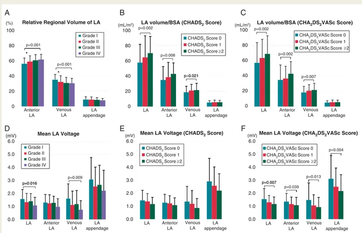
lengths, endocardial LA voltage, LV functions, and recurrence. The proportion of relative volumes of anterior LA was significantly greater (P , 0.001), but posterior venous LA was smaller (P , 0.001) in patients with high-grade remodelling than those
with low-grade remodelling (Figure 2A). The increase in relative
anterior LA volume was more significant in Grade 2 than in
Grade 1 remodelling (P ¼ 0.006, Figure2A). Left atrial endocardial
voltage was generally reduced more in patients with high-grade
remodelling than in those with low-grade remodelling
(P ¼ 0.002), and it was most significant in venous LA (P ¼ 0.001,
Figure2D).
Greater left atrial volume in patients
with higher CHADS
2score
We compared mean and regional LA volumes in terms of CHADS2
score (0, n ¼ 154; 1, n ¼ 124; and≥2, n ¼ 70; Table2) and CHA
2-DS2VASc score (0, n ¼ 146; 1, n ¼ 106; and ≥2, n ¼ 96; Table3).
Mean LA volume/BSA was significantly higher in patients with high
CHADS2score (P ¼ 0.002, Figure2B), and this difference was
sig-nificant especially in the regional volume of the anterior portion of
LA (P ¼ 0.048, Table2). The proportions of PeAF were 34.4% in
CHADS2 score 0, 40.3% in CHADS2 score 1, and 42.9% in
CHADS2 score ≥2 (P ¼ NS). In addition, EF was significantly
lower (P ¼ 0.005), E/E’ was higher (P ¼ 0.002), and the pro-portions of hypertension (P , 0.001) and renal insufficiency (P ¼
0.016) were higher in patients with higher CHADS2 score than
those with lower CHADS2 score in ANOVA analyses (Table2).
When we compared depending on CHA2DS2VASc score
(Table 3), these findings were consistent with the CHADS2
scores, and LA volume/BSA was significantly higher in patients
with high CHA2DS2VASc scores (Figure 2C). In the analyses of
LA endocardial voltage, the mean and regional endocardial
vol-tages tended to be lower in patients with high CHADS2 score
LA volume/BSA (CHADS2 Score)
Relative Regional Volume of LA (mL/m2) LA volume/BSA (CHA2DS2VASc Score)
p=0.002 (%) (mL/m2) 100 p=0.002 80 p=0.008 * p=0.002 60 p<0.001 p=0.021 * p=0.007 p 0.021 40 20 LA 0 100 80 60 40 20 0 100 80 60 40 20 0 Anterior LA Venous LA LA appendage Anterior LA Venous LA LA appendage LA Anterior LA Venous LA LA appendage LA Anterior LA Venous LA LA appendage LA Anterior LA Venous LA LA appendage LA Anterior LA Venous LA LA appendage
Mean LA Voltage (CHA2DS2VASc Score)
Mean LA Voltage (CHADS2 Score)
Mean LA Voltage (mV) (mV) (mV) 6.0 6.0 6.0 5.0 5.0 p=0.004 5.0 4.0 4.0 4.0 3.0 3.0 3.0 p=0 007 p=0.013 p=0 016 p=0.009 2.0 2.0 p=0.007 p=0.039 p=0.016 2.0 1.0 1.0 1.0 0.0 0.0 0.0 p<0.001 Grade I Grade II Grade III Grade IV Grade I Grade II Grade III Grade IV CHADS2 Score 0 CHADS2 Score 1 CHADS2 Score≥2 CHADS2 Score 0 CHADS2 Score 1 CHADS2 Score≥2 CHA 2DS2VAScScore 0
CHA2DS2VAScScore 1 CHA2DS2VAScScore ≥2
CHA2DS2VAScScore 0 CHA 2DS2VAScScore 1 CHA 2DS2VAScScore ≥2
A
B
C
D
E
F
Figure 2 In high-grade remodelled left atrium, portion of anterior left atrium (A) was significantly larger, mean left atrium voltage (D) was significantly lower than those low-grade remodelled left atrium. In higher CHADS2score, left atrium volume/body surface area (B) was
signifi-cantly larger; left atrium voltage (E) was lower than those lower CHADS2score. In higher CHA2DS2VASc score, left atrium volume/body
surface area (C ) was significantly larger; left atrium voltage (F ) was significantly lower than in the case of lower CHADS2score.
at YONSEI UNIVERSITY MEDICAL LIBRARY on November 10, 2013
http://europace.oxfordjournals.org/
than in those with low scores (Figure2E), but it was significant in
terms of CHA2DS2VASc score (P ¼ 0.007, Figure2F).
Low left atrial endocardial voltage and
higher anterior left atrial volume in
patients with stroke
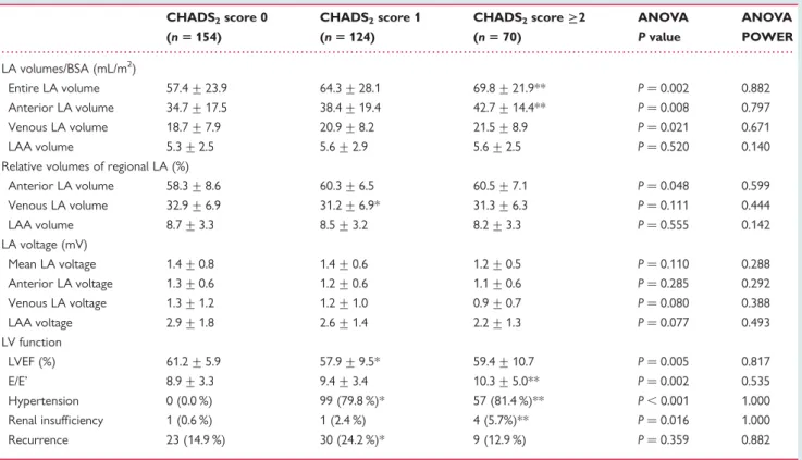
Figure3A and B display the representative examples of CT-merged
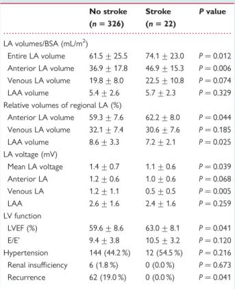
LA voltage maps, and the patients who experienced stroke show low endocardial voltage with enlarged LA volume in comparison with the patient without stroke. We compared them with 326
patients without stroke and summarized this in Table 4. There
were 22 patients who experienced episodes of stroke in this study. The stroke occurred 18.2+18.4 months before catheter ablation, and CHADS2 score was 3.2+0.2 at that time. The type of stroke was ischaemic embolic stroke in all 22 patients, but haemorrhagic transformation occurred during stroke manage-ment in 2 patients. In patients who had experienced a previous stroke, mean LA volume/BSA (P ¼ 0.012), especially anterior LA (P ¼ 0.006), was significantly enlarged, and mean LA endocardial voltage (P ¼ 0.039), especially venous LA voltage (P ¼ 0.005),
was significantly lower than those without stroke (Figure 3C
and D). In the uni-variate analyses (t-test), LA diameter (P ¼ 0.011), left ventricular ejection fraction (LVEF; P ¼ 0.041),
CHADS2 score (P , 0.001), LA volume/BSA (P ¼ 0.026), LAA
volume% (P ¼ 0.012), anterior LA volume% (P ¼ 0.025), and LA
voltage (P ¼ 0.039) were related with the event of stroke. In the multi-variate regression analysis, the factor most related to the
episode of stroke was CHADS2 score (OR ¼ 3.641, CI 2.033 –
6.521, P , 0.001) among them.
Discussion
This is the first study that reports on the relationship between the degree of electroanatomical remodelling of LA and the risk or events of stroke in patients with non-valvular AF. We also eluci-dated disproportional enlargement of the anterior portion of LA in the early stages of structural remodelling and its relationship
with CHADS2 score, and low endocardial voltage of posterior
venous atrium in patients with stroke. Therefore, CHADS2score
represents not only a clinical risk factor for ischaemic stroke, but is also related to atrial substrates for thoromboembolism in patients with AF.
Risks scores and potential mechanisms
of stroke in patients with non-valvular
atrial fibrillation
Atrial fibrillation causes a five-fold increase in the risk of
ischae-mic stroke or transient ischaeischae-mic attack (TIA).15CHADS2score
is a risk stratification scheme for ischaemic stroke in patients with non-valvular AF based on the presence of heart failure, . . . . Table 2 Comparison of left atrial morphology and left ventricular function according to atrial fibrillation -related
CHADS2score
CHADS2score 0 CHADS2score 1 CHADS2score≥2 ANOVA ANOVA
(n 5 154) (n 5 124) (n 5 70) P value POWER LA volumes/BSA (mL/m2) Entire LA volume 57.4 + 23.9 64.3 + 28.1 69.8 + 21.9** P ¼ 0.002 0.882 Anterior LA volume 34.7 + 17.5 38.4 + 19.4 42.7 + 14.4** P ¼ 0.008 0.797 Venous LA volume 18.7 + 7.9 20.9 + 8.2 21.5 + 8.9 P ¼ 0.021 0.671 LAA volume 5.3 + 2.5 5.6 + 2.9 5.6 + 2.5 P ¼ 0.520 0.140 Relative volumes of regional LA (%)
Anterior LA volume 58.3 + 8.6 60.3 + 6.5 60.5 + 7.1 P ¼ 0.048 0.599 Venous LA volume 32.9 + 6.9 31.2 + 6.9* 31.3 + 6.3 P ¼ 0.111 0.444 LAA volume 8.7 + 3.3 8.5 + 3.2 8.2 + 3.3 P ¼ 0.555 0.142 LA voltage (mV) Mean LA voltage 1.4 + 0.8 1.4 + 0.6 1.2 + 0.5 P ¼ 0.110 0.288 Anterior LA voltage 1.3 + 0.6 1.2 + 0.6 1.1 + 0.6 P ¼ 0.285 0.292 Venous LA voltage 1.3 + 1.2 1.2 + 1.0 0.9 + 0.7 P ¼ 0.080 0.388 LAA voltage 2.9 + 1.8 2.6 + 1.4 2.2 + 1.3 P ¼ 0.077 0.493 LV function LVEF (%) 61.2 + 5.9 57.9 + 9.5* 59.4 + 10.7 P ¼ 0.005 0.817 E/E’ 8.9 + 3.3 9.4 + 3.4 10.3 + 5.0** P ¼ 0.002 0.535 Hypertension 0 (0.0 %) 99 (79.8 %)* 57 (81.4 %)** P , 0.001 1.000 Renal insufficiency 1 (0.6 %) 1 (2.4 %) 4 (5.7%)** P ¼ 0.016 1.000 Recurrence 23 (14.9 %) 30 (24.2 %)* 9 (12.9 %) P ¼ 0.359 0.882
BSA, body surface area; LAA, LA appendage; LV, left ventricle; EF, ejection fraction, ANOVA, analysis of variance.
*P , 0.05, CHADS2score 0 vs. CHADS2score 1, **P , 0.05, CHADS2score 0 vs. CHADS2score≥2.
at YONSEI UNIVERSITY MEDICAL LIBRARY on November 10, 2013
http://europace.oxfordjournals.org/
hypertension, age .75 years old, diabetes, previous stroke, or
TIA.16 The 2006 version of the American College of
Cardiol-ogy/American Heart Association Task Force on Practice Guide-lines/European Society of Cardiology Committee for Practice (ACC/AHA/ESC) practical guidelines for AF recommends
war-farin or aspirin for patients depending on their CHADS2
score.5Recently, European Society of Cardiology have suggested
new guidelines for AF management,13and proposed a CHA2DS
2-VASc score that strengthens the importance of age, sex, and combined vascular disease as risk factors for ischaemic stroke
in addition to CHADS2 score. Although both CHADS2 and
CHA2DS2VASc scores are effective predictors for ischaemic
stroke, their pathophysiologic mechanisms remain to be clarified. Atrial contractile remodelling and blood stasis in LA during AF has generally been considered to be a major mechanism of
thor-omboembolism in patients with AF.17–19Blood stasis or
hyper-coagulable states are major contributors for thrombogenesis,
but tissue factors cannot be underestimated. CHADS2 score
and CHA2DS2VASc score include risk factors related to vascular
remodelling, metabolic syndrome, change of cardiac substrates, or tissue factors. The degree of electroanatomical remodelling of AF
is closely related with myocardial fibrosis,20,21matrix remodelling,
and angiotensin II-NADPH oxidase-mediated thrombus
for-mation.22 In this study, we demonstrated the relationship
between LA volume/endocardial voltage and risk scores or events of stroke in patients with AF.
Electroanatomical remodelling process
in atrial fibrillation
The mechanisms for LA remodelling include pressure or volume overload to LA by LV dysfunction, deranged plasma volume control, intensified neurohormonal activation, or atriomyopathy
itself.23In this study, the anterior portion of the LA was enlarged
in the early stages of LA remodelling, and the potential mechanisms are as follows: first, haemodynamic overload may stretch thin the traveculated wall of the anterior portion of LA early by Laplace’s law. Secondly, the posterior venous LA and the anteroseptum abut the fixed rigid structures, the spine, and the ascending aorta, respectively. Therefore, LA volume increased antero-laterally and the volume of anterior LA was enlarged in the early phases of remodelling.
The presence of fibrosis/low-voltage tissue has been postulated as a potential cause of abnormalities in atrial activation that may
underlie the initiation and maintenance of fibrillation.24,25 The
degree of voltage reduction may help grade the severity of tissue
pathology underlying AF before and after catheter ablation.26
Increased fibrosis has also been clearly demonstrated in human
LA tissue specimens of patients with AF,20,21 and correlations
have been observed between serum markers of atrially selective
fibroblasts and clinical AF.27 We also previously reported that
low endocardial voltage of LA is closely related to LA volume
remodelling,11reduced conduction velocity,10and the patterns of
. . . . Table 3 Comparison of left atrial morphology and left ventricular function according to atrial fibrillation-related
CHADS2VASc score
CHA2DS2VASc score 0 CHA2DS2VASc score 1 CHA2DS2VASc score≥2 ANOVA ANOVA
(n 5 146) (n 5 106) (n 5 96) P value POWER LA volumes/BSA (mL/m2) Entire LA volume 57.1 + 26.4 63.5 + 24.4 68.8 + 23.7** P ¼ 0.002 0.885 Anterior LA volume 34.8 + 19.6 36.6 + 16.0 42.8 + 15.9**† P ¼ 0.002 0.873 Venous LA volume 18.4 + 8.0 20.7 + 7.9* 21.7 + 8.7** P ¼ 0.007 0.810 LAA volume 5.2 + 2.4 5.6 + 2.8 5.7 + 2.7 P ¼ 0.256 0.269 Relative volumes of regional LA (%)
Anterior LA volume 58.3 + 8.5 59.6 + 6.9 61.1 + 6.8** P ¼ 0.019 0.701 Venous LA volume 33.0 + 8.3 31.8 + 6.8 30.7 + 6.6 P ¼ 0.050 0.553 LAA volume 8.7 + 3.3 8.6 + 3.1 8.2 + 3.4 P ¼ 0.525 0.170 LA voltage (mV) Mean LA voltage 1.5 + 0.8 1.3 + 0.5 1.2 + 0.5** P ¼ 0.007 0.587 Anterior LA voltage 1.3 + 0.6 1.2 + 0.5 1.1 + 0.6** P ¼ 0.039 0.332 Venous LA voltage 1.5 + 1.3 1.1 + 0.8 0.9 + 0.7** P ¼ 0.013 0.816 LAA voltage 3.1 + 1.8 2.5 + 1.4 2.2 + 1.3** P ¼ 0.004 0.811 LV function LVEF (%) 60.6 + 6.9 57.3 + 9.8* 61.5 + 9.0† P ¼ 0.001 0.924 E/E’ 9.0 + 3.4 8.8 + 3.2 11.0 + 4.5**† P , 0.001 0.968 Hypertension 0 (0.0 %) 80 (75.2 %)* 76 (79.2 %)** P , 0.001 1.000 Renal insufficiency 0 (0.0 %) 2 (1.9 %) 4 (4.2 %) P ¼ 0.051 1.000 Recurrence 24 (16.4 %) 21 (19.8 %)* 17 (17.7 %) P ¼ 0.683 1.000
BSA, body surface area; LAA, LA appendage; LV, left ventricle; EF, ejection fraction, ANOVA, analysis of variance.
*P , 0.05, CHADS2score 0 vs. CHADS2score 1, **P , 0.05, CHADS2score 0 vs. CHADS2score≥2,†P , 0.05, CHADS2score 1 vs. CHADS2score≥2.
at YONSEI UNIVERSITY MEDICAL LIBRARY on November 10, 2013
http://europace.oxfordjournals.org/
complex fractionated atrial electrograms in AF.12In this study, the degree of electroanatomical remodelling of LA was directly related to the risk or events of stroke in patients with AF. Further studies by non-invasive imaging methods, such as fibrosis on magnetic res-onance imaging or stain on echocardiography, will characterize the
degree of LA remodelling anatomically and functionally.28
Clinical implication of left atrial structural
remodelling
Electroanatomical remodelling of the atria has been known to be a
predictor of AF recurrence after cardioversion29or RFCA,11,30and
a risk of stroke, as shown in this study. Therefore, more strict anti-coagulation is warranted in those patients with low-voltage scars and enlarged LA. Longer duration of RF energy delivery is more of a necessity for effective rhythm control in AF patients with remodelled atria than for those with less remodelled atria. However, the operator should keep in mind that profuse RF
abla-tion can increase LA scar or atriomyopathy,26and might raise the
risk of stroke by electroanatomical remodelling. Whether reverse remodelling and LA voltage occurs after successful catheter abla-tion of AF is unclear. The appropriate strategy for anti-coagulaabla-tion
after abolishing AF remains to be studied. New drugs31,32
or devices33 provide more options for AF management, and
customized guidelines will be required according to the symptoms, haemodynamic factors, and risks of stroke.
Study limitations
The patients included in this study were a highly selective group referred for RFCA, and the number of patients was also limited. We also excluded patients with LA size of .50 mm. Because we acquired voltage maps via point-by-point contact mapping, the maps did not reflect a spatiotemporally homogeneous distri-bution. We analysed 3D voltage maps using 2D measurements. Although we strictly followed ACC/AHA/ESC guideline based on
the CHADS2score,5we do not have data of the anti-thrombotic
regimen at the time of ischaemic stroke because most patients included in this study were referred patients for catheter ablation. The patient group in this study was highly selected patients with relatively small LA volume with low risk of stroke. Therefore, the result of this study may not be extrapolated to the patients with permanent AF and significant LA remodelling.
Conclusion
We documented different patterns of CHADS2 score in patients
with structurally remodelled LA. In patients with non-valvular AF,
LA volume was larger in patients with high CHADS2score than
LA voltage (No Stroke) B LA voltage (Stroke)
A 10 mV 5 mV 20 00 /di 20 00 /di 0 mV 20.00 mm/div 20.00 mm/div Mean LA Voltage D LA volume/BSA C (mL/m2) 5.0 (mV) 100 p=0.012 4.0 80 p 0 006 3.0 p=0.006 p=0.039 p=0.005 60 2.0 40 1.0 20 0 0.0 Anterior LA
LA Venous LA LA appendage LA Anterior LA Venous LA LA appendage
10 mV 0 mV 5 mV No Stroke Stroke No Stroke Stroke
Figure 3 Anterior and posterior views of colour-coded left atrium voltage map during ablation. (A) High left atrium voltage in patients without stroke. (B) Low left atrium voltage in patients with stroke. In patients with stroke, mean left atrium volume and relative regional volume of anterior left atrium were larger (C), and mean left atrium voltage and regional left atrium voltage of venous atrium were lower (D) than those without stroke.
at YONSEI UNIVERSITY MEDICAL LIBRARY on November 10, 2013
http://europace.oxfordjournals.org/
those with low CHADS2 score and LA voltage was lower in
patients with stroke. Thus, stroke risk factors may be related to the degree of electroanatomical remodelling of LA and anterior LA might play a role in the early phase of structural remodelling in patients with AF.
Funding
This work was supported by a grant (A085136) from the Korea Health 21 R&D Project, the Ministry of Health and Welfare, and a grant (2010-0010537) from the Basic Science Research Program run by the National Research Foundation of Korea (NRF), which is funded by the Ministry of Education, Science and Technology of the Republic of Korea.
Conflict of interest: none declared.
References
1. Page RL. Clinical practice. Newly diagnosed atrial fibrillation. N Engl J Med 2004; 351:2408 – 16.
2. Stewart S, Hart CL, Hole DJ, McMurray JJ. A population-based study of the long-term risks associated with atrial fibrillation: 20-year follow-up of the Renfrew/ Paisley study. Am J Med 2002;113:359 – 64.
3. Go AS, Hylek EM, Chang Y, Phillips KA, Henault LE, Capra AM et al. Anticoagula-tion therapy for stroke prevenAnticoagula-tion in atrial fibrillaAnticoagula-tion: how well do randomized trials translate into clinical practice? JAMA 2003;290:2685 – 92.
4. Rietbrock S, Heeley E, Plumb J, van Staa T. Chronic atrial fibrillation: incidence, prevalence, and prediction of stroke using the Congestive heart failure, Hyperten-sion, Age .75, Diabetes mellitus, and prior Stroke or transient ischemic attack (CHADS2) risk stratification scheme. Am Heart J 2008;156:57 – 64.
5. Fuster V, Ryden LE, Cannom DS, Crijns HJ, Curtis AB, Ellenbogen KA et al. ACC/ AHA/ESC 2006 Guidelines for the management of patients with atrial fibrillation:
a report of the American College of Cardiology/American Heart Association Task Force on Practice Guidelines and the European Society of Cardiology Com-mittee for Practice Guidelines (Writing ComCom-mittee to Revise the 2001 Guidelines for the management of patients with atrial fibrillation): developed in collaboration with the European Heart Rhythm Association and the Heart Rhythm Society. Circulation 2006;114:e257 – 354.
6. Allessie M, Ausma J, Schotten U. Electrical, contractile and structural remodeling during atrial fibrillation. Cardiovasc Res 2002;54:230 – 46.
7. Frustaci A, Chimenti C, Bellocci F, Morgante E, Russo MA, Maseri A. Histological substrate of atrial biopsies in patients with lone atrial fibrillation. Circulation 1997; 96:1180 – 4.
8. Ausma J, Litjens N, Lenders MH, Duimel H, Mast F, Wouters L et al. Time course of atrial fibrillation-induced cellular structural remodeling in atria of the goat. J Mol Cell Cardiol 2001;33:2083 – 94.
9. Boldt A, Wetzel U, Lauschke J, Weigl J, Gummert J, Hindricks G et al. Fibrosis in left atrial tissue of patients with atrial fibrillation with and without underlying mitral valve disease. Heart 2004;90:400 – 5.
10. Park JH, Pak HN, Kim SK, Jang JK, Choi JI, Lim HE et al. Electrophysiologic charac-teristics of complex fractionated atrial electrograms in patients with atrial fibrilla-tion. J Cardiovasc Electrophysiol 2009;20:266 – 72.
11. Park JH, Pak HN, Choi EJ, Jang JK, Kim SK, Choi DH et al. The relationship between endocardial voltage and regional volume in electroanatomical remo-deled left atria in patients with atrial fibrillation: comparison of three-dimensional computed tomographic images and voltage mapping. J Cardiovasc Electrophysiol 2009;20:1349 – 56.
12. Park JH, Park SW, Kim JY, Kim SK, Jeoung B, Lee MH et al. Characteristics of complex fractionated atrial electrogram in the electroanatomically remodeled left atrium of patients with atrial fibrillation. Circ J 2010;74:1557 – 63. 13. Camm AJ, Kirchhof P, Lip GY, Schotten U, Savelieva I, Ernst S et al. Guidelines for
the management of atrial fibrillation: The Task Force for the Management of Atrial Fibrillation of the European Society of Cardiology (ESC). Eur Heart J 2010;31: 2369 – 429.
14. Douglas YL, Jongbloed MR, Gittenberger-de Groot AC, Evers D, Dion RA, Voigt P et al. Histology of vascular myocardial wall of left atrial body after pulmon-ary venous incorporation. Am J Cardiol 2006;97:662 – 70.
15. Wolf PA, Abbott RD, Kannel WB. Atrial fibrillation as an independent risk factor for stroke: the Framingham Study. Stroke 1991;22:983 – 8.
16. Gage BF, Waterman AD, Shannon W, Boechler M, Rich MW, Radford MJ. Vali-dation of clinical classification schemes for predicting stroke: results from the National Registry of Atrial Fibrillation. JAMA 2001;285:2864 – 70.
17. Fatkin D, Kuchar DL, Thorburn CW, Feneley MP. Transesophageal echocardio-graphy before and during direct current cardioversion of atrial fibrillation: evi-dence for ‘atrial stunning’ as a mechanism of thromboembolic complications. J Am Coll Cardiol 1994;23:307 – 16.
18. Missault L, Jordaens L, Gheeraert P, Adang L, Clement D. Embolic stroke after unanticoagulated cardioversion despite prior exclusion of atrial thrombi by trans-oesophageal echocardiography. Eur Heart J 1994;15:1279 – 80.
19. Dunn MI, Marcum JL. Atrial mechanical performance following internal and exter-nal cardioversion of atrial fibrillation: its relationship to peripheral embolization and acute cerebrovascular accident. Chest 2002;121:1 – 3.
20. Kostin S, Klein G, Szalay Z, Hein S, Bauer EP, Schaper J. Structural correlate of atrial fibrillation in human patients. Cardiovasc Res 2002;54:361 – 79.
21. Nakai T, Chandy J, Nakai K, Bellows WH, Flachsbart K, Lee RJ et al. Histologic assessment of right atrial appendage myocardium in patients with atrial fibrillation after coronary artery bypass graft surgery. Cardiology 2007;108:90 – 6. 22. Goette A, Bukowska A, Lendeckel U, Erxleben M, Hammwohner M, Strugala D
et al. Angiotensin II receptor blockade reduces tachycardia-induced atrial adhesion molecule expression. Circulation 2008;117:732 – 42.
23. Wang TJ, Parise H, Levy D, D’Agostino RB Sr, Wolf PA, Vasan RS et al. Obesity and the risk of new-onset atrial fibrillation. JAMA 2004;292:2471 – 7.
24. Spach MS, Boineau JP. Microfibrosis produces electrical load variations due to loss of side-to-side cell connections: a major mechanism of structural heart disease arrhythmias. Pacing Clin Electrophysiol 1997;20(Part 2):397 – 413.
25. Spach MS, Josephson ME. Initiating reentry: the role of nonuniform anisotropy in small circuits. J Cardiovasc Electrophysiol 1994;5:182 – 209.
26. Verma A, Wazni OM, Marrouche NF, Martin DO, Kilicaslan F, Minor S et al. Pre-existent left atrial scarring in patients undergoing pulmonary vein antrum iso-lation: an independent predictor of procedural failure. J Am Coll Cardiol 2005; 45:285 – 92.
27. Li X, Ma C, Dong J, Liu X, Long D, Tian Y, Yu R. The fibrosis and atrial fibrillation: is the transforming growth factor-beta 1 a candidate etiology of atrial fibrillation. Med Hypotheses. 2008;70:317 – 9.
28. Kuppahally SS, Akoum N, Burgon NS, Badger TJ, Kholmovski EG, Vijayakumar S et al. Left atrial strain and strain rate in patients with paroxysmal and persistent . . . .
Table 4 Degree of left atrial remodelling in patients with and without stroke
No stroke Stroke P value (n 5 326) (n 5 22) LA volumes/BSA (mL/m2) Entire LA volume 61.5 + 25.5 74.1 + 23.0 P ¼ 0.012 Anterior LA volume 36.9 + 17.8 46.9 + 15.3 P ¼ 0.006 Venous LA volume 19.8 + 8.0 22.5 + 10.8 P ¼ 0.074 LAA volume 5.4 + 2.6 5.7 + 2.3 P ¼ 0.329 Relative volumes of regional LA (%)
Anterior LA volume 59.3 + 7.6 62.2 + 8.0 P ¼ 0.044 Venous LA volume 32.1 + 7.4 30.6 + 7.6 P ¼ 0.185 LAA volume 8.6 + 3.3 7.2 + 2.1 P ¼ 0.025 LA voltage (mV) Mean LA voltage 1.4 + 0.7 1.1 + 0.6 P ¼ 0.039 Anterior LA 1.2 + 0.6 1.0 + 0.6 P ¼ 0.068 Venous LA 1.2 + 1.1 0.5 + 0.5 P ¼ 0.005 LAA 2.6 + 1.6 2.4 + 1.6 P ¼ 0.259 LV function LVEF (%) 59.6 + 8.6 63.0 + 8.1 P ¼ 0.041 E/E’ 9.4 + 3.8 10.5 + 3.2 P ¼ 0.120 Hypertension 144 (44.2 %) 12 (54.5 %) P ¼ 0.216 Renal insufficiency 6 (1.8 %) 0 (0.0 %) P ¼ 0.673 Recurrence 62 (19.0 %) 0 (0.0 %) P ¼ 0.041
BSA, body surface area; LAA, LA appendage; LV, left ventricle; EF, ejection fraction.
at YONSEI UNIVERSITY MEDICAL LIBRARY on November 10, 2013
http://europace.oxfordjournals.org/
atrial fibrillation: relationship to left atrial structural remodeling detected by delayed-enhancement MRI. Circ Cardiovasc Imaging 2010;3:231 – 9.
29. Volgman AS, Soble JS, Neumann A, Mukhtar KN, Iftikhar F, Vallesteros A et al. Effect of left atrial size on recurrence of atrial fibrillation after electrical cardiover-sion: atrial dimension versus volume. Am J Card Imaging 1996;10:261 – 5. 30. Tsao HM, Wu MH, Huang BH, Lee SH, Lee KT, Tai CT et al. Morphologic
remo-deling of pulmonary veins and left atrium after catheter ablation of atrial fibrilla-tion: insight from long-term follow-up of three-dimensional magnetic resonance imaging. J Cardiovasc Electrophysiol 2005;16:7 – 12.
31. Connolly SJ, Ezekowitz MD, Yusuf S, Eikelboom J, Oldgren J, Parekh A et al. Dabigatran versus warfarin in patients with atrial fibrillation. N Engl J Med 2009;361:1139 – 51. 32. Hohnloser SH, Crijns HJ, van Eickels M, Gaudin C, Page RL, Torp-Pedersen C
et al. Effect of dronedarone on cardiovascular events in atrial fibrillation. N Engl J Med 2009;360:668 – 78.
33. Holmes DR, Reddy VY, Turi ZG, Doshi SK, Sievert H, Buchbinder M et al. Percu-taneous closure of the left atrial appendage versus warfarin therapy for preven-tion of stroke in patients with atrial fibrillapreven-tion: a randomised non-inferiority trial. Lancet 2009;374:534 – 42.
IMAGES IN ELECTROPHYSIOLOGY
. . . . doi:10.1093/europace/eur237
Online publish-ahead-of-print 21 July 2011
A case of atrioventricular nodal reentrant tachycardia with high take-off
coronary sinus
Seigo Yamashita , Teiichi Yamane *, and Michihiro Yoshimura
Department of Cardiology, The Jikei University School of Medicine, 3-25-8 Minato-ku Nishi-shinbashi, Tokyo 105-8461, Japan
*Corresponding author. Tel:+81 33 433 1111; fax: +81 33 433 1112. E-mail: [email protected]
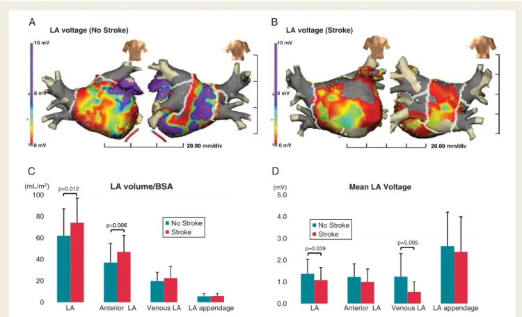
A 73-year-old female with atrioventricular nodal reentrant tachycardia (AVNRT) presented with high-takeoff coronary sinus (CS). Coronary sinus venography revealed that the CS ostium was located at an unusual site where the His-bundle is supposed to be
located (arrows in Figure 1A and B), and the successful ablation site for the slow pathway was located below the CS ostium
(Figures 1A – C). This patient was treated by slow-pathway ablation using only the electrophysiological approach. Therefore, slow pathway ablation for AVNRT should not be guided by only anatomical approaches in cases with an anomaly of the CS.
Conflict of interest: none declared.
Published on behalf of the European Society of Cardiology. All rights reserved.&The Author 2011. For permissions please email: [email protected].
Figure 1 Radiographs demonstrating high-takeoff CS, and intracardiac electrogram.
at YONSEI UNIVERSITY MEDICAL LIBRARY on November 10, 2013
http://europace.oxfordjournals.org/