© 2016 The Korean Ophthalmological Society
This is an Open Access article distributed under the terms of the Creative Commons Attribution Non-Commercial License (http://creativecommons.org/licenses /by-nc/3.0/) which permits unrestricted non-commercial use, distribution, and reproduction in any medium, provided the original work is properly cited.
Original Article
Anterior segment morphometry and intraocular pressure (IOP) can change after intraocular surgery. There are vari-
ous studies reporting these changes using different devices after phacoemulsification and intraocular lens (IOL) im- plantation [1-4]. Examining the anterior segment parame- ters is an important step for planning and evaluating the success of cataract surgery. Many clinical studies have re- ported that cataract extraction causes deepening of the an- terior chamber, widening of the anterior chamber angle
Evaluation of Anterior Segment Parameter Changes Using the Sirius after Uneventful Phacoemulsification
Ali Şimşek1, Burak Bilgin2, Musa Çapkın1, Şemsettin Bilak1, Mete Güler1, Ali Hakim Reyhan1
1Department of Ophthalmology, Adıyaman University School of Medicine, Adiyaman, Turkey
2Department of Ophthalmology, Adıyaman Gozde Hospital, Adiyaman, Turkey
Purpose: To investigate changes in anterior chamber depth (ACD), corneal volume (CV), anterior chamber angle (ACA), anterior chamber volume (ACV), central corneal thickness (CCT), horizontal visible iris diameter (HVID), pupil diameter (PD), and intraocular pressure (IOP) after uneventful phacoemulsification cataract surgery with intraocular lens implantation.
Methods: A total of 132 eyes of 132 patients (87 men and 45 women) that underwent uneventful phacoemulsi- fication cataract surgery and intraocular lens implantation were prospectively studied. The mean age of the patients was 63.68 ± 12.51 years. All patients were evaluated preoperatively and at 1 month postoperatively with the Sirius 3D Rotating Scheimpflug camera topography system. The ACD, CV, ACA, ACV, CCT, HVID, and PD measurements were recorded. IOP was measured using the Goldmann applanation tonometer, which was corrected for CCT of the Sirius device using Ehlers’ formula.
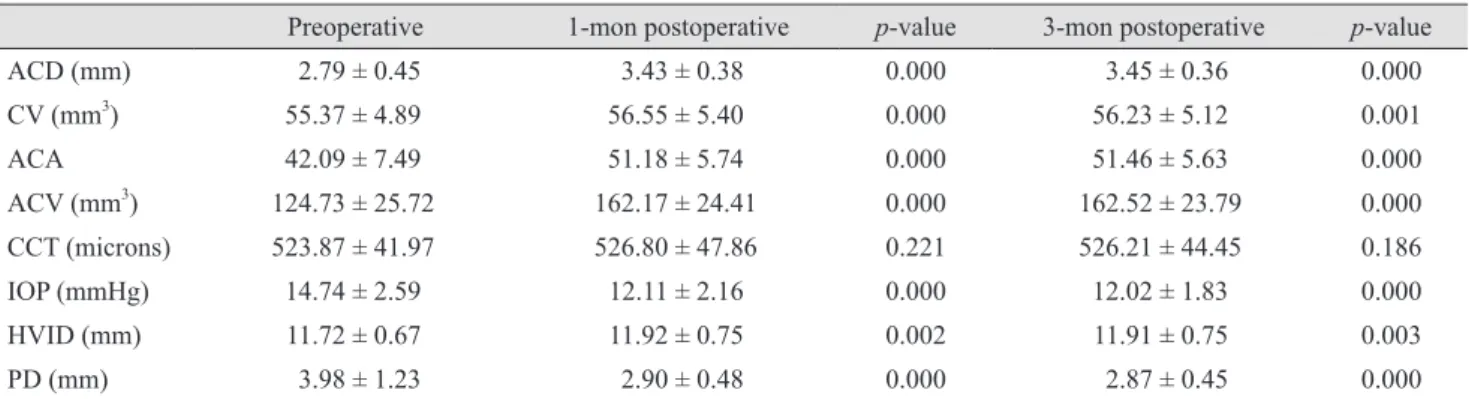
Results: The preoperative mean ACD, ACV, ACA, CCT, CV, PD, HVID, and IOP were 2.79 ± 0.45 mm, 124.73 ± 25.72 mm3, 42.09 ± 7.490, 523.87 ± 41.97 microns, 55.37 ± 4.89 mm3, 3.98 ± 1.23 mm, 11.72 ± 0.67 mm, and 14.74 ± 2.59 mmHg, respectively. Three months postoperatively, the mean ACD, ACV, ACA, CCT, CV, PD, HVID, and IOP were 3.45 ± 0.6 mm, 162.52 ± 23.79 mm3, 51.46 ± 5.630, 526.21 ± 44.45 microns, 56.23 ± 5.12 mm3, 2.87 ± 0.45 mm, 11.91 ± 0.75 mm, and 12.02 ± 1.83 mmHg, respectively. There was a statistically signif- icant increase in mean postoperative ACD, ACV, ACA, CV, and HVID compared with the corresponding pre- operative values (p < 0.05). CCT remained stable after surgery. Postoperative PD and IOP were significantly decreased compared to corresponding preoperative values (p < 0.05).
Conclusions: Preoperative measurements by the Sirius 3D Rotating Scheimpflug camera topography system might help surgeons to predict postoperative changes resulting from phacoemulsification and intraocular lens implantation. This is a noncontact, noninvasive, and comfortable system for patients that is highly reliable and repeatable for anterior segment measurements.
Key Words: Anterior eye segment, Corneal topography, Phacoemulsification
Received: June 15, 2015 Accepted: October 12, 2015
Corresponding Author: Burak Bilgin, MD. Department of Ophthalmolo- gy, Adıyaman Gozde Hospital, Eskisaray mah. 02040, Adiyaman, Turkey.
Tel: 90-533-5601355, Fax: 90-553-4448202, E-mail: [email protected]
(ACA), and a significant decrease in IOP [5-11]. Traditional methods examining the anterior segment are not objective and do not provide quantitative information. Quantifica- tion of these changes has been assessed with different de- vices and methods. These new devices and methods are noncontact, noninvasive, and comfortable for patients in addition to providing highly reliable and repeatable anteri- or segment measurements; options include the Pentacam (Oculus Inc., Wetzlar, Germany) [1-4], anterior segment optical coherence tomography [12,13], and the Sirius sys- tem (Costruzione Strumenti Oftalmici, Florence, Italy) [3- 15].
In this prospective study, we investigated changes in an- terior chamber depth (ACD), corneal volume (CV), ACA, anterior chamber volume (ACV), central corneal thickness (CCT), horizontal visible iris diameter (HVID), pupil di- ameter (PD), and IOP after uneventful phacoemulsification cataract surgery and IOL implantation.
Materials and Methods
A total of 132 eyes of 132 patients (86 men and 46 wom- en) that underwent uneventful phacoemulsification cata- ract surgery and IOL implantation between December 2013 and January 2014 were prospectively studied. The mean age of the patients was 63.68 ± 12.51 years. Exclusion criteria included history of trauma, ocular surgery, corneal pathology, pseudoexfoliation, uveitis, glaucoma, angle-clo- sure, posterior segment pathology, diabetes, or use of topi- cal or systemic medications that might affect anterior seg- ment and IOP measurements. This study was approved by the Adıyaman University ethics committee, and the study protocol adhered to the tenets of the Declaration of Helsin- ki. Informed consent was obtained from each patient prior to any procedures.
All patients were examined preoperatively and 1 and 3 months postoperatively with the Sirius 3D Rotating Scheimpflug camera topography ystem, which was used to measure ACD, CV, ACA, ACV, CCT, HVID, and PD. IOP was measured using the Goldmann applanation tonometer and corrected for CCT of the Sirius device using Ehlers’
formula. The patient was seated with the head positioned with the help of a chinrest and forehead strap during Sirius measurements. All measurements were obtained under standard dim light conditions without dilatation by the
same trained examiner. To minimize diurnal corneal hy- dration variations, all measurements were obtained be- tween 2 p.m. and 5 p.m.
The Sirius obtains topographic and anterior segment measurements using Scheimpflug photography. Its work- ing principle is based on the combination of two rotating Scheimpflug cameras and a Placido disc with 22 rings.
All surgeries were performed by the same experienced surgeon (AŞ) under topical anesthesia with proparacaine drops (Alcaine; Alcon, Istanbul, Turkey). A 2.8-mm clear corneal incision was made in the superior quadrant. A con- tinuous curvilinear capsulorhexis approximately 5.5 mm in diameter was completed after viscoelastic injection (Viscoat; Alcon Surgical, Fort Worth, TX, USA). Cortical cleaving hydrodissection was performed using balanced salt solution (Alcon Laboratories, Hemel Hempstead, UK).
The nucleus was emulsified using torsional phaco technol- ogy (Infiniti, Alcon Laboratories) with the stop and chop technique. After cleaning the residual cortex with irriga- tion and aspiration, a foldable, acrylic, posterior chamber IOL (AcrySof, SA60AT; Alcon) was implanted into the capsular bag. To achieve wound integrity, stromal hydra- tion was performed. Topical antibiotic and steroid drops were used postoperatively in all eyes. Topical antibiotic drops were applied four times a day for 1 week. Topical steroid drops were applied four times each day for 1 week and then tapered over 3 weeks.
Statistical analysis was performed with SPSS ver. 15.0 (SPSS Inc., Chicago, IL, USA). The paired t-test was used to compare variables between the preoperative and postop- erative periods. Independent samples t-test was used to compare variables with a normal distribution between men and women before and after surgery. The Mann-Whitney U-test was used to compare variables without a normal distribution between men and women before and after sur- gery. Pearson’s correlation test was used to evaluate the re- lationships among variables. A p-value less than 0.05 was considered statistically significant.
Results
The mean age of the patients was 63.68 ± 12.51 years (range, 15 to 87 years). Of 132 patients, 86 (65.2%) were men and 46 (34.8%) were women (Table 1). The mean ACD measurement was 2.79 ± 0.45 mm preoperatively,
3.43 ± 0.38 mm 1 month postoperatively, and 3.45 ± 0.6 mm 3 months postoperatively. The mean ACV measure- ment was 124.73 ± 25.72 mm3 preoperatively, 162.17 ± 24.41 mm3 1 month postoperatively, and 162.52 ± 23.79 mm3 3 months postoperatively. The mean ACA measure- ment was 42.09 ± 7.490 preoperatively, 51.18 ± 5.740 1 month postoperatively, and 51.46 ± 5.630 3 months postoperatively.
Mean CCT measurement was 523.87 ± 41.97 microns pre- operatively, 526.80 ± 47.86 microns 1 month postoperative- ly, and 526.21 ± 44.45 microns 3 months postoperatively.
Mean central 10 mm CV measurement was 55.37 ± 4.89 mm3 preoperatively, 56.55 ± 5.40 mm3 1 month postopera- tively, and 56.23 ± 5.12 mm3 3 months postoperatively.
Mean PD measurement was 3.98 ± 1.23 mm preoperative- ly, 2.90 ± 0.48 mm 1 month postoperatively, and 2.87 ± 0.45 mm 3 months postoperatively. Mean HVID measurement was 11.72 ± 0.67 mm preoperatively, 11.92 ± 0.75 1 month postoperatively, and 11.91 ± 0.75 mm 3 months postopera- tively. Mean IOP measurement was 14.74 ± 2.59 mmHg preoperatively, 12.11 ± 2.16 mmHg 1 month postoperative- ly, and 12.02 ± 1.83 mmHg 3 months postoperatively (Table 2).
There was a statistically significant increase in mean
ACD, ACV, ACA, CV, and HVID at 1 and 3 months post- operatively compared with corresponding preoperative values. Postoperative PD and IOP were statistically signifi- cantly decreased compared with corresponding preopera- tive values. There was not a significant change in CCT at 1 or 3 months postoperatively compared to the preoperative values.
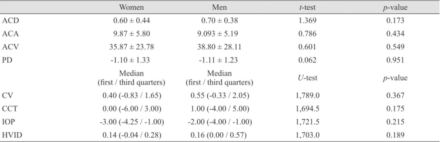
The differences in all characteristics between 3 months postoperatively and preoperatively in women and men were not statistically significant (Table 3).
There was a statistically significant (p < 0.05) negative correlation between the 3-month postoperative IOP values and ACD values (r = -0.204). In addition, there was a nega- tive correlation between the 3-month postoperative IOP values and ACV values (r = -0.223). There was a positive correlation between the 3-month postoperative IOP values and CV values (r = 0.210) and a positive correlation be- tween the 3-month postoperative IOP values and CCT val- ues (r = 0.175). There was no statistically significant cor- relation between the 1- and 3-month postoperative IOP values and other anterior segment parameters (p > 0.05) (Table 4).
Table 1. Demographic characteristics of the patients
Case % Range Mean Standard deviation
Men 86 65.2 29-87 64.34 11.90
Women 46 34.8 15-83 62.46 13.63
Total 132 100 15-87 63.68 12.51
Table 2. Comparison of preoperative and postoperative anterior segment parameters
Preoperative 1-mon postoperative p-value 3-mon postoperative p-value
ACD (mm) 2.79 ± 0.45 3.43 ± 0.38 0.000 3.45 ± 0.36 0.000
CV (mm3) 55.37 ± 4.89 56.55 ± 5.40 0.000 56.23 ± 5.12 0.001
ACA 42.09 ± 7.49 51.18 ± 5.74 0.000 51.46 ± 5.63 0.000
ACV (mm3) 124.73 ± 25.72 162.17 ± 24.41 0.000 162.52 ± 23.79 0.000
CCT (microns) 523.87 ± 41.97 526.80 ± 47.86 0.221 526.21 ± 44.45 0.186
IOP (mmHg) 14.74 ± 2.59 12.11 ± 2.16 0.000 12.02 ± 1.83 0.000
HVID (mm) 11.72 ± 0.67 11.92 ± 0.75 0.002 11.91 ± 0.75 0.003
PD (mm) 3.98 ± 1.23 2.90 ± 0.48 0.000 2.87 ± 0.45 0.000
Values are presented as mean ± standard deviation. Paired t-test was used to compare preoperative and postoperative measurements.
ACD = anterior chamber depth; CV = corneal volume; ACA = anterior chamber angle; ACV = anterior chamber volume; CCT = central corneal thickness; IOP = intraocular pressure; HVID = horizontal visible iris diameter; PD = pupil diameter.
Discussion
Pentacam (Oculus Optikgerate, Wetzlar, Germany) is the first device to use the Scheimpflug camera in ophthalmolo- gy practice. After Pentacam, Galilei (Ziemer Group, Port, Switzerland) introduced a combination of the Scheimpflug camera and Placido-disk topography system. Newer devic- es that use both the Scheimpflug camera and a Placido disk topography system are the Sirius (Costruzione Strumenti Oftalmici) and the TMS-5 (Tomey, Nagoya, Japan).
The Sirius topography device analyzes the anterior seg- ment using a combination of a monochromatic three-di- mensional (3D) rotating Scheimpflug camera and a Placi- do-disk with 22 rings. It obtains 25 radial sections of the cornea and anterior chamber. Scheimpflug cameras pro- vide focused images from the anterior corneal surface to the posterior lens and transform them into a 3D model us- ing their rotating capability [16]. Using a 3D rotating Scheimpflug camera, 21,632 points on the anterior corne- al surface and 16,000 points on the posterior corneal sur- face are measured in less than 1 second. Anterior corneal surface measurement data are derived from both the Scheimpflug camera and Placido-disk images. Data for the posterior corneal surface and internal structures are de- rived only from the Scheimpflug camera.
In this study, we observed significant increase in ACD, ACV, ACA, CV, and HVID and decrease in PD and IOP after uneventful phacoemulsification cataract surgery. Ac-
Table 3. Comparison of 3-month postoperative and preoperative differences in all characteristics between women and men
Women Men t-test p-value
ACD 0.60 ± 0.44 0.70 ± 0.38 1.369 0.173
ACA 9.87 ± 5.80 9.093 ± 5.19 0.786 0.434
ACV 35.87 ± 23.78 38.80 ± 28.11 0.601 0.549
PD -1.10 ± 1.33 -1.11 ± 1.23 0.062 0.951
Median
(first / third quarters) Median
(first / third quarters) U-test p-value
CV 0.40 (-0.83 / 1.65) 0.55 (-0.33 / 2.05) 1,789.0 0.367
CCT 0.00 (-6.00 / 3.00) 1.00 (-4.00 / 5.00) 1,694.5 0.175
IOP -3.00 (-4.25 / -1.00) -2.00 (-4.00 / -1.00) 1,721.5 0.215
HVID 0.14 (-0.04 / 0.28) 0.16 (0.00 / 0.57) 1,703.0 0.189
Independent samples t-test was used for the variables with a normal distribution (ACD, ACA, ACV, and PD) and Mann-Whitney U-test was used for the variables that did not have a normal distribution (CV, CCT, IOP, and HVID).
ACD = anterior chamber depth; ACA = anterior chamber angle; ACV = anterior chamber volume; PD = pupil diameter; CV = corneal volume; CCT = central corneal thickness; IOP = intraocular pressure; HVID = horizontal visible iris diameter.
Table 4. Correlations between anterior segment parameters and intraocular pressure before operation and 1 and 3 months postoperatively (n = 132)
Intraocular pressure Preoperative 1-mon
postoperative 3-mon postoperative
ACD r* -0.015 -0.254 -0.204
p† 0.861 0.003 0.019
CV r 0.213 0.211 0.210
p 0.014 0.015 0.016
ACV r -0.062 -0.188 -0.223
p 0.478 0.031 0.010
ACA r -0.062 -0.175 -0.136
p 0.482 0.045 0.121
CCT r 0.145 0.255 0.175
p 0.096 0.003 0.044
HVID r -0.069 -0.138 -0.156
p 0.430 0.114 0.074
PD r 0.073 0.011 0.025
p 0.408 0.898 0.778
Pearson correlation analysis was used to investigate the relation- ship between variables.
ACD = anterior chamber depth; CV = corneal volume; ACV = anterior chamber volume; ACA = anterior chamber angle; CCT = central corneal thickness; HVID = horizontal visible iris diame- ter; PD = pupil diameter.
*Pearson’s correlation coefficient; †A p-value less than 0.05 was considered statistically significant.
curate measurement of anterior chamber parameters is critical when planning surgery. In modern cataract sur- gery, to achieve desired refractive outcomes, correct IOL calculation with precise biometry is crucial. Accurately determining ACD is essential to achieve precise biometry.
There are several methods to measure ACD [17,18]. Stan- dard ultrasound devices define ACD as the distance be- tween the anterior surface of the cornea and the anterior surface of the lens. Ultrasound devices are the most com- mon method to measure ACD; however, as a contact de- vice, ultrasound has some disadvantages such as corneal abrasions, infections, and off-axis measurement [18]. Sirius imaging uses Scheimpflug technology to measure ACD and is a relatively new and non-contact automatic optical technique.
There have been a number of studies using different de- vices, reporting marked anterior chamber changes follow- ing cataract surgery with phacoemulsification and foldable IOL implantation [6,9,19,20]. In our study, ACD and ACV increased 1.22- and 1.30-fold, respectively, compared to preoperative measurements. The results of our study are comparable with those of previous studies. Using Sirius, Takmaz et al. [3] reported a significant increase in ACD and ACV. The clinical meaning of these increases in ACD and ACV is more considerable for angle closure glaucoma patients who have a shallow anterior chamber.
Evaluation of ACA is critical in glaucoma patients. The conventional method used to evaluate ACA is gonioscopic examination. The major disadvantage of this method is subjectivity. ACA width can be quantitatively measured by Sirius. Using Sirius, Takmaz et al. [3] have reported a sig- nificant increase in ACA after uneventful phacoemulsifi- cation surgery and IOL implantation. Our study also showed a significant increase in ACA.
The effects of cataract surgery and IOL implantation on IOP have been shown in various studies. There are several studies in both normotensive eyes and glaucomatous eyes, including open angle glaucoma and angle closure glauco- ma, reporting significant decreases in IOP after cataract surgery with phacoemulsification and foldable IOL im- plantation [8,19,21]. Altan et al. [9] reported that, in non-glaucomatous eyes with preoperative open ACA, un- eventful phacoemulsification surgery reduced IOP, in- creased ACD, and widened the ACA.
The mechanism of the decrease in IOP resulting from phacoemulsification surgery is controversial. There are
multiple possible mechanisms such as increased uveoscler- al outflow, decreased resistance to conventional aqueous humour outflow, and hyposecretion of aqueous humour [22-25]. In our study, the mean IOP measured at 1 month postoperatively was significantly lower than mean preop- erative value.
There are many recent methods to measure CCT, but ul- trasound pachymetry is the older, gold standard method.
Various studies comparing Pentacam measurements and ultrasound pachymetry have reported that Pentacam mea- surements are similar to but thinner than ultrasound pa- chymetry [26,27]. In our study, the mean CCT value was 523 μm preoperatively. In a study by Doganay et al. [1] us- ing Pentacam, the mean CCT was found to be 534 μm pre- operatively. Using Sirius, Takmaz et al. [3] found a mean CCT of 545 μm preoperatively.
CCT is affected by corneal water content, which is maintained by endothelium pump and barrier functions.
Corneal endothelial cell density and morphology can be assessed using specular microscopy in clinical practice, but the function of the endothelium pump, which is crucial for CCT and CV, cannot be assessed. Also, specular mi- croscopic assessment can be performed in a limited corne- al area. Therefore, it is difficult to statistically evaluate the entire cornea. The Sirius offers CV measurement in a cen- tral 10-mm area of the cornea. The numerical value ob- tained can be used for statistical assessment.
To our knowledge, no previous study has compared CV changes before and after phacoemulsification surgery us- ing The Sirius. Suzuki et al. [28] used Pentacam to evalu- ate CV changes in the central 3-mm and 10-mm areas after phacoemulsification surgery. They reported that the 3-mm area returned to preoperative values at 3 months after sur- gery, whereas the central 10-mm area remained signifi- cantly higher than it had been preoperatively. In contrast to a previous study, Doganay et al. [1] reported no signifi- cant alterations in the 3-, 5- and 7-mm CV values after 1, 3, or 6 months of uneventful phacoemulsification using Pentacam. In our study, we found a mean central 10-mm CV of 55.37 mm3 preoperatively, 56.55 mm3 1 month post- operatively, and 56.23 mm3 3 months postoperatively.
There was a significant increase in CV after uneventful phacoemulsification surgery.
Pupil size is important for visual acuity and depends on the amount of light that enters the eye. This factor is par- ticularly important in patients who have undergone
phacoemulsification cataract surgery and multifocal IOL implantation. In their study using Pentacam, Doganay et al. [1] reported a mean preoperative pupil size of 2.6 mm and no significant change after surgery. In their study with Sirius, Takmaz et al. [3] reported a mean preoperative pu- pil size of 3.4 mm; in accordance with a previous study, they did not find a significant difference between preoper- ative and postoperative values. In our study, mean PD was 3.98 mm preoperatively, 2.90 mm 1 month postoperatively, and 2.87 mm 3 months postoperatively. The mean postop- erative PD was significantly lower than preoperative val- ues.
In conclusion, after uneventful phacoemulsification cat- aract surgery and IOL implantation, significant increase in ACD, ACV, ACA, CV, and HVID and significant decrease in PD and IOP were recorded compared to preoperative measurements. There was not a significant change in CCT between preoperative and postoperative values. The Sirius allows for quick, easy, and reliable demonstration of ante- rior segment changes before and after phacoemulsification surgery and IOL implantation.
Conflict of Interest
No potential conflict of interest relevant to this article was reported.
References
1. Doganay S, Bozgul Firat P, Emre S, Yologlu S. Evaluation of anterior segment parameter changes using the Pentacam after uneventful phacoemulsification. Acta Ophthalmol 2010;88:601-6.
2. Cho YK, Chang HS, La TY, et al. Anterior segment param- eters using Pentacam and prediction of corneal endothelial cell loss after cataract surgery. Korean J Ophthalmol 2010;
24:284-90.
3. Takmaz T, Kosekahya P, Kurkcuoglu PZ. Anterior segment morphometry and intraocular pressure change after un- eventful phacoemulsification. Turk J Med Sci 2013;43:289- 93.
4. Dooley I, Charalampidou S, Malik A, et al. Changes in in- traocular pressure and anterior segment morphometry after uneventful phacoemulsification cataract surgery. Eye
(Lond) 2010;24:519-26.
5. Issa SA, Pacheco J, Mahmood U, et al. A novel index for predicting intraocular pressure reduction following cata- ract surgery. Br J Ophthalmol 2005;89:543-6.
6. Kurimoto Y, Park M, Sakaue H, Kondo T. Changes in the anterior chamber configuration after small-incision cata- ract surgery with posterior chamber intraocular lens im- plantation. Am J Ophthalmol 1997;124:775-80.
7. Arai M, Ohzuno I, Zako M. Anterior chamber depth after posterior chamber intraocular lens implantation. Acta Oph- thalmol (Copenh) 1994;72:694-7.
8. Cekic O, Batman C, Totan Y, et al. Changes in anterior chamber depth and intraocular pressure after phacoemulsi- fication and posterior chamber intraocular lens implanta- tion. Ophthalmic Surg Lasers 1998;29:639-42.
9. Altan C, Bayraktar S, Altan T, et al. Anterior chamber depth, iridocorneal angle width, and intraocular pressure changes after uneventful phacoemulsification in eyes with- out glaucoma and with open iridocorneal angles. J Cata- ract Refract Surg 2004;30:832-8.
10. Memarzadeh F, Tang M, Li Y, et al. Optical coherence to- mography assessment of angle anatomy changes after cata- ract surgery. Am J Ophthalmol 2007;144:464-5.
11. Kashiwagi K, Kashiwagi F, Tsukahara S. Effects of small-in- cision phacoemulsification and intraocular lens implanta- tion on anterior chamber depth and intraocular pressure. J Glaucoma 2006;15:103-9.
12. Leung CK, Palmiero PM, Weinreb RN, et al. Comparisons of anterior segment biometry between Chinese and Cauca- sians using anterior segment optical coherence tomography.
Br J Ophthalmol 2010;94:1184-9.
13. Kim M, Park KH, Kim TW, Kim DM. Changes in anterior chamber configuration after cataract surgery as measured by anterior segment optical coherence tomography. Korean J Ophthalmol 2011;25:77-83.
14. Jorge J, Rosado J, Diaz-Rey J, Gonzalez-Meijome J. Cen- tral corneal thickness and anterior chamber depth mea- surement by Sirius(R) Scheimpflug tomography and ultra- sound. Clin Ophthalmol 2013;7:417-22.
15. Savini G, Barboni P, Carbonelli M, Hoffer KJ. Repeatabili- ty of automatic measurements by a new Scheimpflug cam- era combined with Placido topography. J Cataract Refract Surg 2011;37:1809-16.
16. Wegener A, Laser-Junga H. Photography of the anterior eye segment according to Scheimpflug’s principle: options and limitations. A review. Clin Experiment Ophthalmol
2009;37:144-54.
17. Nemeth G, Vajas A, Kolozsvari B, et al. Anterior chamber depth measurements in phakic and pseudophakic eyes:
Pentacam versus ultrasound device. J Cataract Refract Surg 2006;32:1331-5.
18. Koranyi G, Lydahl E, Norrby S, Taube M. Anterior cham- ber depth measurement: a-scan versus optical methods. J Cataract Refract Surg 2002;28:243-7.
19. Hayashi K, Hayashi H, Nakao F, Hayashi F. Changes in anterior chamber angle width and depth after intraocular lens implantation in eyes with glaucoma. Ophthalmology 2000;107:698-703.
20. Pereira FA, Cronemberger S. Ultrasound biomicroscopic study of anterior segment changes after phacoemulsifica- tion and foldable intraocular lens implantation. Ophthal- mology 2003;110:1799-806.
21. Nonaka A, Kondo T, Kikuchi M, et al. Angle widening and alteration of ciliary process configuration after cataract surgery for primary angle closure. Ophthalmology 2006;
113:437-41.
22. Miyake K, Asakura M, Kobayashi H. Effect of intraocular lens fixation on the blood-aqueous barrier. Am J Ophthal-
mol 1984;98:451-5.
23. Alpar JJ. Glaucoma after intraocular lens implantation:
survey and recommendations. Glaucoma 1985:7:241-5.
24. Meyer MA, Savitt ML, Kopitas E. The effect of phacoemul- sification on aqueous outflow facility. Ophthalmology 1997;104:1221-7.
25. Kerstetter JR, Brubaker RF, Wilson SE, Kullerstrand LJ.
Prostaglandin F2 alpha-1-isopropylester lowers intraocular pressure without decreasing aqueous humor flow. Am J Ophthalmol 1988;105:30-4.
26. O’Donnell C, Maldonado-Codina C. Agreement and re- peatability of central thickness measurement in normal corneas using ultrasound pachymetry and the OCULUS Pentacam. Cornea 2005;24:920-4.
27. Amano S, Honda N, Amano Y, et al. Comparison of central corneal thickness measurements by rotating Scheimpflug camera, ultrasonic pachymetry, and scanning-slit corneal topography. Ophthalmology 2006;113:937-41.
28. Suzuki H, Takahashi H, Hori J, et al. Phacoemulsification associated corneal damage evaluated by corneal volume.
Am J Ophthalmol 2006;142:525-8.