© 2015 The Korean Ophthalmological Society
This is an Open Access article distributed under the terms of the Creative Commons Attribution Non-Commercial License (http://creativecommons.org/licenses /by-nc/3.0/) which permits unrestricted non-commercial use, distribution, and reproduction in any medium, provided the original work is properly cited.
Original Article
Structural Analysis of Different Incision Sizes and Stromal Hydration in Cataract Surgery Using Anterior Segment Optical Coherence
Tomography
Jong-Wook Bang, Jong-Hyun Lee, Jin-Hyoung Kim, Do-Hyung Lee
Department of Ophthalmology, Ilsan Paik Hospital, Inje University College of Medicine, Goyang, Korea
Purpose: To analyze healing changes of corneal wounds of different corneal incision sizes with or without stro- mal hydration in cataract surgery using anterior segment optical coherence tomography.
Methods: Cataract surgeries were performed by a single surgeon and 2.2- and 2.8-mm corneal incisions were made using a diamond blade (ME-759; Meyco, Biel-Bienne, Swiss). Patients were divided into four groups according to incision size (2.2 and 2.8 mm), and with/without stromal hydration. Fifteen eyes were assigned to each group and incision wounds were measured using anterior segment optical coherence tomography at 2 hours, 1 day, 1 week, 1 month, and 3 months postoperatively. Corneal thickness, incision length and incision angle were measured and existence of epithelial, endothelial gaping and Descemet’s membrane detachment was evaluated.
Results: Incision thickness was greater in the group with stromal hydration than in the group without on opera- tion day (p < 0.05). Stromal hydration exerted greater influence in the 2.2-mm incision group than in the 2.8- mm incision group. Corneal thickness decreased more rapidly in the stromal hydration group than in the group with no hydration (p = 0.022). Endothelial gaping was greater in the 2.2-mm incision group than in the 2.8-mm incision group 1 day, 1 month, and 3 months after surgery (p = 0.035, p = 0.009, and p = 0.008, respectively).
No other statistical significance was observed between the two groups (2.2 and 2.8 mm) during follow-up re- garding corneal thickness, epithelial gaping and Descemet’s membrane detachment.
Conclusions: Corneal wounds with a smaller incision could be more vulnerable to external stimuli such as stro- mal hydration and are less stable than those with a larger incision.
Key Words: Corneal pachymetry, Corneal stroma, Wounds and injuries
Currently, microincision coaxial cataract surgery (MICS) is a state-of-the-art cataract removal treatment with re- duced rates of wound leaks, astigmatism and postoperative
infection. However, when compared with conventional co- axial cataract surgery, its overall advantage remains ques- tionable. Phacoemulsification of microincisions may re- quire longer and more intense ultrasound exposure, create difficulty in handling, and may inflict greater loss of endo- thelial cells [1-4]. Furthermore, several reports indicate that surgically-induced astigmatism does not differ whether MICS or a conventional approach is used [1-5].
Clear corneal incision increases the risk of endophthal-
Received: May 15, 2014 Accepted: July 7, 2014
Corresponding Author: Do-Hyung Lee, MD, PhD. Department of Oph- thalmology, Ilsan Paik Hospital, Inje University College of Medicine,
#170 Juhwa-ro, Ilsanseo-gu, Goyang 411-706, Korea. Tel: 82-31-910-7240,
Fax: 82-31-911-7241, E-mail: [email protected]
mitis, which is countered by corneal stromal hydration.
The latter is applied to enhance the wound sealing and to prevent inflow of ocular surface fluid. Nevertheless, the tendency for endothelial misalignment is increased with stromal hydration in the early postoperative period [6]
thus, incisional size and the use of stromal hydration are key variables affecting configuration and healing of clear corneal incisions.
Anterior segment optical coherence tomography (AS- OCT) is a sophisticated imaging technique that allows visu- alization of the incisions in real time and qualitative analysis of structural changes in the cornea [6]. In the present study, we determined differences in wound structure and changes in dynamic healing in variably-sized corneal incisions (with and without stromal hydration) using AS-OCT.
Materials and Methods
In this retrospective case study, the clinical records of patients who underwent cataract surgery at our hospital from August, 2012 to December, 2013 were reviewed.
A total of 60 eyes undergoing standard cataract surgery were evaluated. Patients were divided into four groups ac- cording to incision size (2.2 or 2.8 mm) and with/without stromal hydration; 15 eyes were assigned to each group.
The procedures were performed under topical anesthesia by a single surgeon at Inje University Ilsan Paik Hospital.
Inclusion criteria were no ocular surgery history, absence of biomicroscopic signs of pseudoexfoliation, normal fun- dus examination and endothelial cell count of at least 2,000 cells/mm
2. Exclusion criteria were the presence of preex- isting corneal disease or glaucoma.
Phacoemulsification and intraocular lens implantation were performed as follows: A side-port incision was creat- ed and an ophthalmic viscosurgical device was injected into the anterior chamber and then the main clear corneal incision was made using a 2.2- or a 2.8-mm diamond blade in each group. All clear corneal incisions were made tem- porally. Phacoemulsification was performed using OZil (Alcon, Fort Worth, TX, USA) torsional technology with an ultrasleeve on an Infiniti phacoemulsification platform (Alcon). An intraocular lens (NY-60; HOYA, Tokyo, Ja- pan) was implanted using the cartilage and injector. Cor- neal stromal hydration was applied in 15 randomly selected eyes in the 2.2- and 2.8-mm incision groups by placing the
tip of a 25-gauge cannula in the side walls of the incision and gently irrigating balanced salt solution into the stroma.
The remaining 15 eyes in each group were not hydrated.
Incision wounds were measured using AS-OCT (3D-OCT 2000; Topcon, Tokyo, Japan) at 2 hours, 1 day, 1 week, 1 month, and 3 months postoperatively. Anterior module of the machine was used for corneal imaging.
During the processes, the patients were asked to look straight ahead to the opposite side of the corneal incisions.
Complete transverse scans were taken and the high-defini- tion images were recorded. Corneal thickness at the inci- sion was measured using the anterior OCT module caliper.
Incision length and incision angle were also measured and presence of epithelial gaping, endothelial gaping and De- scemet’s membrane detachment was evaluated.
Epithelial gaping was defined as crack of the limbal edge of the superior corneal wound surface, endothelial gaping as crack of the limbal edge of posterior corneal wound sur- face and Descemet’s membrane detachment as separation of Descemet’s membrane from the stroma (Fig. 1).
Statistical analyses of endothelial gaping, corneal thick- ness and incision length were performed using SPSS ver.
12 (SPSS Inc., Chicago, IL, USA). The independent sample t-tests were used to compare parameters between the two groups (2.2 vs. 2.8 mm, hydration vs. no hydration). Paired t-tests were used to compare the series of postoperative values. A p-value <0.05 was considered to indicate a sig- nificant difference. The 95% confidence intervals of the incidence of Descemet’s membrane detachment were cal- culated. The chi-square test was used to compare the inci- dence values.
Results
In this study 60 eyes of 48 patients were enrolled and each group was composed of 15 eyes that were randomly assigned. The average patient age was 64.26 ± 12.68 years (range, 50 to 77 years). No incidence of postoperative com- plications such as wound leaking or endophthalmitis was observed in the no stromal hydration group.
2.2-mm incision group
In eyes with stromal hydration, corneal thickness at the
clear corneal incision was 1,098.50 ± 67.01, 827.00 ± 85.49,
and 722.00 ± 23.20 µm on operation day, 1 week, and 1 month after surgery, respectively. In eyes without stromal hydration, corneal thickness at the clear corneal incision was 1,003.17 ± 22.44, 929.14 ± 67.10, and 797.71 ± 64.01 µm on operation day, 1 week, and 1 month after surgery, re- spectively.
On operation day, corneal thickness in the 2.2 mm hy- dration group was statistically significantly thicker than in the 2.2 mm no-hydration group (p = 0.002). However, at 1 week and 1 month postoperatively, corneal thickness in the 2.2 mm no-hydration group was statistically significantly thicker than in the 2.2 mm hydration group (p = 0.035, p = 0.022, respectively) (Table 1 and Fig. 1).
Endothelial gaping was statistically significantly larger in the 2.2 mm stromal hydration group than in the 2.2 mm no-hydration group only on operation day (p = 0.041).
There was no difference between the two groups postoper- atively.
2.8-mm incision group
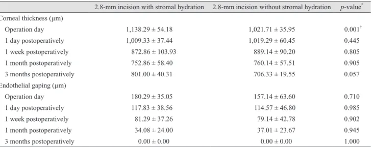
In eyes with stromal hydration, corneal thickness at the clear corneal incision was 1,138.29 ± 54.18, 872.86 ± 103.93, and 752.86 ± 58.40 µm on operation day, 1 week, and 1 month after surgery, respectively. In eyes without stromal hydration, corneal thickness at the clear corneal incision was 1,021.71 ± 35.95, 889.14 ± 90.20, and 760.14 ± 57.51 µm on operation day, 1 week, and 1 month after surgery, re- spectively. Corneal thickness was statistically significantly larger in the 2.8 mm stromal hydration group than in the 2.8 mm no-hydration group only on operation day (p = 0.001). There was no difference between the two groups postoperatively (Table 2).
Incision size
Corneal thickness was not significantly different be- tween the two groups (2.2 and 2.8 mm) on operation day, 1 day, 1 week, 1 month, and 3 months after surgery.
In eyes with the 2.2-mm incision, endothelial gaping was 169.58 ± 72.72, 80.46 ± 59.73, and 61.00 ± 47.82 at 1 day, 1 month, and 3 months after surgery, respectively. In eyes with the 2.8-mm incision, endothelial gaping was 116.08 ± 41.06, 33.93 ± 23.85, and 0.00 ± 0.00 µm at 1 day, 1 month, and 3 months after surgery, respectively. At 1 day, 1 month, and 3 months postoperatively, endothelial gaping
was statistically significantly larger in the 2.2-mm incision group (p = 0.035, p = 0.009, and p = 0.008, respectively).
There was no significant difference between the two groups on operation day and 1 week after surgery (p = 0.131, p = 0.116) (Table 3).
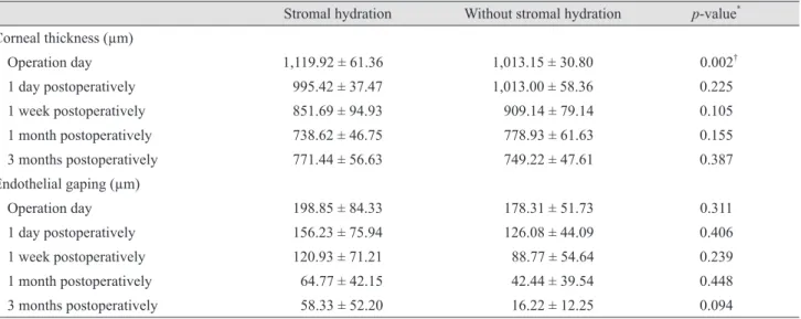
Stromal hydration (integrating 2.2- and 2.8-mm inci- sion groups)
In eyes with stromal hydration, corneal thickness at the clear corneal incision was 1,119.92 ± 61.36, 851.69 ± 94.93, and 738.62 ± 46.75 µm on operation day, 1 week, and 1 month after surgery, respectively. In eyes without stromal
A
B
C
Fig. 1. (A) Thickness of corneal incision, (B) endothelial gape, and (C) Descemet’s membrane detachment.
942
285
A
hydration, corneal thickness at the clear corneal incision was 1013.15 ± 30.80, 909.14 ± 19.14, and 778.93 ± 61.63 µm on operation day, 1 week, and 1 month after surgery, re- spectively. Only on operation day, corneal thickness was statistically significantly larger in the stromal hydration group than the no-hydration group (p = 0.002). Significant
difference in corneal thickness with or without hydration was not observed postoperatively.
Other parameters showed no significant difference be- tween the two groups (with or without stromal hydration) on operation day, 1 day, 1 week, 1 month, and 3 months af- ter surgery (Table 4).
Table 1. Corneal thickness and endothelial gaping in the 2.2-mm incision group with and without stromal hydration
2.2-mm incision with stromal hydration 2.2-mm incision without stromal hydration p-value
*Corneal thickness (µm)
Operation day 1,098.50 ± 67.01 1,003.17 ± 22.44 0.002
†1 day postoperatively 981.50 ± 34.96 1,005.67 ± 60.58 0.240
1 week postoperatively 827.00 ± 85.49 929.14 ± 67.10 0.035
†1 month postoperatively 722.00 ± 23.20 797.71 ± 64.01 0.022
†3 months postoperatively 747.80 ± 60.19 770.67 ± 42.64 0.429
Endothelial gaping (µm)
Operation day 247.50 ± 83.30 176.00 ± 70.27 0.041
‡1 day postoperatively 204.83 ± 77.12 134.33 ± 51.99 0.093
1 week postoperatively 162.71 ± 71.28 97.50 ± 73.00 0.073
1 month postoperatively 100.00 ± 65.88 57.67 ± 46.87 0.295
3 months postoperatively 87.50 ± 36.02 29.20 ± 42.18 0.082
Values are presented as mean ± standard deviation.
*
Paired t-test;
†In the 2.2-mm incision group, corneal thickness showed statistical significance with or without stromal hydration on opera- tion day, 1 week, and 1 month postoperatively. On operation day, corneal thickness was thicker in the stromal hydration group, but 1 week and 1 month postoperatively, corneal thickness was thinner in the stromal hydration group;
‡In the 2.2-mm incision group, endothelial gaping showed statistical significance with or without stromal hydration on operation day.
Table 2. Corneal thickness and endothelial gaping in the 2.8-mm incision group with and without stromal hydration
2.8-mm incision with stromal hydration 2.8-mm incision without stromal hydration p-value
*Corneal thickness (µm)
Operation day 1,138.29 ± 54.18 1,021.71 ± 35.95 0.001
†1 day postoperatively 1,009.33 ± 37.44 1,019.29 ± 60.45 0.445
1 week postoperatively 872.86 ± 103.93 889.14 ± 90.20 0.805
1 month postoperatively 752.86 ± 58.40 760.14 ± 57.51 0.905
3 months postoperatively 801.00 ± 40.31 706.33 ± 19.55 0.057
Endothelial gaping (µm)
Operation day 180.29 ± 35.05 157.14 ± 63.60 0.710
1 day postoperatively 117.83 ± 38.56 114.57 ± 46.80 0.985
1 week postoperatively 81.29 ± 37.26 79.14 ± 42.78 0.902
1 month postoperatively 34.08 ± 24.00 37.01 ± 23.67 0.945
3 months postoperatively 0.00 ± 0.00 0.00 ± 0.00 1.000
Values are presented as mean ± standard deviation.
*
Paired t-test;
†In the 2.8-mm incision group, corneal thickness showed statistical significance with or without stromal hydration on opera-
tion day.
No stromal hydration
Corneal thickness was only significantly different be- tween the 2 groups (2.2 and 2.8 mm) at 3 months after sur- gery (770.67 vs. 706.33 µm, p = 0.048).
In eyes with the 2.2-mm incision, endothelial gaping was 247.50 ± 83.30, 204.83 ± 77.12, and 100.00 ± 65.88 µm on operation day, 1 day, and 1 month after surgery, respec-
tively. In eyes with the 2.8-mm incision, endothelial gaping was 157.14 ± 63.60, 114.57 ± 46.80, and 37.01 ± 23.6 7 µm on operation day, 1 day, and 1 month after surgery, respec- tively. In the 2.8-mm no-hydration group, endothelial gap- ing was statistically smaller on operation day and 1 day and 1 month postoperatively (p = 0.035, p = 0.035, and p = 0.035, respectively) (Table 5).
Table 3. Corneal thickness and endothelial gaping in the different incision group (2.2- and 2.8-mm)
2.2-mm incision 2.8-mm incision p-value
*Corneal thickness (µm)
Operation day 1,050.83 ± 68.91 1,080.00 ± 74.90 0.297
1 day postoperatively 993.58 ± 48.81 1,014.69 ± 49.38 0.270
1 week postoperatively 882.00 ± 90.03 881.00 ± 93.87 0.943
1 month postoperatively 762.77 ± 61.78 756.50 ± 55.81 0.981
3 months postoperatively 760.27 ± 50.01 760.43 ± 59.16 0.930
Endothelial gaping (µm)
Operation day 211.75 ± 82.42 168.71 ± 50.77 0.131
1 day postoperatively 169.58 ± 72.72 116.08 ± 41.06 0.035
†1 week postoperatively 132.62 ± 76.85 80.21 ± 38.56 0.116
1 month postoperatively 80.46 ± 59.73 33.93 ± 23.85 0.009
†3 months postoperatively 61.00 ± 47.82 0.00 ± 0.00 0.008
†Values are presented as mean ± standard deviation.
*
Paired t-test;
†Endothelial gaping was significantly thicker in the 2.2-mm incision group 1 day, 1 month, and 3 months postoperatively.
Table 4. Corneal thickness and endothelial gaping in groups with and without stromal hydration
Stromal hydration Without stromal hydration p-value
*Corneal thickness (µm)
Operation day 1,119.92 ± 61.36 1,013.15 ± 30.80 0.002
†1 day postoperatively 995.42 ± 37.47 1,013.00 ± 58.36 0.225
1 week postoperatively 851.69 ± 94.93 909.14 ± 79.14 0.105
1 month postoperatively 738.62 ± 46.75 778.93 ± 61.63 0.155
3 months postoperatively 771.44 ± 56.63 749.22 ± 47.61 0.387
Endothelial gaping (µm)
Operation day 198.85 ± 84.33 178.31 ± 51.73 0.311
1 day postoperatively 156.23 ± 75.94 126.08 ± 44.09 0.406
1 week postoperatively 120.93 ± 71.21 88.77 ± 54.64 0.239
1 month postoperatively 64.77 ± 42.15 42.44 ± 39.54 0.448
3 months postoperatively 58.33 ± 52.20 16.22 ± 12.25 0.094
Values are presented as mean ± standard deviation.
*
Paired t-test;
†Corneal thickness was significantly thicker in the stromal hydration group on operation day.
Descemet’s membrane detachment
Descemet’s membrane detachment was observed in 66.7% (10 / 15 eyes) and 40.0% (6 / 15 eyes) on operation day and 1 day postoperatively, respectively and gradually decreased in the 2.2-mm incision stromal hydration group.
In the 2.2-mm no-hydration group, Descemet’s membrane detachment was observed in 40% (6 / 15 eyes), 26.7% (4 / 15 eyes), 13.3% (2 / 15 eyes), 0% (0 / 15 eyes), and 0% (0 / 15 eyes) on operation day, 1 day, 1 week, 1 month, and 3 months postoperatively, respectively. In the 2.8-mm inci- sion with stromal hydration group, Descemet’s membrane detachment was observed in 40% (6 / 15 eyes), 26.7% (4 / 15 eyes), 26.7% (4 / 15 eyes), 13.3% (2 / 15 eyes), and 0% (0 / 15 eyes) on operation day, 1 day, 1 week, 1 month, and 3 months postoperatively, respectively. In the 2.8-mm no-hy- dration group, Descemet’s membrane detachment was ob- served in 26.7% (4 / 15 eyes), 26.7% (4 / 15 eyes), 13.3% (2 / 15 eyes), 0% (0 / 15 eyes), and 0% (0 / 15 eyes) on operation day, 1 day, 1 week, 1 month, and 3 months postoperatively, respectively.
In the stromal hydration group (integrating 2.2- and 2.8- mm incision groups), Descemet’s membrane detachment was observed in 53.3% (16 / 30 eyes), 33.3% (10 / 30 eyes), 20% (6 / 30 eyes), 13.3% (4 / 15 eyes) and 0% (0 / 15 eyes) on operation day, 1 day, 1 week, 1 month, and 3 months postoperatively, respectively. In the no-hydration group
(integrating 2.2- and 2.8-mm incision groups), Descemet’s membrane detachment was observed in 33.3% (10 / 30 eyes), 26.7% (8 / 30 eyes), 13.3% (4 / 30 eyes), 0% (0 / 15 eyes) and 0% (0 / 15 eyes) on operation day, 1 day, 1 week, 1 month, and 3 months postoperatively, respectively.
Descemet’s detachment gradually decreased in all groups but without statistically significant difference be- tween the two groups including all data from both incision sizes and with or without stromal hydration.
Discussion
AS-OCT has been used to evaluate shape and thickness of cataract incisions, investigate related problems such as epithelial gaping, endothelial gaping and Descemet’s mem- brane detachment and assess early postoperative outcomes [3,6-10]. In our study, AS-OCT was utilized to monitor pa- tients for up to 3 months after surgery.
In the immediate postoperative period, the structural in- tegrity of a clear corneal incision is critical as a barrier against entry of contaminants into the anterior chamber via the tear film. For this reason, incisional stromal hydra- tion is widely used [6]. Fine et al. [9] reported that incision- al stromal swelling persists for at least 24 hours after sur- gery, but the duration of stromal swelling after cataract Table 5. Corneal thickness and endothelial gaping in the 2.2- and 2.8-mm incision groups without stromal hydration
2.2-mm incision without stromal hydration 2.8-mm incision without stromal hydration p-value
*Corneal thickness (µm)
Operation day 1,003.17 ± 22.44 1,021.71 ± 35.95 0.366 1 day postoperatively 1,005.67 ± 60.58 1,019.29 ± 60.45 0.628
1 week postoperatively 929.14 ± 67.10 889.14 ± 90.20 0.456
1 month postoperatively 797.71 ± 64.01 760.14 ± 57.51 0.165
3 months postoperatively 770.67 ± 42.64 706.33 ± 19.55 0.048
†Endothelial gaping (µm)
Operation day 247.50 ± 83.30 157.14 ± 63.60 0.035
‡1 day postoperatively 204.83 ± 77.12 114.57 ± 46.80 0.035
‡1 week postoperatively 162.71 ± 71.28 79.14 ± 42.78 0.073
1 month postoperatively 100.00 ± 65.88 37.01 ± 23.67 0.035
‡3 months postoperatively 87.50 ± 36.02 0.00 ± 0.00 0.024
‡Values are presented as mean ± standard deviation.
*