• 검색 결과가 없습니다.

The Incidence and Risk Factors of Lens-iris Diaphragm Retropulsion Syndrome during Phacoemulsification

N/A
N/A
Protected

Academic year: 2022

Share "The Incidence and Risk Factors of Lens-iris Diaphragm Retropulsion Syndrome during Phacoemulsification"

Copied!
7
0
0

로드 중.... (전체 텍스트 보기)

전체 글

(1)

© 2017 The Korean Ophthalmological Society

This is an Open Access article distributed under the terms of the Creative Commons Attribution Non-Commercial License (http://creativecommons.org/licenses /by-nc/3.0/) which permits unrestricted non-commercial use, distribution, and reproduction in any medium, provided the original work is properly cited.

Original Article

The Incidence and Risk Factors of Lens-iris Diaphragm Retropulsion Syndrome during Phacoemulsification

Dong Hui Lim

1,2

, Dong Hoon Shin

1

, Gyule Han

1

, Eui-Sang Chung

1

, Tae-Young Chung

1

1

Department of Ophthalmology, Samsung Medical Center, Sungkyunkwan University School of Medicine, Seoul, Korea

2

Department of Preventive Medicine, Graduate School, The Catholic University of Korea, Seoul, Korea

Purpose: In the present study, the incidence and risk factors of lens-iris diaphragm retropulsion syndrome (LIDRS) were evaluated.

Methods: Patients who underwent cataract surgery using phacoemulsification between June 2014 and Decem- ber 2014 were included in the study. The preoperative ocular biometric and intraoperative surgical parameters were examined. The incidence of LIDRS and various risk factors were analyzed using an independent t-test, Pearson’s chi-square test, and univariable and multivariable logistic regression analyses.

Results: Among 124 eyes of 124 patients, 100 (80.6%) had no LIDRS and 24 (19.4%) had LIDRS. LIDRS oc- curred in 13 of 31 vitrectomized eyes (41.9%) and 11 of 93 non-vitrectomized eyes (11.8%). Based on uni- variable analysis, age (odds ratio [OR], 0.920; p = 0.001), vitrectomized eye (OR, 5.038; p = 0.001), spherical equivalent (OR, 0.778; p < 0.001), axial length (OR, 1.716; p < 0.001), anterior chamber depth (OR, 3.328; p

= 0.037), and 3.0 mm vs. 2.2 mm incision size (OR, 4.964; p = 0.001) were statistically significant risk factors associated with the development of LIDRS. Conditional multivariable logistic regression showed that vitrecto- mized eye (OR, 3.865; 95% confidence interval [CI], 1.201 to 12.436; p = 0.023), long axial length (OR, 1.709;

95% CI, 1.264 to 2.310; p = 0.001), and 3.0 vs. 2.2 mm incision size (OR, 3.571; 95% CI, 1.120 to 11.393; p = 0.031) were significant independent risk factors associated with LIDRS.

Conclusions: LIDRS is a relatively common occurrence and was found to be associated with vitrectomized eye, long axial length, and larger incision size. Evaluating risk factors prior to cataract surgery can help reduce associated morbidity.

Key Words: Iris, Lens-iris diaphragm retropulsion syndrome, Phacoemulsification

Iris stability is an important factor for successful cataract surgery. Poor preoperative or intraoperative mydriasis or

damaged iris during surgery may induce pseudophakic macular edema, which can lead to poor outcomes follow- ing cataract surgery [1]. Several previous reports on intra- operative floppy-iris syndrome have indicated that this con- dition can affect the results of cataract surgery and can be associated with systemic sympathetic alpha-1A antagonists such as tamsulosin [2-4].

In 1992, Zauberman [5] first described a phenomenon

Received: May 16, 2016 Accepted: September 2, 2016

Corresponding Author: Tae-Young Chung, MD, PhD. Department of Ophthalmology, Samsung Medical Center, Sungkyunkwan University School of Medicine, #81 Irwon-ro, Gangnam-gu, Seoul 06351, Korea.

Tel: 82-2-3410-3568, Fax: 82-2-3410-0074, E-mail: taeyoung15.chung@

samsung.com

(2)

that occurs during phacoemulsification and that is charac- terized by excessive anterior chamber deepening, retropul- sion of the iris, and extreme widening of the pupil. In 1994, Wilbrandt and Wilbrandt [6] designated this phenomenon as lens-iris diaphragm retropulsion syndrome (LIDRS) and described its mechanism. Posterior movement of the lens- iris diaphragm causes significant discomfort and pain un- der topical or intracameral anesthesia, and an excessively deep anterior chamber renders phacoemulsification more difficult for the operating surgeon.

Wilbrandt and Wilbrandt [6] and Cionni et al. [7] sug- gested possible etiologies of LIDRS such as myopic eyes and reverse pupillary block. Management recommenda- tions for LIDRS have also been suggested in several stud- ies [7-11]. Ghosh et al. [12] reported the incidence and asso- ciation of LIDRS in patients who underwent vitrectomy.

However, no prior reports have discussed the prevalence or risk factors of LIDRS in the general population. Therefore, we determined the prevalence and attributing factors for LIDRS and evaluated the efficacy of a bottle-lowering pro- cedure for the management of LIDRS.

Materials and Methods

In this retrospective comparative study, the incidence and risk factors of LIDRS were evaluated in patients who underwent phacoemulsification cataract surgery. The pa- tients received information regarding the procedure and provided informed consent. The study was performed at Samsung Medical Center, Seoul, Korea, in accordance with the Declaration of Helsinki and was approved by the iInstitutional review board of Samsung Medical Center.

The medical records of patients having unilateral cata- ract and who visited the outpatient department at Samsung Medical Center between June 2014 and December 2014, were retrospectively reviewed. Patients were excluded if they had a history of iris surgery or iris pathology such as iridocyclitis or iris neovascularization or other ocular pro- cedures other than vitrectomy such as trabeculectomy, stra- bismus surgery, scleral buckling, or intravitreal injection.

Exclusion criteria also included traumatic cataract, zonuly- sis, or any condition that would obstruct the exact evalua- tion of LIDRS, such as general or retrobulbar anesthesia.

All patients underwent routine phacoemulsification cata- ract surgery without complications; all surgeries were per-

formed by the same skilled surgeon (ESC).

Preoperative ocular biometric parameters

Before the surgery, patients underwent full ophthalmo- logical examinations including uncorrected distance visual acuity, corrected distance visual acuity, manifest and cy- cloplegic refractions, slit lamp evaluation, tonometry, go- nioscopy, keratometry, corneal pachymetry and topogra- phy (Orbscan II; Bausch & Lomb, Rochester, NY, USA), noncontact specular microscopy (SP-8000; Konan Medical, Nishinomiya, Japan), and binocular indirect ophthalmos- copy with the pupils dilated. Information regarding preop- erative risk factors was obtained and evaluated, including sex, age, history of diabetes or hypertension, history of vitrectomy, spherical equivalent, and axial length and an- terior chamber depth (defined as corneal epithelium to an- terior crystalline lens surface) measured using IOLMaster (Carl Zeiss Meditec, Jena, Germany).

Surgical procedures and intraoperative surgical param- eters

A standard dilating regimen consisting of three rounds of 2.5% topical phenylephrine and 1% tropicamide (Mydrin-P; Santen Pharmaceuticals, Osaka, Japan) fol- lowed by instillation of proparacaine HCl 0.5% (Alcaine;

Alcon Laboratories, Fort Worth, TX, USA) every 5 min- utes was used. In all cases, cataract surgeries were per- formed using a 2.2- or 3.0-mm-sized clear corneal incision, continuous curvilinear capsulorrhexis, phacoemulsification using the Infiniti system (Alcon Laboratories), cortex re- moval using a bimanual irrigation/aspiration tip, and pos- terior chamber foldable intraocular lens implantation. Vis- coelastics were used to maintain the anterior chamber and mechanically hold the pupil open.

Additional intraoperative factors including incision site (superior vs. temporal), incision size (3.0 mm vs. 2.2 mm), and ophthalmic viscosurgical device (Healon 5; Advanced Medical Optics, Santa Ana, CA, USA vs. Discovisc, Alcon Laboratories) were investigated.

The presence of LIDRS was evaluated three times,

during the phacoemulsification, bimanual irrigation and

aspiration of residual cortex, and bimanual irrigation and

aspiration of ophthalmic viscosurgical devices. LIDRS was

graded on a scale from 0 to 3: grade 0, no LIDRS; grade 1,

(3)

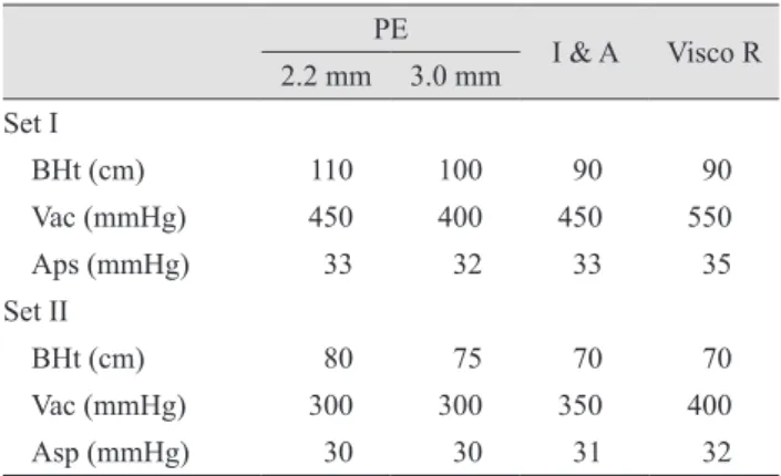
mild pupil dilatation; grade 2, pupil dilatation and patient discomfort; grade 3, abrupt pupil dilatation and patient pain. When LIDRS occurred, the bottle height, vacuum pressure, and aspiration pressure were reduced from set I to set II, and the effect on LIDRS was recorded (Table 1).

Statistical analysis

The data analysis was performed using PASW ver. 18.0 (SPSS Inc., Chicago, IL, USA). The absolute frequency (n) and relative frequency (%) were computed for qualitative variables, and the mean and standard deviations were computed for quantitative variables. The clinical charac- teristics of patients with and without LIDRS were com- pared using Pearson’s chi-square test for categorical vari- ables and the independent t-test for continuous parameters.

The 95% confidence intervals (CIs) for odds ratios (ORs) were calculated. Univariable simple logistic regression analysis was used to examine the associations between LIDRS and the variables. Factors with a p-value <0.2 were considered associated with LIDRS and were included as candidates in multivariable analysis. Stepwise conditional multivariable logistic regression analysis was performed to evaluate meaningful risk factors affecting LIDRS. A p-value <0.05 was considered statistically significant.

Results

This study consisted of 124 eyes of 124 patients, includ- ing 80 females and 44 males, with a mean age of 62.5 ± 11.1 years (range, 31 to 92 years). All surgeries were un- eventful. Twenty-four eyes (19.4%) developed LIDRS, which were 13 of 31 vitrectomized eyes (41.9%), and 11 of 93 non-vitrectomized eyes (11.8%). Table 2 shows the inci- dence and most severe grade of LIDRS during the phacoemulsification, irrigation and aspiration of the resid- ual cortex, and residual viscoelastic removal.

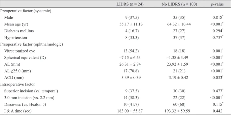
Table 3 shows the associations between LIDRS and clin- ical characteristics. Table 4 shows the ORs for clinical characteristics associated with LIDRS based on univari- able logistic regression analysis. In brief, statistically sig- nificant associations with LIDRS were found for age (OR, 0.920; 95% CI, 0.877 to 0.965; p = 0.001), vitrectomized eye (OR, 5.038; 95% CI, 1.957 to 12.971; p = 0.001), spherical equivalent (OR, 0.778; 95% CI, 0.693 to 0.875; p < 0.001),

axial length (OR, 1.716; 95% CI, 1.326 to 2.222; p < 0.001), anterior chamber depth (OR, 3.328; 95% CI, 1.078 to 10.279; p = 0.037), and 3.0 mm incision size (vs. 2.2 mm;

OR, 4.964; 95% CI, 1.940 to 12.699; p = 0.001). Multivari- able data analysis using conditional stepwise logistic re- gression analysis of the variables showed that vitrecto- mized eye (OR, 3.865; 95% CI, 1.201 to 12.436; p = 0.023), long axial length (OR, 1.709; 95% CI, 1.264 to 2.310; p = 0.001), and 3.0 mm incision size (vs. 2.2 mm; OR, 3.571;

95% CI, 1.120 to 11.393; p = 0.031) were significant inde- pendent factors associated with LIDRS (Table 5).

Among the 24 eyes that experienced LIDRS, bottle height, vacuum pressure, and aspiration pressure were re- duced from set I to set II in 15 eyes, nine eyes (60%) showed a decrease in the grade of LIDRS, and six eyes (40%) showed no interval change.

Discussion

LIDRS is an emerging problematic condition that can Table 1. Intraoperative hydrodynamic parameters

PE I & A Visco R 2.2 mm 3.0 mm

Set I

BHt (cm) 110 100 90 90

Vac (mmHg) 450 400 450 550

Aps (mmHg) 33 32 33 35

Set II

BHt (cm) 80 75 70 70

Vac (mmHg) 300 300 350 400

Asp (mmHg) 30 30 31 32

PE = phacoemulsification; I & A = irrigation and aspiration of residual cortex; Visco R = irrigation and aspiration of residual viscoelastics; BHt = bottle height; Vac = vacuum pressure; Asp = aspiration pressure.

Table 2. Incidence and LIDRS grade among 124 eyes of 124 patients

Incidence

LIDRS grade 1 7 (5.6)

LIDRS grade 2 13 (10.5)

LIDRS grade 3 4 (3.2)

Total 24 (19.4)

Values are presented as number (%).

LIDRS = lens-iris diaphragm retropulsion syndrome.

(4)

increase surgical challenges and patient morbidity. Al- though this condition has been attributed to an excessively deep anterior chamber, iris retropulsion, and extreme wid-

ening of the pupil during small incision cataract surgery, the incidence and factors that contribute to this condition remain unclear, and no strategy for management of pa- Table 3. Comparison of clinical factors between LIDRS and controls

LIDRS (n = 24) No LIDRS (n = 100) p-value Preoperative factor (systemic)

Male 9 (37.5) 35 (35) 0.818

*

Mean age (yr) 55.17 ± 11.13 64.32 ± 10.44 <0.001

Diabetes mellitus 4 (16.7) 27 (27) 0.294

*

Hypertension 8 (33.3) 37 (37) 0.737

*

Preoperative factor (ophthalmologic)

Vitrectomized eye 13 (54.2) 18 (18) 0.001

*

Spherical equivalent (D) –7.15 ± 6.53 –1.38 ± 3.49 <0.001

AL (mm) 26.31 ± 2.74 23.92 ± 1.59 <0.001

AL ≥25.0 (mm) 17 (70.8) 21 (21) <0.001

*

ACD (mm) 3.39 ± 0.39 3.19 ± 0.42 0.033

Intraoperative factor

Superior incision (vs. temporal) 9 (37.5) 30 (30) 0.477

*

3.0 mm incision (vs. 2.2 mm) 14 (58.3) 22 (22) <0.001

*

Discovisc (vs. Healon 5) 10 (41.7) 60 (60) 0.115

*

I & A time (sec) 183.00 ± 55.87 193.32 ± 59.59 0.442

Values are presented as number (%) or mean ± standard deviation.

LIDRS = lens-iris diaphragm retropulsion syndrome; D = diopters; AL = axial length; ACD = anterior chamber depth; I & A = irrigation and aspiration.

*

Pearson’s chi-square test;

Independent t-test.

Table 4. The results of univariable logistic regression for potential risk factors of LIDRS during phacoemulsification

Variable Regression coefficient OR 95% CI p-value

Preoperative factor (systemic)

Age –0.083 0.920 0.877–0.965 0.001

Male 0.108 1.114 0.443–2.804 0.818

Diabetes mellitus –0.615 0.541 0.169–1.726 0.299

Hypertension –0.161 0.851 0.332–2.181 0.737

Preoperative factor (ophthalmologic)

Vitrectomized eye 1.617 5.038 1.957–12.971 0.001

Spherical equivalent –0.250 0.778 0.693–0.875 <0.001

AL 0.540 1.716 1.326–2.222 <0.001

ACD 1.202 3.328 1.078–10.279 0.037

Intraoperative factor

Superior incision vs. temporal 0.336 1.400 0.552–3.550 0.478

3.0 mm incision size vs. 2.2 mm 1.602 4.964 1.940–12.699 0.001

Discovisc vs. Healon 5 –0.742 0.476 0.193–1.177 0.108

I & A time (sec) –0.003 0.997 0.989–1.005 0.439

LIDRS = lens-iris diaphragm retropulsion syndrome; OR = odds ratio; CI = confidence interval; AL = axial length; ACD = anterior

chamber depth; I & A = irrigation and aspiration.

(5)

tients has been clearly defined. LIDRS increases surgical challenges and may be associated with significant discom- fort for patients receiving phacoemulsification under topi- cal or intracameral anesthesia [7].

LIDRS is a well-recognized condition in myopic eyes and eyes that undergo vitrectomy [12]. However, previous clinical reports have not discussed the presentation of LIDRS, and the incidence and risk factors of LIDRS in the non-vitrectomized general population remain unknown. In the present study, the incidence of LIDRS and associated risk factors were evaluated.

The incidence of LIDRS in eyes that previously under- went vitrectomy has been reported in a few studies, with rates ranging from 0% to 100% [12-18]. In our study, among 31 vitrectomized eyes, 13 (41.9%) experienced some degree of LIDRS during phacoemulsification surgery. Bar- doloi et al. [14] evaluated the occurrence of LIDRS in my- opic patients and found that it occurred in eight of 52 non-vitrectomized myopic eyes (15.4%). The present study is the first report on the incidence of LIDRS in the non-vit- rectomized general population of patients that underwent phacoemulsification cataract surgery; we observed an inci- dence of 11.8%.

Only patients of Korean ethnicity were enrolled in this study. Most Koreans have a dark brown iris with light brown eyes; individuals without a brown iris are extremely rare. Studies have indicated that iris color may contribute to iris stability; hence, the incidence of LIDRS can vary based on genetic differences in iris color [19].

Predicting LIDRS before phacoemulsification cataract surgery is important for both cataract surgeons and pa- tients. Univariable analysis showed that the variables sig- nificantly correlated with or associated with LIDRS (p <

0.2) were age, vitrectomized eye, spherical equivalent, axi- al length, anterior chamber depth, 3.0 mm incision size (vs.

2.2 mm), and use of Discovisc as an ophthalmic viscosurgi- cal device (vs. Healon 5). Among these, the best fit condi- tional multiple logistic regression model showed that vit-

rectomized eye, long axial length, and 3.0 mm incision size (vs. 2.2 mm) were statistically significant at a 0.05 signifi- cance level.

Previous vitrectomy and axial myopia are well-known risk factors of LIDRS [12,19,20]. Ghosh et al. [12] reported the LIDRS severity was significantly greater in extensive vitrectomy than in limited vitrectomy (adjusted OR, 4.42;

95% CI, 1.19 to 16.45; p = 0.027) and with increasing axial length (adjusted OR, 3.05; 95% CI, 1.86 to 5.01; p < 0.001).

In our study, the significance of both risk factors were demonstrated using multivariable logistic regression anal- ysis, with ORs of 3.865 (95% CI, 1.201 to 12.436; p = 0.023) in the vitrectomized eye and 1.709 (95% CI, 1.264 to 2.310;

p = 0.001) in the long axial length. Ghosh et al. [12] pro- posed that low-viscosity vitreous fluid that leaked into the anterior chamber during the initial stage of surgery in pa- tients with axial myopia, vitreous degeneration, and a his- tory of vitrectomy resulted in a relative volume deficit in the vitreous cavity.

Our multivariate analysis indicated that the incidence of LIDRS increased in eyes with previously known risk fac- tors such as previous vitrectomy and long axial length. In addition, new associations were identified, including a rel- atively large corneal incision width of 3.0 mm compared with 2.2 mm. The direction and size of the clear corneal incision were determined based on the axis and amount of corneal astigmatism. We measured corneal astigmatism using manual keratometry, automated keratometry with optical biometry (IOLMaster), and corneal topography; all three methods gave comparable results. An incision was made on either the temporal or superior side, depending on which had a steeper axis. If the corneal astigmatism was greater than 0.75 D, a 3.0 mm incision was made with a 3.0 mm angled diamond keratome. If the corneal astigmatism was equal to or less than 0.75 D, a 2.2 mm incision was made with a 2.2 mm angled diamond keratome.

Two possible hypotheses can explain the increase in LIDRS with a relatively larger clear corneal incision. In Table 5. The result of multivariable logistic regression for significant risk factors of LIDRS during phacoemulsification

Variable Regression coefficient OR 95% CI p-value

Vitrectomized eye 1.352 3.865 1.201–12.436 0.023

Axial length 0.536 1.709 1.264–2.310 0.001

3.0 mm vs. 2.2 mm incision 1.273 3.571 1.120–11.393 0.031

LIDRS = lens-iris diaphragm retropulsion syndrome; OR = odds ratio; CI = confidence interval.

(6)

the present study, most LIDRS during phacoemulsification occurred during the initial phase, especially after insertion of the phacoemulsification tip. A greater phacoemulsifica- tion sleeve volume through a larger incision may cause rel- ative overload of the volume and pressure in the anterior chamber compared to the vitreous cavity. The differences may also be explained by iris stability depending on inci- sion size. Corneal incisions are characterized by three pa- rameters: construction, location, and size. All of the sur- geries were performed by the same surgeon using the same technique; therefore, significant differences in clear corne- al incision wound construction were unlikely. Hence, wound stability may be associated with wound size. Small- er wounds reduce iris prolapse, allow less leakage during surgery, and increase chamber stability. Therefore, the rel- ative instability of a larger wound may explain the occur- rence of LIDRS during the middle phacoemulsification and irrigation/aspiration stages.

Several guidelines have been suggested for managing LIDRS based on the characterization by Wilbrandt and Wilbrandt [6]. Cionni et al. [7] reported that mechanical separation of the iris and capsule is helpful for managing reverse pupillary block, an essential mechanism of LIDRS.

Nahra Saad et al. [8], Lee et al. [9], and Vishwanath [10]

used an iris hook retractor to lift the iris and relieve the pupillary block. Packard introduced a series of maneuvers, bottle lowering, and a side-port instrument to prevent LIDRS in high-risk patients [11]. Ghosh et al. [12] suggest- ed that lowering of parameters, slight bottle height reduc- tion, and a corresponding reduction in maximum aspira- tion rate could be applied to avoid anterior chamber collapse in cases at risk of LIDRS. We evaluated the ef- fects of bottle- and pressure-lowering procedures in sever- al patients and observed these techniques were effective in nine of 15 eyes (60%).

This study was limited by the retrospective design and the insufficient evaluation of various management tech- niques. Although all specific events such as LIDRS or in- traoperative floppy iris syndrome during surgery were re- corded, there is a possibility that cases were missed. We managed all cases without complications but were unable to assess all possible risk factors or evaluate the effects of bottle- and pressure-lowering procedures in all cases. Ad- ditional prospective interventional large-scale studies are needed to verify the application of our proposed manage- ment guidelines.

Our study results indicate the incidence of LIDRS is one fifth (20%) in the general population. LIDRS occurred in 41.9% of vitrectomized eyes and 11.5% of non-vitrecto- mized eyes. The results indicated that young age, history of vitrectomy, myopic spherical equivalent, long axial length, deep anterior chamber depth, and large corneal in- cision size (3.0 mm vs. 2.2 mm) were associated with LIDRS; specifically, vitrectomized eye, long axial length, and 3.0 mm incision size (vs. 2.2 mm) were determined to be significant independent factors.

Conflict of Interest

No potential conflict of interest relevant to this article was reported.

Acknowledgements

Involved in conception and design (DHL, ESC, TYC) and conducted the study (DHS, DHL, GH); collection, management, and interpretation of data (DHL, DHS, GH);

data analysis (DHL, DHS, GH); writing the manuscript (DHL, DHS); preparation, review, and approval of the man- uscript (DHL, ESC, TYC).

References

1. Ray S, D’Amico DJ. Pseudophakic cystoid macular edema.

Semin Ophthalmol 2002;17:167-80.

2. Chang DF, Campbell JR. Intraoperative floppy iris syn- drome associated with tamsulosin. J Cataract Refract Surg 2005;31:664-73.

3. Neff KD, Sandoval HP, Fernandez de Castro LE, et al. Fac- tors associated with intraoperative floppy iris syndrome.

Ophthalmology 2009;116:658-63.

4. Lim DH, Lee MG, Chung TY, Chung ES. Korean patients taking α1-adrenergic receptor antagonists show lower inci- dence of intraoperative floppy iris syndrome than Western patients. Br J Ophthalmol 2014;98:479-83.

5. Zauberman H. Extreme deepening of the anterior chamber during phacoemulsification. Ophthalmic Surg 1992;23:555-6.

6. Wilbrandt HR, Wilbrandt TH. Pathogenesis and manage-

ment of the lens-iris diaphragm retropulsion syndrome during

(7)

phacoemulsification. J Cataract Refract Surg 1994;20:48-53.

7. Cionni RJ, Barros MG, Osher RH. Management of lens-iris diaphragm retropulsion syndrome during phacoemulsifica- tion. J Cataract Refract Surg 2004;30:953-6.

8. Nahra Saad D, Castilla Cespedes M, Martinez Palmer A, Pazos Lopez M. Phacoemulsification and lens-iris dia- phragm retropulsion syndrome. Ophthalmic Surg Lasers Imaging 2005;36:512-3.

9. Lee VY, Liu DT, Chan WM, Lam DS. Management of lens-iris diaphragm retropulsion syndrome. J Cataract Re- fract Surg 2005;31:1273-4.

10. Vishwanath MR. Lens-iris diaphragm retropulsion syn- drome and iris hooks. J Cataract Refract Surg 2006;32:708.

11. Packard RB. Avoiding lens-iris diaphragm retropulsion syndrome. J Cataract Refract Surg 2010;36:1245.

12. Ghosh S, Best K, Steel DH. Lens-iris diaphragm retropul- sion syndrome during phacoemulsification in vitrecto- mized eyes. J Cataract Refract Surg 2013;39:1852-8.

13. Ahfat FG, Yuen CH, Groenewald CP. Phacoemulsification and intraocular lens implantation following pars plana vit- rectomy: a prospective study. Eye (Lond) 2003;17:16-20.

14. Bardoloi N, Barukial J, Borgohain MM. Iris lens retropul- sion syndrome: a study. In: Bhattacharya D, editor. Cata- ract III: 102-3. The 67th All India Ophthalmological Con- ference; 2009 February 5-8; Jaipur, India.

15. McDermott ML, Puklin JE, Abrams GW, Eliott D.

Phacoemulsification for cataract following pars plana vit- rectomy. Ophthalmic Surg Lasers 1997;28:558-64.

16. Grusha YO, Masket S, Miller KM. Phacoemulsification and lens implantation after pars plana vitrectomy. Ophthal- mology 1998;105:287-94.

17. Diaz Lacalle V, Orbegozo Garate FJ, Martinez Alday N, et al. Phacoemulsification cataract surgery in vitrectomized eyes. J Cataract Refract Surg 1998;24:806-9.

18. Osher RH, Osher JM, Cionni RJ. Multifocal iris sphincter ruptures: new sign of the lens-iris diaphragm retropulsion syndrome. J Cataract Refract Surg 2010;36:170-2.

19. Smiddy WE, Stark WJ, Michels RG, et al. Cataract ex- traction after vitrectomy. Ophthalmology 1987;94:483-7.

20. Diehl DL, Robin AL, Wand M. The influence of iris pig-

mentation on the miotic effect of thymoxamine. Am J Oph-

thalmol 1991;111:351-5.

수치

Table 3 shows the associations between LIDRS and clin- clin-ical characteristics. Table 4 shows the ORs for clinclin-ical  characteristics associated with LIDRS based on  univari-able logistic regression analysis
Table 4. The results of univariable logistic regression for potential risk factors of LIDRS during phacoemulsification

참조

관련 문서

2. The finite element is calculating using tension test of uni-direction 0° and 90°, compression test of uni-direction 0° and 90° and shear test results and, the results

Conclusion : The results reveal that it is not unusual that pedestrians are injured by automobiles in daily life. Awareness of risk factors would be useful in

3-38 Data processing area for estimating the crack length .... 3-39 Relationship between the crack length and the sum

These results indicated that KS28 could inhibit MAPK pathway signaling and that inhibition of the MAPK pathway could affect the suppression of

This study was conducted to analyze the effects of obesity on the core stability and metabolic syndrome risk factors of middle - aged women.. The effect

The study results indicated the role and importance of illustration that was newly recognized, and confirmed the contribution of illustration to motivation

The lateral size of the defect obtained from the deformation amount and the defect detection test using the shearing interferometer, width and depth of

The Dynamic Mechanical Analysis is a high precision technique for measuring the viscoelastic properties of materials. It consists in applying a sinusoidal deformation to a