Pentacam을 이용한 한국 청소년 집단에서 각막의 중심두께와 전방깊이 분석
김덕훈1*, 배한용2
1*마산대학교 안경광학과
2부산대학교 병원 안과
Analysis of the Central Corneal Thickness and Anterior Chamber Depth of Eyes in Korean Young Population using the Pentacam
Douk-Hoon Kim1*, Han-Yong Bae2
4)
1*Department of Optometry, Masan University
2Department of Ophthalmology, Busan National, University Hospital (Received June 3, 2013: Revised June 10, 2013: Accepted June 19, 2013)
Abstract
Purpose. This study was to investigate the analysis of the central corneal thickness and anterior chamber depth of eyes in Korean Young Population using the Pentacam topography.
Methods. The subjects consisted of 126 eyes from normal cornea of Korean aged 7-12(8.857±1.501) years during 2009. The thinnest location, pachy apex, and pupil central region of the cornea thickness was measured using the Pentacam pachymetry. The anterior chamber depth of normal corneas was measured with Pentacam.
Results. The thinnest location of corneal thickness was 547.413±37.893um. The pachy apex of cornea thickness was 552.103±36.016um. Also, the pupil center of corneal thickness was measured 552.143±36.418um. On the other hand, the mean thickness in the central cornea was a statically significant relationship(t-test, p=0.0002) between pachy apex and thinnest zone. Also the mean thickness of the central cornea was a statically significant correlation(pearson p=0.0001) among the pupil center, pachy apex and thinnest zone. The anterior chamber depth was 3.137±0.308mm. The mean depth in the anterior chamber of cornea was a statically relationship(t-test, p>0.05) between OD and OS.
Conclusions. The results of this clinical study suggested that the analysis of the central
* Corresponding Author : [email protected]
corneal thickness and anterior chamber depth using Pentacam in Korean young population can provide the useful morphological information in diagnosis of cornea for the contact lens fitting and corneal refraction surgery.
Key Words : cornea thickness, pachy apex, pentacam, pupil center, thinnest location
1. 서 론
각막은 시각에서 눈 전체 굴절의 약 2/3를 차지하는 중요한 굴절기관이다. 인간 눈의 굴절교정을 통한 시력개선이 대부분 각막의 형태적 변화를 가지는 기능적 효과로서 시 행되고 있다. 각막의 굴절 교정을 통한 방 법은 광학적인 방법과 의학적인 것이 있다.
최근에는 콘택트렌즈 착용과 각막 교정 수 술을 하는 방법이 주로 시행되고 있다. 특 히 원추각막에 대한 시력 교정은 초기에 주 로 광학적 방법인 RGP 콘택트렌즈를 착용 하는데, 이 경우 각막에 대한 정확한 진단 은 아주 중요하다. 조기에 정확한 진단을 통한 처방은 증상의 진행을 느리게 할 수 있고, 처방 후 환자가 보다 좋은 시 생활을 할 수 있게 된다. 이와 같이 각막 굴절 교 정을 통한 시력개선은 가장 중요한 것이 각 막에 대한 정확한 진단이 우선적이다.
각막의 진단에는 OCT1), 초음파2), 각막 지 형도3-4) 등 여러 가지 검사 도구가 이용되어 오고 있다.
최근에는 각막진단에 대한 방법으로 각막 지형도가 많이 이용되는데 이 가운데도 360 도 회전이 가능한 Scheimpflug 카메라의 부 착을 가진 Pentcam system을 임상에서 많 이 사용되고 있다.
이 검사 특징은 비접촉성으로서 각막의 지 형을 전면과 후면의 높이, 각막 굴절력, 곡 률반경, 전방 깊이, 각막중심 부위의 두께 측정, 동공 크기 등 다양한 자료를 3차원적 으로 얻을 수 있다 5).
이 같은 장점 때문에 각막 질환6), 녹내장7), 원추각막8), 각막의 생리적 변화9) 등의 진단
에 이용하고 있다.
본 연구는 한국인 청소년을 대상으로 Pentacam을 사용해서 각막지형도에서 각막 중심부의 두께와 전방의 깊이에 대해 분석 을 통해서 임상적 자료를 제공하고자 한다.
2. 방법
2.1. 연구대상
본 연구는 2009년 1월부터 12월 사이에 부 산광역시와 경상남도 지역에 거주하는 한국 인 청소년 63명의 126개 눈의 각막을 대상 으로 하였다. 눈에 이학적으로 소견으로 외 안부 질환, 눈 질환, 눈 외상, 약물 복용, 눈 알 수술, 약시 등이 있는 사람은 연구 대상 에서 제외 하였다.
2.2. 연구 방법
각막 검사는 오전 10시부터 12시 사이에 하였다. 검사 도구는 Pentacam(Bausch &
Lomb, USA)를 사용하였다. 검사과정에서 피검사는 머리와 턱은 턱받이에 고정시키고 눈 높이는 검사 동안 주시 광을 보도록 적 당한 위치에 고정하고, 눈을 깜박거리지 않 도록 하였다. 검사자는 피검사자가 컴퓨터 의 모니터를 보고 화면에 각막의 중심과 동 공 중앙부가 오도록 한 후 스켄 하였다. 측 정된 자료는 자동으로 컴퓨터에 입력하였으 며, 분석도구는 SPSS 20과 Excel 프로그램 을 사용하였다. 분석은 T-test 및 Pearson 상관관계를 사용해서 유의성 검증과 통계처 리를 하였다.
3. 결과
피검사자는 7세부터 12세(평균 8.857±1.501 세) 사이인 남녀 63명의 126개 안구를 대상 으로 실시하였다(Table 1).
Table 1. The age and sex distribution of the cornea test in Korean young population.
Age(Years) Number
Total(%) Male Female
7 3 11 14(22.22)
8 5 9 14(22.22)
9 6 12 18(28.57)
10 3 4 7(11.11)
11 1 5 6(9.52)
12 1 3 4(6.35)
Total(%) 19(30.16) 44(69.84) 63(100)
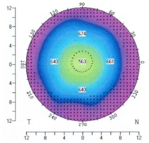
한국청소년 각막의 전형적인 모습은 대게 각막의 중심에서 주변으로 갈수록 두터위지 는 원반형 모습을 가지며, 각 지역별 두께 의 분포도 나르게 나타났다(Fig. 1).
Figure. 1. Typical corneal thickness in Korean young children using Pentacam topography.
양안의 각막 중심두께와 전방 깊이에서 가장 얇은 각막부위 평균두께는 오른눈 548.238±
34.793um, 왼눈 546.587±41.025um이며, 각막 첨부 평균 두께는 오른 눈 551.226±34.997um, 왼눈 552.921±37.269um이며, 동공 중앙부 평균 두께는 오른눈 551.175±35.202um, 왼 눈 553.111±37.853um 였으나, 양안 평균에 대한 유의성은 없었다(Table 2). 한편 전방 각 깊이의 평균에서 오른눈 3.161±0.294, 왼눈 3.113±0.323mm이며, 양안은 유의성 p<0.05로 나타났다(Table 2).
오른 눈과 왼눈의 pearson 상관관계에서는 각막 중심부 두께 및 전방각 깊이에서 상관 성( p< 0.01)이 있는 것으로 나타났다(Table 2).
Table. 2. Compare to the central cornea thickness(um) and anterior chamber depth(mm) using Pentacam in Korea Young Children. Mean±S.D, N=126
Classification Right Eye Left Eye p-value (paired t-test)
Pearson correlation
r p
Thinnest location
548.238
±34.793
546.587
±41.025 0.563 0.886*** 0.0001
Pachy apex
551.286
±34.997
552.921
±37.269 0.478 0.878*** 0.0001
Pupil center
551.175
±35.202
553.111
±37.853 0.390 0.839*** 0.0001
ACD 3.161
±0.294
3.113
±0.323 0.035* 0.836*** 0.0001
p-values: *p< 0.05, ** p< 0.01,*** p< 0.001
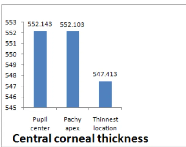
각막중심부 전체 평균 두께에서 가장 얇은 지역은 평균 두께가 547.413±37.893(최저 470um, 최대 622um)이며, 각막 첨부는 552.103±36.016(최저 472um,최대628um)이며, 동공중심부는 552.143±36.418(최저 472um, 최고 626um)um이며, 전방의 평균 깊이는 3.137±0.308mm(최저 2.22mm, 최대 3.71mm) 를 나타내었다( Table 3; Figure 2).
Table. 3. The central cornea thickness (um) and anterior chamber depth(mm) using Pentacam in Korea Young Children. Mean±
S.D, N=126
classification Minimum Maximum Mean ±S.D.
Thinnest
location 470 622 547.413
±37.893 Pachy
apex 472 628 552.103
±36.016 Pupil
center 472 626 552.143
±36.418
ACD 2.22 3.71 3.137±0.308
Figure 2. The central cornea thickness (um) using Pentacam in Korea Young Children.
Mean±S.D, N=126
각막중심부 두께에서 얇은 부위와 첨부는 유의성(p< 0.001)가지나, 다른 부위는 유의성 을 가지지 않았( Table 3). 한편 각막 중심부 두께의 부위별 상관관계 검증에서는 모두 양 성 방향으로 높은 상관성을 가지고 있다 (Table 4).
Table 4. Comparison of the cornea thickness (um) between 3 different central cornea zone using Pentacam in Korean young population. Mean±S.D, N=126
Classification Thickness (Mean ±S.D.) p-value (paired t-test)
Pearson correlation
r p
Thinnest Apex
547.413±37.893
552.103±36.016 0.0002 0.931
** 0.0001
Thinnest Pupil
547.413±37.893
552.143±36.418 3.319 0.943** 0.0001
Apex Pupil
552.103±36.016
552.143±36.418 0.885 0.996** 0.0001
4. 고찰
Pentacam 각막지형도는 기존의 각막검사 장비에 비해서 비침습적인 방법으로 각막 굴절력, 전방의 깊이, 각막 두께, 동공의 크 기, 각막 굴절력, 각막 곡룰반경 등의 다양 한 정보를 3차원적이로 제공하여 임상에서 진단을 위한 검증도구로 많이 사용되고 있
다. 더욱이 Pentacam pachymetry는 각막의 여러 부위의 두께를 측정하는 것으로 각막 질환6), 녹내장7), 원추각막8), 각막의 생리적 변화9) 등의 진단에 이용되고 있다.
각막의 중심부 두께의 측정에 대한 Orbscan을 사용한 한국인 대상에서 4세에서 9세는 남성 562.382±36.852um, 여성 548.883±28.794um 이며, 10세에서 19세 사이는 550.621±31.620um, 여성 556.276±50.344um로 보고한 것이 있다
10). 이는 본 연구의 Pentacam을 사용한 각 막의 얇은 지역 547.413±37.893 um, 첨부지 역 552.103± 36.016 um, 동공 중심부 552.143±36.418um 결과 값과 차이가 거의 없음을 알 수 있다. 즉 이는 두 검사장비의 결과가 유사함을 알 수 있다.
Fam HB 등11)은 근시를 가진 중국성인을 대상으로 각막 중심부의 측정에서 평균 534.5um, Tong L등 12)은 싱가폴 어린이를 대상으로 543.6um, Foster 13)은 싱가폴 성인 을 대상으로 547.1um을 보고하였다. Price 등14)는 Caucasian 사람을 대상으로 각막 중 심부는 533um, Vijaya 등15)는 정상 인도 시 골 집단에서는 평균 505.9um를 보고하였다.
이와 같이 각 국가간의 근시를 가진 아시아 인과 Causian 사람들 사이의 각막중심 두께 의 차이는 종족의 차이로 Fam HB11)는 제 시하였다. 본 연구의 대상이 한국인으로서 다른 아시아 및 유럽인들과는 차이가 있음 을 알 수 있다. 이 같은 차이는 종족 및 종 족과 근시와의 관계로 인해서 차가가 발생 될 수 있다고 여겨진다.
한편 Chang 등16)과 Tokoro 등17)은 근시를 가진 사람은 정시를 가진 사람에 비해서 각 막 중심부가 얇으며 이는 근시와 각막중심 부는 상관관계가 있다고 하였다. 그러나 Fam HB11)과 Price 등14) 및 Pederson 등18) 는 근시와 각막 중심부와는 관계가 없다고 보고하였다. 본 연구에서 연구 대상이 어린 청소년으로서 대부분 근시를 가진 것으로 예상되며 이들의 각막 중심부와 근시와의 관계는 연관성을 가진 것으로 추측하나 추 후 연구가 필요한 실정이다.
Auffarth 등19)은 원추각막 피검사자의 각막 중심부 두께는 평균 457um으로, Kawana 등20)는 약 449um, Auffarth et al 19)은 중 심 각막의 두께가 약 457um를 원추각막의 진단으로 제시하였다. 본 연구에서는 각막 중심부는 모두 550um 이상으로 원추각막에 해당되는 피검사자는 없는 것으로 확인 되 었다.
본 연구에서 오른눈과 왼눈의 평균 두께 비교에서 각막중심부의 각 지역별 비교 즉 얇은 지역과 동공중심, 얇은 지역과 첨부, 첨부와 동공 중심 사의의 유의성은 없는 것 으로 나타났으나, 양안의 총합의 평균 두께 의 비교에서는 가장 얇은 지역과 첨부지역 은 상관관계는 있는 것으로 나타났다. 한편 오른눈과 왼눈의 각막 중심부 비교 및 양안 을 총합한 각막중심부의 각 지역별 비교에 서는 모두 상관성을 가졌다. 이 같은 결과 는 각막 중심부 두께측정의 진단에 유용한 자료가 될 수 있다고 여겨진다.
Pentacam을 사용한 Lam과 Tse21)의 전방 깊이를 중국인을 대상으로 측정한 결과 20 대 청소년은 3.60±0.40 mm는 40대의 3.24
±0.34 mm 보다 약간 깊은 차이를 보고하 였다. 한국인 청소년을 대상으로 한 본 연 구에서는 3.137±0.308mm로 나타났다. 이는 중국인에 비해서 다소 낮은 값을 가지는 것 이다. 이는 종족의 차이라 생각된다.
한편 본연구의 오른 눈과 왼눈의 전방각 깊이의 평균값은 유의성을 가졌으며 또한 상관관계도 있는 것으로 나타났다.
이상에서 Pentacam을 이용한 한국 청소년 을 대상으로 한 연구 결과에서 각막 중심부 의 두께 측정에서 얇은 부위, 첨부 및 동공 중심부는 지역별 상관관계를 가지고 있었으 나, 평균 값에 대한 유의성을 가장얇은 지 역과 첨부만 가졌다. 한편 각막 전방 깊이 의 측정에서 양안은 평균값에 대한 유의성 과 상관관계를 모두 가지고 있었다. 본 연 구의 연구 결과는 향후 임상에서 한국 청소 년의 각막 진단 및 연구에 중요한 자료가 될 것으로 여겨진다. 그러나 앞으로 더욱 다양한 연령층을 대상으로 각막 진단에 연 관되는 다양한 요소들의 상호관계 분석이 있어야 된다고 여겨진다.
5. 결론
Pentacam을 이용한 한국인 청소년의 각막 중심부와 전방 깊이의 분석에서 연구 결과 는 다음과 같다. 첫째, 각막 중심부 두께는 가장 얇은 지역의 값이 가장 낮았다. 둘째, 각막 중심부 두께의 오른 눈과 왼눈의 차이 는 거의 없었다. 셋째, 각막 중심부의 평균 두께에서 각막 첨부지역과 가장 얇은 지역 은 유의성을 가졌다. 넷째 양안에서 각막중 심부의 각 지역별 두께는 상관관계가 깊었 다. 다섯째 각막의 전방깊이에서 오른 눈과 왼눈은 평균 두께에서 유의성을 가졌으며, 또한 상관관계도 있었다. 이상의 연구에서 한국인 청소년의 각막 중심부의 지역별두께 및 전방의 깊이는 일부 유의성과 상관관계 가 있음을 알 수 있었다.
Reference
1. Fuduka S, Kawana K, Yasuno Y, et al.
Anterior ocular biography using 3- dimensional optical coherence tomography.
Ophthalmology, 2009: 116: 882-889.
2. Cavanagh HD, El-Agha MS, Petroll WM, et al. Specular microscopy, confocal microscopy, and ultrasound biomicroscopy diagnostic tools of the past quarter century. Cornea, 2009; 19:712-722.
3. Gonen T, Cosar CB, Sener B, Keskinbora KH.
Comparison of keratometric data obtained by automated keratometer. Dicon CT 200, Allegro Topolyzer, and Pentacam. J Refract Surg, 2012 ;28(8):557-61. .
4. Wilson SE, Lin DTC, Klyce SD. Corneal topography of keratconus, Cornea, 1991;
10: 2-8.
5. Laszlo T, Eszter S, Gabor N, Andras B.
Reliability of the corneal thickness measurement with the Pentacam HR imaging system and ultrasound pachymetry. Cornea, 2011; 30:
561-566.
6. Elflein HM, Hofherr T, Berisha-Ramadani F, Weyer V, Lampe C, Beck M, Pitz S.
Measuring corneal clouding in patients suffering from mucopolysaccharidosis with the Pentacam densitometry programme. Br J Ophthalmol, 2013; 97(7):829-33.
7. Pakravan M, Sharifipour F, Yazdani S, Koohestani N, Yaseri M. Scheimpflug imaging criteria for identifying eyes at high risk of acute angle closure. J Ophthalmic Vis Res, 2012; 7(2):111-7.
8. Henriquez MA, Izquierdo L Jr, Mannis MJ.
Intereye asymmetry detected by Scheimpflug imaging in subjects with normal corneas and keratoconus. Cornea, 2013; 32(6):779-82.
9. Mohammed HN, Amirhossein M, Mohammad HM, Javad R. Effect of Ramadan fasting in tropical summer months on ocular refractive and biometric characteristics. Clin Exp Optom 2012; 95; 173-176.
10. Lee DH, Kim DH, Park SH. Age and sex related changes in corneal thickness and anterior corneal curvature in Korean young population with Orbscan II topography system. Journal of the Optical Society of Korea, 2011; 15(1): 68-73.
11. Fam HB, How ACS, Baskaran M, Lim KL, Chan YH, Aung T. Central corneal thickness and its relationship to myopia in Chinese adults. Br, J Ophthalmol, 2006; 90(12);
1451-1453.
12. Tong L, Saw SM, Siak JK, et al. Corneal thickness determinination and correlation in Singaporean. Invest Ophthalmol Vis Sci, 2004; 454004-454009.
13. Foster PJ, Michin D, Wong TY, et al.
Determinants of intraocular pressure and its association with glaucomatous optic neuropathy in Chinese Singaporeans: The Tanjong Pagar Study. Invest Ophthalmol
Vis Sci, 2003; 443885-443891.
14. Price FW, Koller DL, Price MO. Central pachymetry in patients undergoing laser in situ keratomilieusis. Ophthalmology, 2000;
1062216-1062220.
15. Vijaya L, George R, Paul PG, et al.
Prevalence of open -angle glaucoma in a rural South Indican population. Invest Ophthalmol Vis Sci, 2005; 464461-464467.
16. Chang SW, Tsai IL, Hu FR, et al. The cornea in young myopic adults. Br J Ophthalmol, 2001; 85916-85920.
17. Tokoro T, Hyashi K, Muto M, et al. Central corneal thickness in high myopia. Folia Ophthalmol Jpn, 1976; 27610.
18. Pederson L, Hjortdal J, Ehlers N. Center
corneal thickness in high myopia. Acta Ophthalmol Scand; 2005: 83539-83542.
19 . Auffarth GU, Wang L, Volcker HE.
Keratoconus evaluation usign the ORBscan topography system. J Cataract Refract Surg, 2000; 26:222-228.
20. Kawana K, Miuta K, Tokunaga T, et al.
Central thickness measurements using Orbscan II scanning slit topography, non contact specular microscopy, and ultrasonic pachymetry in eyes with keratoconus.
Cornea, 2005; 24:967-971.
21. Lam AK, Tse JS.Pentacam anterior chamber parameters in young and middle-aged Chinese. Clin Exp Optom, 2013; 96(1): 85-91.