Original Article
Changes in Anterior Chamber Configuration after Cataract Surgery as Measured by Anterior Segment Optical
Coherence Tomography
Martha Kim1, Ki Ho Park2, Tae-Woo Kim1, Dong Myung Kim2
1Department of Ophthalmology, Seoul National University Bundang Hospital, Seongnam, Korea
2Department of Ophthalmology, Seoul National University Hospital, Seoul National University College of Medicine, Seoul, Korea
Purpose: To evaluate the changes in anterior chamber depth (ACD) and angle width induced by phacoemulsifica- tion and intraocular lens (IOL) implantation in normal eyes using anterior segment optical coherence tomography (AS-OCT).
Methods: Forty-five eyes (45 patients) underwent AS-OCT imaging to evaluate anterior chamber configuration be- fore and 2 days after phacoemulsification and IOL implantation. We analyzed the central ACD and angle width using different methods: anterior chamber angle (ACA), trabecular-iris angle (TIA), angle opening distance (AOD), and trabecular iris surface area (TISA) in the nasal and temporal quadrants. Comparison between pre- operative and postoperative measurement was done using paired t-tests and each of the angle parameters was analyzed with Pearson correlation testing. Subgroup analyses according to the IOL and axial length were per- formed with a general multivariate linear model adjusted for age.
Results: Before surgery, the mean anterior chamber angle widths were 23.21 ± 6.70° in the nasal quadrant and 24.89 ± 7.66° in the temporal quadrant. The mean central ACD was 2.75 ± 0.43 mm. After phacoemulsification and IOL implantation, the anterior chamber angle width increased significantly to 35.16 ± 4.65° in the nasal quad- rant (p = 0.001) and 36.03 ± 4.86° in the temporal quadrant (p = 0.001). Also, central ACD increased to 4.14 ± 0.31 mm (p = 0.001). AOD, TISA, and TIA increased significantly after cataract surgery and showed positive cor- relation with ACA.
Conclusions: After cataract surgery, the ACD and angle width significantly increased in eyes with cataract. AS-OCT is a good method for obtaining quantitative data regarding anterior chamber configuration.
Key Words: Anterior chamber, Cataract extraction, Optical coherence, Tomography
ⓒ2011 The Korean Ophthalmological Society
This is an Open Access article distributed under the terms of the Creative Commons Attribution Non-Commercial License (http://creativecommons.org/licenses /by-nc/3.0/) which permits unrestricted non-commercial use, distribution, and reproduction in any medium, provided the original work is properly cited.
Received: February 28, 2009 Accepted: August 26, 2010
Corresponding Author: Ki Ho Park, MD. Department of Ophthalmology, Seoul National University College of Medicine, #103 Daehak-ro, Jongno-gu, Seoul 110-799, Korea. Tel: 82-2-2072-2438, Fax: 82-2-741-3187, E-mail:
Anterior segment cross-sectional imaging is essential in evaluating the anterior chamber configuration with quantita- tive data. Several methods have been studied and used for the diagnosis of glaucoma or other anterior segment pathology and quantification of angle configuration [1-11]. Ultrasound biomicroscopy (UBM) provides high-resolution images of the anterior chamber and is able to penetrate opaque media.
With UBM, detailed images of the iridotrabecular angle and
ciliary body can be obtained; these structures are not visible with a gonioscope [12-14]. However, the UBM transducer requires the eye to be immersed in a water bath of saline with the patient supine; the procedure is uncomfortable for the pa- tient and requires a skilled examiner. Scheimpflug photog- raphy is a non-invasive and repeatable technique that uses a slit beam and a camera. However, because of its optical and acoustic characteristics, images must undergo processing and do not allow visualization of the actual angle recess [9,11,14].
Recently, anterior segment optical coherence tomography (AS-OCT) has emerged as a new imaging technique for the anterior segment that is non-invasive and non-contact.
AS-OCT provides high resolution images by using a long
A
B
Fig. 1. Anterior segment optical coherence tomography image of the right eye of a 50-year-old woman before (A) and after (B) cata- ract surgery. Anterior chamber depth (ACD) was defined as the dis- tance from the endothelium at the center of the cornea to the anterior pole of the lens or intraocular lens. ACA = anterior chamber angle.
A B
Fig. 2. (A) The angle-opening distance (AOD) at 500 μm anterior to the scleral spur (AOD500) and the AOD at 750 μm anterior to the scleral spur (AOD750) were defined as the distance from the cor- neal endothelium to the anterior iris perpendicular to a line drawn along the trabecular meshwork at 500 μm or 750 μm from the scler- al spur. The trabecular-iris space areas (TISA) at 500 μm or 750 μm from the scleral spur (TISA500, TISA750) were defined as the areas bounded by the corneal endothelium, trabecular meshwork, and an- terior iris surface out to a distance of 500 μm or 750 μm from the scleral spur. (B) The trabecular-iris angle (TIA), defined as the an- gle measured with the apex in the scleral spur and the arms of the an- gle passing through a point on the trabecular meshwork 500 μm from the scleral spur and a perpendicular point on the iris.
wavelength (1,310 nm) of light; it offers rapid and easy quan- titative analysis of various structures [12,15-17]. AS-OCT has exhibited good repeatability and reproducibility with low intraobserver and interobserver variability [18-21]. One limi- tation of AS-OCT is that it has incomplete penetration through the pigmented epithelium of the iris, thus making it difficult to obtain accurate images of the ciliary body, lens, and zonules behind the pigmented iris [17].
Cataract extraction with intraocular lens (IOL) implantation causes the anterior chamber angle to widen and the depth to deepen in glaucomatous and normal eyes [2,3,5,22-26].
Previous studies have used several methods to evaluate the changes before and after cataract surgery.
The object of this study was to analyze changes in the ante- rior chamber depth and angle width after phacoemulsifica- tion and IOL implantation in normal eyes using AS-OCT. We obtained quantitative data from normal Korean eyes as meas- ured by AS-OCT and compared standardized parameters of anterior chamber configuration.
Materials and Methods
This prospective study comprised 45 eyes from 45 consec- utive patients with cataract who underwent cataract surgery from February 2008 to November 2008. Patients with glau- coma, previous intraocular surgery, or other intraocular path- ology were excluded.
All patients underwent routine ophthalmic examinations including visual acuity, Goldmann tonometry, slit-lamp bio- microscopy, and funduscopy. Refractive errors were meas- ured as manifest refraction. Axial lengths were obtained us-
ing a Humphrey 820 model A-scan ultrasound unit (Humphrey Systems, Dublin, CA, USA). AS-OCT (Visante; Carl Zeiss Meditec, Dublin, CA, USA) was performed preoperatively and 2 days postoperatively. One examiner obtained all im- ages under identical lighting conditions. For measurement, the pupil was not dilated and the patient was asked to sit and fixate on an indicator in the AS-OCT. Images of the nasal and temporal angle quadrants (0° and 180° meridians) were cap- tured until the centration and quality were sufficient for analyzation.
The same surgeon (PKH) performed all phacoemulsifica- tion and IOL implantation under topical anesthesia.
Phacoemulsification and foldable IOL implantation were performed through a 2.75 mm temporal clear corneal incision. The corneal incision was not sutured. Nine eyes re- ceived an SA60AT (Acrysof; Alcon, Fort Worth, TX, USA) IOL, 14 eyes received a Biovue (OII, Ontario, CA, USA) IOL, and 22 eyes received an IQ (Acrysof) IOL.
Data analysis
The best image was selected and analyzed using custom software (Iridocorneal Module; Carl Zeiss Meditec). The an- terior chamber angle was calculated using two different defi- nitions: 1) anterior chamber angle (ACA) - the angle between the iris tangential line and that of the posterior corneal sur- face with its apex in the angle recess (Fig. 1); and 2) tra- becular-iris angle (TIA) - the angle between the arms passing
Fig. 3. (A,B) Preoperative anterior chamber angle (ACA) measurements and changes in the angle following cataract sur- gery showed negative correlations in the nasal (A) and tempo- ral (B) quadrants. (C) Preoperative anterior chamber depth (ACD) and postoperative changes in ACD also showed a negative correlation.
through a point on the trabecular meshwork 500 μm from the scleral spur and a point perpendicularly opposite on the iris (Fig. 2) [7]. ACA width was also analyzed with standardized angle parameters after manual identification of the scleral spur: 1) angle opening distance (AOD) at 500 μm (AOD500) and AOD at 750 μm (AOD750) - distance of a perpendicular from the trabecular meshwork on the iris at a point 500 μm or 750 μm from the scleral spur; and 2) trabecular iris space area (TISA) up to 500 μm (TISA500) or 750 μm (TISA750) - the area bounded by the corneal endothelium, trabecular mesh- work, and anterior iris surface out to a distance of 500 μm or 750 μm from the scleral spur (Fig. 2). ACD was defined as the distance from the endothelium at the center of the cornea to the anterior pole of the lens or IOL (Fig. 1).
Patients were divided into 3 groups according to the axial length of the globe: group 1 (axial length <23 mm); group 2 (axial length 23 to 26 mm); and group 3 (axial length >26 mm).
All statistical analyses were performed using SPSS ver. 12 (SPSS Inc., Chicago, IL, USA). Comparison between pre- operative and postoperative angle parameters and ACD was done using a paired t-test. Analysis of each angle parameter was performed using Pearson correlation testing. Comparison
according to the axial length and IOL type were performed using a general multivariate linear model adjusted for age.
All p-values were 2-sided and were considered statistically significant when <0.05.
Results
Of the 45 patients, 16 were men and 29 were women. The mean age was 67.8 ± 9.7 years (range, 32 to 83 years). All pa- tients were diagnosed with senile cataract except for two middle-aged patients who had used corticosteroids for sys- temic diseases.
The mean preoperative ACD was 2.75 ± 0.43 mm. The mean postoperative ACD was 4.14 ± 0.31 mm at 2 days after cataract surgery, approximately 50.5% deeper than before surgery (p < 0.001). Changes in ACD and preoperative ACD showed a negative correlation (r = -0.680, p < 0.01) (Fig. 3).
All angle parameters analyzed with AS-OCT showed a significant increase after cataract surgery for both the nasal and temporal angles. The mean preoperative ACA was 23.21
± 6.70° at the nasal angle and 24.89 ± 7.66° at the temporal angle. After cataract surgery, ACA increased to 35.16 ± 4.65° at the nasal angle and 36.03 ± 4.86° at the temporal an-
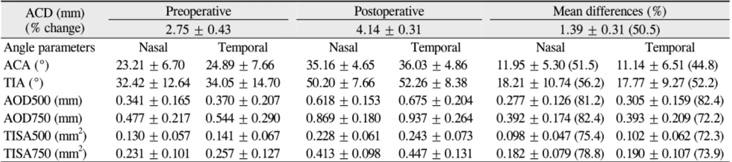
Table 1. Changes in anterior chamber parameters before and after cataract surgery (n = 45) ACD (mm)
(% change) Preoperative Postoperative Mean differences (%)
2.75 ± 0.43 4.14 ± 0.31 1.39 ± 0.31 (50.5)
Angle parameters Nasal Temporal Nasal Temporal Nasal Temporal
ACA (°) 23.21 ± 6.70 24.89 ± 7.66 35.16 ± 4.65 36.03 ± 4.86 11.95 ± 5.30 (51.5) 11.14 ± 6.51 (44.8) TIA (°) 32.42 ± 12.64 34.05 ± 14.70 50.20 ± 7.66 52.26 ± 8.38 18.21 ± 10.74 (56.2) 17.77 ± 9.27 (52.2) AOD500 (mm) 0.341 ± 0.165 0.370 ± 0.207 0.618 ± 0.153 0.675 ± 0.204 0.277 ± 0.126 (81.2) 0.305 ± 0.159 (82.4) AOD750 (mm) 0.477 ± 0.217 0.544 ± 0.290 0.869 ± 0.180 0.937 ± 0.264 0.392 ± 0.174 (82.4) 0.393 ± 0.209 (72.2) TISA500 (mm2) 0.130 ± 0.057 0.141 ± 0.067 0.228 ± 0.061 0.243 ± 0.073 0.098 ± 0.047 (75.4) 0.102 ± 0.062 (72.3) TISA750 (mm2) 0.231 ± 0.101 0.257 ± 0.127 0.413 ± 0.098 0.447 ± 0.131 0.182 ± 0.079 (78.8) 0.190 ± 0.107 (73.9) All differences p < 0.001.
ACD = anterior chamber depth; ACA = anterior chamber angle; TIA = trabecular-iris angle; AOD = angle-opening distance; TISA = trabecular-iris space area.
Table 2. Correlation coefficients of preoperative angle parameters in the nasal and temporal quadrants
ACA TIA AOD500 AOD750 TISA500
Nasal Temporal Nasal Temporal Nasal Temporal Nasal Temporal Nasal Temporal
TIA 0.845 0.912
AOD500 0.816 0.892 0.807 0.981
AOD750 0.832 0.902 0.783 0.955 0.925 0.964
TISA500 0.712 0.811 0.732 0.926 0.933 0.935 0.847 0.900
TISA750 0.784 0.872 0.778 0.967 0.975 0.978 0.936 0.966 0.978 0.981
All values are statistically significant (p < 0.01).
ACA = anterior chamber angle; TIA = trabecular-iris angle; AOD = angle-opening distance; TISA = trabecular-iris space area.
gle, widening to 51.5% and 44.8%, respectively. An increase in ACA exhibited a significant negative correlation with pre- operative ACA at both the nasal and temporal angles (r = -0.723, p < 0.01 at the nasal angle; r = -0.777, p < 0.01 at the temporal angle) (Fig. 3).
The mean preoperative TIA was 32.42 ± 12.64° at the nasal angle and 34.05 ± 14.68° at the temporal angle. Postoperatively, the TIA increased to 50.20 ± 7.66° at the nasal angle and 52.26
± 8.38° at the temporal angle, widening 56.2% and 52.2%, respectively.
Standardized angle parameters were also significantly increased. Preoperative AOD500 was 0.341 ± 0.165 mm at the nasal quadrant and 0.370 ± 0.207 mm at the temporal quadrant. Postoperative mean AOD500 was 0.618 ± 0.153 mm at the nasal angle and 0.675 ± 0.204 mm at the temporal angle. AOD750 also increased from 0.477 ± 0.217 mm to 0.869 ± 0.180 mm at the nasal angle and from 0.544 ± 0.290 mm to 0.937 ± 0.264 mm at the temporal angle following cataract surgery. TISA500 at the nasal angle increased from 0.130 ± 0.057 mm2 to 0.228 ± 0.061 mm2 and TISA500 at the temporal angle increased from 0.141 ± 0.067 mm2 to 0.243 ± 0.073 mm2. TISA750 also increased from 0.231 ± 0.101 mm2 to 0.413 ± 0.098 mm2 at the nasal angle and from 0.257 ± 0.127 mm2 to 0.447 ± 0.131 mm2 at the temporal angle (Table 1).
This study demonstrated a statistically significant positive correlation between ACA and standardized angle parameters (TIA, AOD, and TISA) in the nasal and temporal quadrants (Table 2).
The changes in ACD and angle variables were not statisti- cally different according to the type of implanted IOL when adjusted for age and axial length.
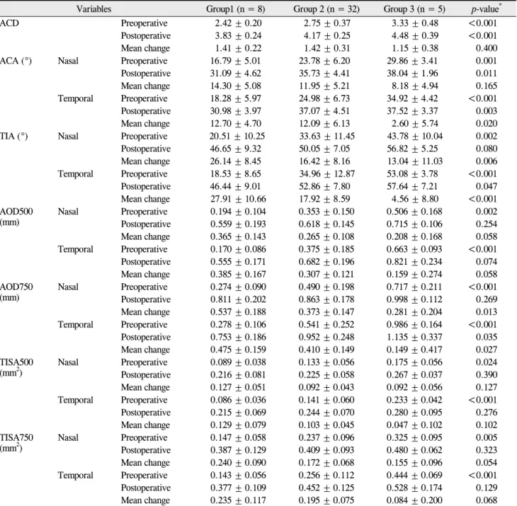
Patients were divided into three groups according to axial length. The mean axial length was 24.26 ± 2.15 mm (range, 21.77 to 31.90). The mean axial length for each group was 22.31 ± 0.33 mm in group 1 (n = 8), 24.00 ± 0.94 mm in group 2 (n = 32), and 29.32 ± 1.88 mm in group 3 (n = 5).
Table 3 shows the subgroup analyses performed according to axial length. Patients with a short axial length (group 1) ex- hibited a shallow ACD and a narrow ACA both preoperatively and postoperatively. All other angle parameters were also smaller in group 1 before surgery. However, postoperative values were not significantly different between the groups, except for AOD750 at the temporal angle (p = 0.035). The mean changes after surgery between the groups were sig- nificantly different for ACA and TISA750 at the temporal angle and for TIA and AOD750 at both angles. Other values also showed a trend toward larger changes in group 1. This analysis was adjusted for age using a general linear model.
Discussion
AS-OCT is a light-based system that rapidly provides high-resolution images. Its non-contact nature ensures pa- tient comfort and allows for rapid image acquisition in the sitting position, without risk of mechanical distortion of the angle. It also allows quantitative and dynamic data analysis with high reproducibility and repeatability [2,3,12,14,16,
Table 3. Anterior chamber variables according to axial length
Variables Group1 (n = 8) Group 2 (n = 32) Group 3 (n = 5) p-value*
ACD Preoperative 2.42 ± 0.20 2.75 ± 0.37 3.33 ± 0.48 <0.001
Postoperative 3.83 ± 0.24 4.17 ± 0.25 4.48 ± 0.39 <0.001
Mean change 1.41 ± 0.22 1.42 ± 0.31 1.15 ± 0.38 0.400
ACA (°) Nasal Preoperative 16.79 ± 5.01 23.78 ± 6.20 29.86 ± 3.41 0.001
Postoperative 31.09 ± 4.62 35.73 ± 4.41 38.04 ± 1.96 0.011
Mean change 14.30 ± 5.08 11.95 ± 5.21 8.18 ± 4.94 0.165
Temporal Preoperative 18.28 ± 5.97 24.98 ± 6.73 34.92 ± 4.42 <0.001
Postoperative 30.98 ± 3.97 37.07 ± 4.51 37.52 ± 3.37 0.003
Mean change 12.70 ± 4.70 12.09 ± 6.13 2.60 ± 5.74 0.020
TIA (°) Nasal Preoperative 20.51 ± 10.25 33.63 ± 11.45 43.78 ± 10.04 0.002
Postoperative 46.65 ± 9.32 50.05 ± 7.05 56.82 ± 5.25 0.080
Mean change 26.14 ± 8.45 16.42 ± 8.16 13.04 ± 11.03 0.006
Temporal Preoperative 18.53 ± 8.65 34.96 ± 12.87 53.08 ± 3.78 <0.001
Postoperative 46.44 ± 9.01 52.86 ± 7.80 57.64 ± 7.21 0.047
Mean change 27.91 ± 10.66 17.92 ± 8.59 4.56 ± 8.80 <0.001 AOD500
(mm) Nasal Preoperative 0.194 ± 0.104 0.353 ± 0.150 0.506 ± 0.168 0.002
Postoperative 0.559 ± 0.193 0.618 ± 0.145 0.715 ± 0.106 0.254 Mean change 0.365 ± 0.143 0.265 ± 0.108 0.208 ± 0.168 0.058 Temporal Preoperative 0.170 ± 0.086 0.375 ± 0.185 0.663 ± 0.093 <0.001 Postoperative 0.555 ± 0.171 0.682 ± 0.196 0.821 ± 0.234 0.074 Mean change 0.385 ± 0.167 0.307 ± 0.121 0.159 ± 0.274 0.058 AOD750
(mm) Nasal Preoperative 0.274 ± 0.090 0.490 ± 0.198 0.717 ± 0.211 <0.001
Postoperative 0.811 ± 0.202 0.863 ± 0.178 0.998 ± 0.112 0.269 Mean change 0.537 ± 0.188 0.373 ± 0.147 0.281 ± 0.204 0.013 Temporal Preoperative 0.278 ± 0.106 0.541 ± 0.252 0.986 ± 0.164 <0.001 Postoperative 0.753 ± 0.186 0.952 ± 0.248 1.135 ± 0.337 0.035 Mean change 0.475 ± 0.159 0.410 ± 0.149 0.149 ± 0.417 0.027 TISA500
(mm2) Nasal Preoperative 0.089 ± 0.038 0.133 ± 0.056 0.175 ± 0.056 0.024
Postoperative 0.216 ± 0.081 0.225 ± 0.058 0.267 ± 0.037 0.390 Mean change 0.127 ± 0.051 0.092 ± 0.043 0.092 ± 0.056 0.127 Temporal Preoperative 0.086 ± 0.036 0.141 ± 0.060 0.233 ± 0.042 <0.001 Postoperative 0.215 ± 0.069 0.244 ± 0.070 0.280 ± 0.095 0.276 Mean change 0.129 ± 0.079 0.103 ± 0.045 0.047 ± 0.102 0.102 TISA750
(mm2) Nasal Preoperative 0.147 ± 0.058 0.237 ± 0.096 0.325 ± 0.095 0.005
Postoperative 0.387 ± 0.129 0.409 ± 0.093 0.480 ± 0.062 0.323 Mean change 0.240 ± 0.090 0.172 ± 0.068 0.155 ± 0.096 0.054 Temporal Preoperative 0.143 ± 0.056 0.256 ± 0.112 0.444 ± 0.069 <0.001 Postoperative 0.377 ± 0.109 0.452 ± 0.125 0.528 ± 0.174 0.129 Mean change 0.235 ± 0.117 0.195 ± 0.075 0.084 ± 0.200 0.068 ACD = anterior chamber depth; ACA = anterior chamber angle; TIA = trabecular-iris angle; AOD = angle-opening distance; TISA = trabecular-iris space area.
*Based on a general linear model with all variables adjusted for age.
18-23,27-30].This study demonstrates changes in anterior segment configuration after phacoemulsification and IOL implantation in normal eyes as measured quantitatively by AS-OCT. We provided normative data of anterior segment parameters in normal eyes and also compared these parame- ters with each other.
This study confirmed angle widening of up to 51.5% (at the nasal angle) and chamber deepening of up to 50.5% after cataract surgery, as other studies have previously demon-
strated [3,22,23,25,27]. Our data also revealed a negative correlation between preoperative ACD and the amount of in- crease of ACD and between preoperative angle parameters and the changes of these angle parameters following cataract surgery.
The mean preoperative ACD was 2.75 ± 0.43 mm and the mean preoperative ACA was 23.21 ± 6.70° at the nasal angle and 24.89 ± 7.66° at the temporal angle. These results are similar to results from other studies which have analyzed
changes in ACD and ACA following cataract surgery [3].
Yi et al. [31] gathered ACD and ACA data in a normal Korean population without cataract. They included 81 healthy volunteers with a mean age of 22.3 ± 3.5 years (range, 18 to 33 years). The ACA in the nasal and temporal quadrants was 45.13 ± 5.89° and 46.18 ± 5.50° in the right eyes and 44.90 ± 5.94° and 46.67 ± 5.98° in the left eyes, respectively. The mean ACD was 3.32 ± 0.26 mm in the right eyes and 3.31 ± 0.28 mm in the left eyes [31]. These results were larger than our results. The patients in our study were considered to represent the normal population without intraocular abnormalities. However, the patients enrolled in this study were primarily elderly patients with cataractous lenses; the mean age of the patients was 67.8 ± 9.7 years (range, 32 to 83 years). Lens thickness increases with age and the anterior lens surface migrates toward the cornea [32]. Furthermore, many cataractous lenses are greater in volume and thickness compared to normal lenses [33]. As such, we believe this accounts for why the ACD and ACA in eyes with cataractous lenses in this study were smaller than those with normal lenses.
Two different methods were used in this study to measure the anterior chamber angle width. The first method, ACA, is simple and unrelated to scleral spur localization. However, due to the iris configuration it is sometimes difficult to de- termine the angle recession and to draw a tangential line to the iris surface. The other method, TIA, is regarded as a standardized parameter for measuring the trabecular mesh- work opening. However, to obtain an exact value it is essen- tial to localize the appropriate scleral spur. ACA and TIA are inherently different parameters, but they demonstrate a high association (r = 0.845, p < 0.01 at the nasal quadrant; r = 0.912, p < 0.01 at the temporal quadrant). The other parame- ters which measure the anterior chamber angle are AOD500 and AOD750. Localization of the scleral spur is essential for these parameters and manipulation of the scale bar increases the frequency of errors.
We obtained data for these different angle parameters and compared them amongst each other. All of the angle parame- ters showed a high degree of correlation (Table 2); we con- firmed that these different angle parameters are highly asso- ciated with each other. Furthermore, it is not possible to de- termine which parameter is superior. Quantitative measure- ment of the angle is difficult due to its non-linear, 3-dimensional, dynamic configuration. Standardized angle parameters were defined artificially to allow quantitative measurement and to compare the angle configurations. As such, each parameter possesses merits and faults. Many studies have explored dif- ferent methods for quantifying angle measurements. Definitions of the angle are only used to quantitatively measure the an- gle, so it is not necessary to determine superior methods of angle standardization. Our results confirmed that all of the methods we measured in this study showed a high correlation with each other, which suggests that results from studies us- ing different angle measurement techniques likely have sim-
ilar meanings.
Nasal and temporal angle parameters showed no statisti- cally significant differences (data not shown). Some studies have shown that the temporal ACA was significantly larger than the nasal ACA as measured by AS-OCT and pentacam [3,31]. However, our data demonstrate that the temporal and nasal angle width and the amount of change were similar.
In this study we used three different kinds of IOLs and ana- lyzed the data according to IOL type. There was no statisti- cally significant difference in the studied variables among the three types of IOLs when adjusted for age and axial length (data not shown). This is concurrent with previous studies [3,34]. This result may be related to the similar prop- erties of the IOLs we used; all of the IOLs in this study were acrylic, single-piece IOLs with an optic diameter of 6.0 mm.
We performed subgroup analysis according to axial length to investigate a possible association with anterior chamber parameters. As expected and in line with a previous report [35], ACD and angle parameters were significantly smaller in group 1 (axial length <23 mm) preoperatively. ACD and ACA remained significantly different after cataract surgery between the groups; however, other angle parameters were not significantly different, with the exception of AOD750 at the temporal quadrant. Changes between groups were stat- istically significant for ACA and TISA750 at the temporal angle and for TIA and AOD 750 at both angles; however, the other values demonstrated a similar trend. A larger number of patients is necessary to confirm these findings.
In accordance with Pavlin et al. [36], ACD was defined as the distance from the posterior surface of the center of the cornea to the anterior surface of the lens or IOL. Definitions of ACD can vary according to the reference structures (angle, pupil, and lens). A previous study revealed that lens-refer- enced ACD was more practical and precise [3]. This parame- ter is limited in that it includes some portion of the posterior chamber when measured in eyes with an IOL. As such, in this study the deepening of the anterior chamber could have been exaggerated.
To the best of our knowledge, this study is the largest re- port in a Korean population regarding anterior chamber con- figuration in cataractous eyes using AS-OCT before and after cataract surgery. Widening of the anterior angle and deep- ening of the anterior chamber depth in normal eyes has been well established in previous studies [3,23,25,27]. This study provides data on a relatively large number of patients regard- ing the comparison of many parameters used to quantita- tively analyze anterior segment configuration; these parame- ters were found to be highly correlated with each other. Axial length is an important factor in anterior chamber measure- ments; it is associated not only with preoperative anterior chamber parameters, but also with changes in certain param- eters such as TIA and AOD750.
Conflict of Interest
No potential conflict of interest relevant to this article was reported.
References
1. Boker T, Sheqem J, Rauwolf M, Wegener A. Anterior chamber angle biometry: a comparison of Scheimpflug photography and ultrasound biomicroscopy. Ophthalmic Res 1995;27 Suppl 1:104-9.
2. Dada T, Sihota R, Gadia R, et al. Comparison of anterior seg- ment optical coherence tomography and ultrasound biomicro- scopy for assessment of the anterior segment. J Cataract Refract Surg 2007;33:837-40.
3. Kucumen RB, Yenerel NM, Gorgun E, et al. Anterior segment optical coherence tomography measurement of anterior cham- ber depth and angle changes after phacoemulsification and in- traocular lens implantation. J Cataract Refract Surg 2008;34:
1694-8.
4. Lam AK, Chan R, Woo GC, et al. Intra-observer and inter-ob- server repeatability of anterior eye segment analysis system (EAS-1000) in anterior chamber configuration. Ophthalmic Physiol Opt 2002;22:552-9.
5. Memarzadeh F, Tang M, Li Y, et al. Optical coherence tomog- raphy assessment of angle anatomy changes after cataract surgery. Am J Ophthalmol 2007;144:464-5.
6. Pavlin CJ, Foster FS. Plateau iris syndrome: changes in angle opening associated with dark, light, and pilocarpine adminis- tration. Am J Ophthalmol 1999;128:288-91.
7. Pavlin CJ, Ritch R, Foster FS. Ultrasound biomicroscopy in plateau iris syndrome. Am J Ophthalmol 1992;113:390-5.
8. Pavlin CJ, Sherar MD, Foster FS. Subsurface ultrasound mi- croscopic imaging of the intact eye. Ophthalmology 1990;97:
244-50.
9. Richards DW, Russell SR, Anderson DR. A method for im- proved biometry of the anterior chamber with a Scheimpflug technique. Invest Ophthalmol Vis Sci 1988;29:1826-35.
10. Sakuma T, Sawada A, Yamamoto T, Kitazawa Y. Appositional angle closure in eyes with narrow angles: an ultrasound bio- microscopic study. J Glaucoma 1997;6:165-9.
11. Shibata T, Sasaki K, Sakamoto Y, Takahashi N. Quantitative chamber angle measurement utilizing image-processing techniques. Ophthalmic Res 1990;22 Suppl 1:81-4.
12. Nolan W. Anterior segment imaging: ultrasound biomicro- scopy and anterior segment optical coherence tomography.
Curr Opin Ophthalmol 2008;19:115-21.
13. Radhakrishnan S, Goldsmith J, Huang D, et al. Comparison of optical coherence tomography and ultrasound biomicroscopy for detection of narrow anterior chamber angles. Arch Ophthalmol 2005;123:1053-9.
14. Radhakrishnan S, Huang D, Smith SD. Optical coherence to- mography imaging of the anterior chamber angle. Ophthalmol Clin North Am 2005;18:375-81.
15. Nolan W. Anterior segment imaging: identifying the landmarks. Br J Ophthalmol 2008;92:1575-6.
16. Radhakrishnan S, Rollins AM, Roth JE, et al. Real-time opti- cal coherence tomography of the anterior segment at 1310 nm.
Arch Ophthalmol 2001;119:1179-85.
17. Lee R, Ahmed II. Anterior segment optical coherence tomog- raphy: non-contact high resolution imaging of the anterior chamber. Tech in Ophthalmol 2006;4:120-7.
18. Li H, Leung CK, Cheung CY, et al. Repeatability and reprodu- cibility of anterior chamber angle measurement with anterior segment optical coherence tomography. Br J Ophthalmol
2007;91:1490-2.
19. Mohamed S, Lee GK, Rao SK, et al. Repeatability and re- producibility of pachymetric mapping with Visante anterior segment-optical coherence tomography. Invest Ophthalmol Vis Sci 2007;48:5499-504.
20. Muller M, Dahmen G, Porksen E, et al. Anterior chamber an- gle measurement with optical coherence tomography: intra- observer and interobserver variability. J Cataract Refract Surg 2006;32:1803-8.
21. Radhakrishnan S, See J, Smith SD, et al. Reproducibility of anterior chamber angle measurements obtained with anterior segment optical coherence tomography. Invest Ophthalmol Vis Sci 2007;48:3683-8.
22. Hayashi K, Hayashi H, Nakao F, Hayashi F. Changes in ante- rior chamber angle width and depth after intraocular lens im- plantation in eyes with glaucoma. Ophthalmology 2000;107:
698-703.
23. Nolan WP, See JL, Aung T, et al. Changes in angle config- uration after phacoemulsification measured by anterior seg- ment optical coherence tomography. J Glaucoma 2008;17:
455-9.
24. Nonaka A, Kondo T, Kikuchi M, et al. Angle widening and al- teration of ciliary process configuration after cataract surgery for primary angle closure. Ophthalmology 2006;113:437-41.
25. Pereira FA, Cronemberger S. Ultrasound biomicroscopic study of anterior segment changes after phacoemulsification and foldable intraocular lens implantation. Ophthalmology 2003;110:1799-806.
26. Dawczynski J, Koenigsdoerffer E, Augsten R, Strobel J.
Anterior segment optical coherence tomography for evalua- tion of changes in anterior chamber angle and depth after intra- ocular lens implantation in eyes with glaucoma. Eur J Ophthalmol 2007;17:363-7.
27. Chang DH, Lee SC, Jin KH. Changes of anterior chamber depth and angle after cataract surgery measured by anterior segment OCT. J Korean Ophthalmol Soc 2008;49:1443-52.
28. Leung CK, Chan WM, Ko CY, et al. Visualization of anterior chamber angle dynamics using optical coherence tomography.
Ophthalmology 2005;112:980-4.
29. Leung CK, Cheung CY, Li H, et al. Dynamic analysis of dark-light changes of the anterior chamber angle with anterior segment OCT. Invest Ophthalmol Vis Sci 2007;48:4116-22.
30. Nemeth G, Vajas A, Tsorbatzoglou A, et al. Assessment and reproducibility of anterior chamber depth measurement with anterior segment optical coherence tomography compared with immersion ultrasonography. J Cataract Refract Surg 2007;33:443-7.
31. Yi JH, Lee H, Hong S, et al. Anterior chamber measurements by pentacam and AS-OCT in eyes with normal open angles.
Korean J Ophthalmol 2008;22:242-5.
32. Dubbelman M, van der Heijde GL, Weeber HA. The thickness of the aging human lens obtained from corrected Scheimpflug images. Optom Vis Sci 2001;78:411-6.
33. Williams DL. Lens morphometry determined by B-mode ul- trasonography of the normal and cataractous canine lens. Vet Ophthalmol 2004;7:91-5.
34. Kim JS, Shyn KH. Periodic biometry in three types of posteri- orly implanted IOLs: PMMA, silicone, and acrylic soft, by EAS-1000 Scheimpflug photography. J Korean Ophthalmol Soc 2000;41:2205-10.
35. Hosny M, Alio JL, Claramonte P, et al. Relationship between anterior chamber depth, refractive state, corneal diameter, and axial length. J Refract Surg 2000;16:336-40.
36. Pavlin CJ, Harasiewicz K, Foster FS. Ultrasound biomicro- scopy of anterior segment structures in normal and glaucoma- tous eyes. Am J Ophthalmol 1992;113:381-9.