DOI : 10.3341/jkos.2009.50.1.128
= 증례보고 =
샤임플러그 사진기를 이용한 폐쇄각녹내장 환자의 앞방계측치 비교
권상민⋅오현철⋅이동준⋅정우진⋅노세현 동아대학교 의과대학 안과학교실
목적: 샤임플러그 사진기를 이용하여 폐쇄각녹내장의 전안부 계측 후 원발개방각녹내장 및 정상인과 비교 분석하고자 하였다.
대상과 방법: 폐쇄각녹내장 환자 92명(93안)과 원발개방각녹내장 환자 87명(90안) 및 정상인 88명(91안)을 대상으로 하였으며 A‐scan (Tomey, Japan)과 PentacamⓇ Schiempflug camera (Oculus inc., Germany)로 안축장, 수정체두께, 중심각막두께, 앞방각, 앞방깊이, 앞방부피를 측정 후 비교하였다.
결과: 폐쇄각녹내장의 평균 중심각막두께와 안축장, 수정체두께는 원발개방각녹내장 및 정상과 의미 있는 차이를 보였으며(p<0.05), 평균 앞방깊이와 앞방각, 앞방부피도 의미 있는 차이를 보였다(p<0.05). 폐쇄각녹내장의 앞방각, 앞방깊이, 앞방부피간 상관관계는 의 미 있게 높음을 알 수 있었고 이 중 급성폐쇄각녹내장에서 제일 높았다(P<0.05).
결론: 샤임플러그 사진기로 녹내장 환자의 앞방을 평가하여 앞방각, 앞방깊이, 앞방부피간의 상관관계를 알 수 있으며, 급성폐쇄각녹내 장에서 상관관계가 가장 높아 앞방폐쇄에 대한 상호 보완적인 정보를 제공할 수 있다.
<대한안과학회지 2009:50(1):128-134>
■ 접 수 일: 2008년 1월 14일 ■ 심사통과일: 2008년 12월 9일
■ 통 신 저 자: 노 세 현
부산시 서구 동대신동 3가 1 동아대학교병원 안과
Tel: 051‐240‐5227, Fax: 051‐254‐1987 E-mail: [email protected]
* 본 논문의 요지는 2007년 대한안과학회 제97회 춘계학술대회에서 구연으로 발표되었음.
안과 영역에 있어 정확한 생체 측정은 매우 중요하며, 특 히 전안부의 여러 측정치는 백내장 및 굴절수술을 비롯한 안과수술에 중요한 자료를 제공해 준다. 이 중 앞방깊이는 인공수정체의 도수 결정, 유수정체 인공수정체의 삽입시 매 우 중요하며,1폐쇄각녹내장에서도 앞방각경검사와 함께 폐 쇄각녹내장의 진단에 중요한 단서를 제공해 준다.2‐4이러한 앞방깊이를 알아보기 위해 여러가지 측정방법이 개발되어 있으며 최근 생체개측 및 광학적인 방법의 발달로 정확하 고 정량적인 측정이 가능하게 되었다.
초음파를 이용한 방법과 광학적 방법 그리고 세극등을 이용한 계측 등이 주로 임상에서 쓰여지고 있으며,5,6 접촉 식인 초음파(A‐scan)의 경우 탐침자를 각막에 접촉시킴 으로 각막 함입에 의해 앞방깊이가 얕게 측정되거나 검사 자에 따라 중심축에서 벗어나 오차가 발생할 수 있으며 검사도중 각막에 손상을 줄 수도 있다.7,8비 접촉식인 광 학적 방법에는 scanning silt topography system을 사용하 는 Orbscan (Orbtek inc., USA)과 rotating Scheimpflug camera를 이용하는 Pentacam® (Oculus inc., Germany), 그리고 partial coherence interferometry의 원리를 이용한
AC‐Master (Zeiss Meditec., Germany)와 optical section 에 기초한 IOL‐Master (Zeiss Meditec., Germany) 등이 있으며,6,9,10 이들 중 Pentacam®은 기존의 방식과 다르게 360도 회전하는 Scheimpflug camera를 이용하며 2초의 짧 은 시간에 3차원적 전안부 영상을 촬영하여 각막 앞면의 굴 절력 뿐만 아니라 각막 후면의 모양과 굴절력 그리고 각막 의 두께와 앞방의 깊이를 정량적으로 측정할 수 있으며, Scheimpflug tomography를 이용하여 수정체의 단면, 밀도 까지 알아 낼 수 있다.11-13또한 고가의 장비인 AC‐Master 와 IOL‐Master보다는 상대적으로 Orbscan과 Pentacam®의 국내 보급율이 높은 편이다. 그리고 세극등에 Jaeger attachment 등을 부착하여 앞방을 계측하는 방법은 측정방 법이 복잡하고 각막직경에 따라 오차가 발생할 수 있으며 검사자에 따라 재현성이 낮아 최근에는 잘 사용되지 않고 있다.14
녹내장 환자에서 전안부 계측시 앞방각경검사를 이용한 앞방각 측정 또한 매우 중요하며 앞방 폐쇄에 대한 중요한 정보를 제공하지만 단순한 앞방각경검사 만으로는 폐쇄각 녹내장에서 앞방폐쇄를 진단하기엔 부족하며 정량적인 측 정에도 많은 제한을 가지고 있다.15,16그래서 본 연구에서 는 중심각막두께와 전안부사진, 그리고 앞방의 깊이를 정 량적으로 측정할 수 있으며 안과수술영역 및 녹내장 분야 에서도 널리 쓰이고 있는 샤임플러그 사진기(Schiempflug camera, Pentacam®, Oculus inc., Germany)를 이용하여 전안부를 3차원적으로 분석하고자 앞방폐쇄의 중요한 요소 인 앞방깊이와 앞방각, 앞방부피 등을 정량적으로 계측하고
각 변수간의 상관관계를 조사하여 원발개방각녹내장 환자 및 정상인과 비교 하였다.
대상과 방법
2003년 5월부터 2007년 6월까지 본원 내원한 환자를 후 향적으로 분석하여 현성굴절검사, 세극등현미경검사, 골드 만 압평안압계를 이용한 안압측정, 안저검사, 앞방각경검 사, 시신경 유두의 모양, the Swedish Interactive Test Algorithm (SITA‐FAST 24‐2)를 이용한 Humphrey 표준 자동 시야계(Carl Zeiss Meditec, Dublin, CA, USA) 등의 결과를 바탕으로 폐쇄각녹내장 환자 92명(93안)과 연령 및 성비, 굴절이상이 비슷한 원발개방각녹내장 환자 87명 (90안) 및 정상인 88명(91안)으로 나누었으며, 폐쇄각녹 내장 환자는 다시 증상별로 급성, 아급성, 만성으로 분류 하 였고 이 중 급성폐쇄각녹내장은 한쪽 눈에 급성발작으로 높은 안압(>40 mmHg), 안통, 충혈, 시력저하, 오심, 구토 등을 호소하며, 동공은 수직 난원형(oval)으로 산대되고 앞 방각경검사에서 앞방각이 폐쇄되어 있는 경우로 각막부종 이 호전되고 모두에서 레이저홍채절개술(laser iridotomy)을 시행하였으며, 아급성폐쇄각녹내장은 급성앞방각폐쇄의 기 왕력은 없으나 앞방각경검사에서 앞방각저(angle recess) 가 20도 미만으로 협소하고 가파른 주변부 홍채(steep peripheral iris configuration)의 모양을 가지며 색소침착이 있거나 주변홍채앞유착(peripheral anterior synechia)이 있으며 안압상승의 기간이 존재하지만 각각 다른 시기에 측정된 3회의 안압측정에서 21 mmHg 이하이며 앞방폐쇄 증상이 존재할 수 있지만 특별한 치료 없이 30분 이내로 사 라지는 경우로 하였으며, 만성폐쇄각녹내장은 만성적으로 안압이 21 mmHg이상 증가되어 있으며 앞방각경검사에서 180도 이상 주변홍채앞유착이 있고 시신경 및 Humphrey 표준 자동 시야검사에서 녹내장성 변화가 관찰될 때로 하 였으며 급성폐쇄각녹내장이 만성으로 변화된 경우와 수정 체팽대녹내장, 신생혈관녹내장, 홍채섬모체염으로 인한 속 발폐쇄각녹내장은 제외하였다. 원발개방각녹내장은 평균 안압이 21 mmHg 이상이며 앞방각경검사에서 정상소견을 보이고 녹내장성 시야 결손 및 시신경 유두의 녹내장성 변 화가 관찰되며, 망막 시신경 섬유층 결손이 있는 경우로, 녹 내장성 변화를 초래할 수 있는 신경계 질환이나 기타 전신 적 질환이 있는 경우는 제외하였다. 정상인은 세극등검사에 서 다른 특이 소견이나 안질환이 없으며 Humphrey 표준 자동 시야계 검사결과 정상이고, 정상 시신경 유두 모양과 정상 망막 시신경 섬유층을 보이는 경우로 평균 안압이 11 mmHg 이하 또는 21 mmHg 이상인 경우와 교정시력이 1.0
미만 혹은 -4 디옵터 이상의 근시는 대상에서 제외하였다.
평균 연령은 55.11±8.27세, 남녀 비는 5:6 이었으며 모든 대상자에게 연구의 목적과 방법에 대하여 설명을 한 후 동 의를 얻었으며, 윤리위원회의 승인을 받아 시행하였다.
중심각막두께, 앞방각, 앞방깊이, 앞방부피는 샤임플러그 사진기를 이용한 Pentacam® (Oculus Inc., Germany)으로 측정하였으며 단색형색각의 475 nm blue LED 를 광원으 로 사용하여 광축(optical axis)을 중심으로 2초간 180도 회전 후 25개의 영상을 재현할 수 있도록 하였고 검사 도중 환자가 눈을 깜빡이거나 움직이지 않도록 하고 안구가 눌 리지 않도록 주의하였다. 중심각막두께측정과 앞방각의 측 정시 수평면(180도)을 사용하였으며 앞방각 측정시 홍채 주변부에서 각막과 홍채면에 가상의 접선을 두 개 그어 이 루는 각도를 측정하였고 앞방깊이는 중심 각막 후면에서 수정체 앞 표면까지의 길이(corrected anterior chamber depth)로 정하였으며 앞방부피는 Pentacam®에 의해 계산 되어진 결과값을 사용하였다(software v1.14r27). 안축장과 수정체두께는 AL 100A‐scan ultrasound (Tomey, Japan) 을 이용하여 측정하였고 Pentacam®시행 후 A‐scan을 측정 하여 접촉식인 A‐scan의 탐침자에 의해 각막 표면이 손상 되어 광학적 방법으로 측정할 경우 오차가 발생할 가능성 을 줄였으며, 각막직경은 밀림자 (calipers)를 이용하여 수 평과 수직 길이를 측정한 후 평균값을 이용하였다. 모든 검 사는 동일한 검사자에 의해 5회 측정 후 의미 있는 3회 값 의 평균을 취하였으며 검사자에 의한 변이를 줄이고자 노 력하였다.
통계적인 분석은 SPSS 15.0 for Window를 이용하였으 며 ANOVA 후 Tukey and Duncan post-hoc analysis를 통 해 각 군의 측정값 사이를 비교하였으며, P<0.05를 유의한 차이가 있다고 보았고, 상관분석은 Pearson 상관관계 및 Spearman rank correlation을 이용하였으며 P<0.05가 유 의한 상관관계가 있다고 보았다.
결 과
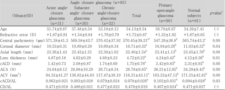
폐쇄각녹내장 환자와 원발개방각녹내장 환자 및 정상인 에서 측정된 전안부 계측치는 Table 1에 나와 있으며 통계 적인 차이를 알기 위해 일원분산분석(one‐way ANOVA) 후 사후검증을 시행하였다. 평균 중심각막두께와 각막직경 그리고 안축장과 수정체두께 비교에서 폐쇄각녹내장 환자 는 원발개방각녹내장 환자 및 정상인과 의미 있는 차이를 보였으며(p<0.05), 원발개방각녹내장 환자와 정상인의 비 교에서는 중심각막두께를 제외하곤 의미 있는 차이를 보이 지 않았다(p<0.05). 평균 앞방깊이와 앞방각, 앞방부피 및
Table 1. Ocular parameters in the subtypes of angle closure‐glaucoma, primary open‐angle glaucoma, and normal subjects
Angle closure glaucoma (n=93)
Primary open‐angle
glaucoma (n=90)
Normal subjects (n=91)
p‐value* (Mean±SD)
Acute angle‐
closure glaucoma
(n=31)
Subacute angle‐ closure
glaucoma (n=30)
Chronic angle‐closure
glaucoma (n=32)
Total
Age 51.74±9.87 57.48±8.54 53.18±8.12 54.13±9.54 56.79±6.67 54.39±7.41 (‐)
Refractive error (D) +1.87±0.91 +1.54±0.84 +1.76±0.79 +1.72±0.87 +1.32±1.02 +1.67±0.65 (‐) Central pachymetry (µm) 571.38±41.3 569.58±43.7 570.42±37.92 570.45±39.21a† 547.26±36.8b 565.74±43.2c 0.00 Corneal diameter (mm) 10.53±0.31 10.89±0.28 10.69±0.34 10.71±0.32a 10.94±0.26b 11.03±0.32b 0.04 Axial length (mm) 22.36±1.43 22.42±1.51 22.38±1.62 22.40±1.54a 23.41±1.13b 23.45±1.76b 0.00 Lens thickness (mm) 4.87±0.18 4.62±0.20 4.68±0.23 4.72±0.22a 4.24±0.42b 4.12±0.36b 0.01 cACD (mm) 1.42±0.73 2.08±0.87 1.74±0.69 1.75±0.76a 2.42±0.83b 2.31±0.92b 0.00 ACA (0) 24.64±9.12 28.98±10.28 26.71±8.34 26.78±9.85a 36.21±12.05b 34.29±9.43b 0.00 ACV (mm3) 94.32±41.27 136.82±44.83 117.47±38.19 116.21±43.11a 163.23±47.13b 171.25±42.82b 0.00 cACD/AL 0.063±0.021 0.093±0.028 0.079±0.024 0.078±0.026a 0.102±0.031b 0.094±0.028b 0.03 CD/AL 0.471±0.018 0.486±0.021 0.477±0.023 0.479±0.019 0.467±0.024‡ 0.471±0.027 (‐)
*By one‐way ANOVA among the all groups (ACG, POAG, Normal), P<0.05; †Values followed by equal letters (a, b, c) in the same row do not differ among them according to the Tukey and Duncan post‐hoc analysis, p<0.05; ‡In primary open‐angle glaucoma, CD/AL value was lower than in angle‐closure glaucoma and normal subjects. cACD=corrected anterior chamber depth (mm); ACA=anterior chamber angle; ACV=anterior chamber volume; AL=axial length; CD=corneal diameter; SD=standard deviation; D=diopter; ACG=angle‐closure glaucoma; POAG=primary open‐angle glaucoma.
안축장으로 보정된 앞방깊이의 비교에서도 폐쇄각녹내장 환자는 다른 두 군과 의미 있는 차이를 보였으며(p<0.05), 원발개방각녹내장 환자와 정상인의 비교에서는 의미 있는 차이를 보이지 않았다(p<0.05). 폐쇄각녹내장 환자 중 급 성에서는 레이저홍채절개술 후 안압이 정상화 되고 각막부 종이 사라지고 난 뒤 각막두께 측정을 시행하였으며, 앞방 깊이의 측정에서도 각막두께에 따른 오차를 줄이고자 각막 후면에서 수정체 사이의 거리(corrected anterior chamber depth)로 정하였다.
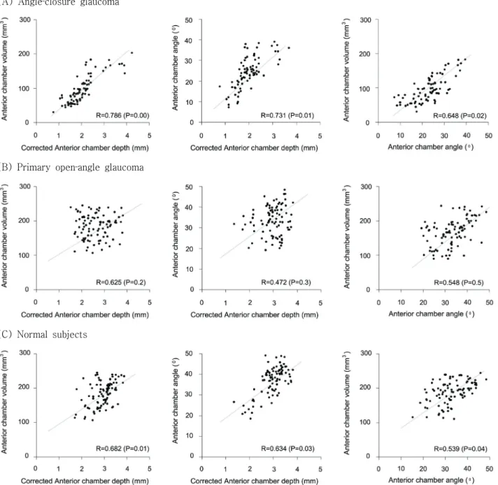
폐쇄각녹내장 환자와 원발개방각녹내장 환자 및 정상인 에서 앞방깊이와 앞방부피, 앞방각의 상관관계를 살펴보면 폐쇄각녹내장 환자에서 앞방깊이와 앞방부피는 제일 높은 상관관계(R=0.786)를 보였으며, 앞방깊이와 앞방각, 앞방 각과 앞방부피도 유의한 상관관계(R=0.731, R=0.648)를 보임을 알 수 있었다(P<0.05)(Fig. 1). 정상인에서도 앞방 깊이와 앞방부피는 높은 상관관계(R=0.682)를 보였으며, 앞방깊이와 앞방각, 앞방각과 앞방부피도 유의한 상관관계 (R=0.634, R=0.539)를 보였다(P<0.05). 그러나 원발개 방각녹내장 환자에서는 앞방깊이, 앞방각, 앞방부피간 유의 한 상관관계를 보이지 않았다(P>0.05).
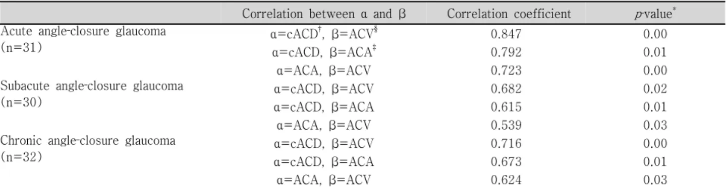
폐쇄각녹내장의 증상별 세 군에서도 비모수적 방법 (Spearman rank correlation)을 이용하여 상관관계를 알아 보았으며 급성폐쇄각녹내장에서 앞방깊이와 앞방부피는 제일 높은 상관관계(R=0.847)를 보였으며 앞방깊이와 앞 방각, 앞방각과 앞방부피도 유의한 상관관계(R=0.792,
R=0.723)를 보임을 알 수 있었다(P<0.05)(Table 2). 만 성폐쇄각녹내장과 아급성폐쇄각녹내장에서도 앞방깊이와 앞방부피는 제일 높은 상관관계(R=0.716, R=682)를 보였 다(P<0.05).
고 찰
성인에서 폐쇄각녹내장은 주로 양안에서 발생하며 적어 도 한 눈에 부분 또는 전체적인 앞방각의 폐쇄가 동반되며, 원인은 다양하지만 크게 동공의 기능적인 폐쇄(functional papillary block)로 인하여 발생하거나 홍채근부(iris root) 가 앞으로 치우쳐진 고원홍채(Plateau iris)에서 섬유주로 의 방수의 흐름을 막아 발생하는 것으로 알려져 있다.17-19 이미 많은 연구에서 이러한 폐쇄각녹내장과 정상안의 해부 학적 차이를 보고 하였으며 저자들 또한 앞방의 여러 계측 치를 구하여 비교해 보았다. 먼저 폐쇄각녹내장 환자의 중 심각막두께와 수정체두께, 각막직경과 안축장은 원발개방 각녹내장 환자와 정상인보다 중심각막두께와 수정체두께에 선 두껍게 측정되었고 각막직경과 안축장은 짧게 측정되어 모두 유의한 차이를 나타내었고 기존에 보고된 문헌과 같 았다.20‐23
저자들은 급성폐쇄각녹내장의 경우 중심각막두께 측정 에서 레이저홍채절개술 후 안압이 정상화 되며 각막부종이 사라지고 급성발작 후 2주에서 4주 사이에 검사를 시행하 였으며 각막두께의 따른 오차가 발생할 가능성이 남아있어
(A) Angle‐closure glaucoma
(B) Primary open‐angle glaucoma
(C) Normal subjects
Figure 1.Correlation plots between each pair of three anterior segment parameters for angle‐closure glaucoma (A), primary open‐angle glaucoma (B), and normal subjects (C). (Pearson’s correlation coefficients (R)and p‐value were determined for each pair of parameters)
앞방깊이의 측정에서는 중심각막두께를 제외하였다.
폐쇄각녹내장의 위험요소로는 인종, 나이, 성별 등과 짧 은 각막직경, 중심각막두께증가, 수정체 두께증가, 짧은 안 축장 뿐 아니라 앞방깊이와 앞방각, 앞방부피 또한 중요한 위치를 차지하며 앞방 폐쇄에 대한 유용한 정보를 제공해 줄 수 있다. Oka et al24은 폐쇄각녹내장 환자와 원발개방각 녹내장 환자의 주변부 앞방깊이 및 앞방부피를 Pentacam® 을 이용하여 측정한 후 폐쇄각녹내장에서 주변부 앞방깊이 및 앞방부피가 유의하게 작음을 보고 하였으며, Lee et al25
은 273명의 폐쇄각녹내장 환자를 급성과 아급성 그리고 만 성으로 분류한 후 앞방직경, 앞방깊이, 앞방부피를 측정하 여 정상인과 비교 하였고 폐쇄각녹내장 환자 모두에서 의 미 있게 작음을 보고 하였다.
본 연구에서도 Pentacam®을 이용하여 폐쇄각녹내장 환 자의 앞방각, 앞방깊이, 앞방부피를 정량적으로 측정 한 후 원발개방각녹내장 환자 및 정상인과 비교 하였으며 앞방각 은 좁고, 앞방깊이는 얕으며 그리고 앞방부피에서는 작아 의미 있는 차이가 있었다(P<0.05).
Table 2. Correlation analysis between anterior segment parameters in the subgroups of angle‐closure glaucoma Correlation between α and β Correlation coefficient p‐value* Acute angle‐closure glaucoma
(n=31)
α=cACD†, β=ACV§ 0.847 0.00
α=cACD, β=ACA‡ 0.792 0.01
α=ACA, β=ACV 0.723 0.00
Subacute angle‐closure glaucoma
(n=30) α=cACD, β=ACV 0.682 0.02
α=cACD, β=ACA 0.615 0.01
α=ACA, β=ACV 0.539 0.03
Chronic angle‐closure glaucoma
(n=32) α=cACD, β=ACV 0.716 0.00
α=cACD, β=ACA 0.673 0.01
α=ACA, β=ACV 0.624 0.03
*In angle‐closure glaucoma subgroups, each two of anterior segment parameters undertaken Spearman rank correlation (p<0.05);
†cACD=corrected anterior chamber depth (mm); ‡ACA=anterior chamber angle (0); §ACV=anterior chamber volume (mm3).
또한 폐쇄각녹내장 환자에서 앞방각, 앞방깊이, 앞방부피 의 상관관계를 분석한 결과 앞방깊이와 앞방부피는 매우 높은 상관관계를 보였으며(R=0.786), 앞방깊이와 앞방각, 앞방각과 앞방부피도 유의한 상관관계를 보임을 알 수 있 었다(P<0.05). 정상인에서도 앞방깊이와 앞방부피는 높은 상관관계(R=0.682)를 보이고 있지만 폐쇄각녹내장에서 더 높은 상관관계를 나타내었으며 본 연구에서는 폐쇄각녹 내장과 연령 및 성비, 굴절력이 보정된 정상인으로 다른 문 헌의 정상군과 비교해 폐쇄각녹내장의 위험요인을 다소 포 함하고 있을 가능성이 있다.26‐28
원발개방각녹내장 환자에서는 앞방각, 앞방깊이, 앞방부 피간 유의한 상관관계를 알 수 없었으며 안축장은 정상과 비슷하지만 상대적으로 각막직경이 짧고 앞방각과 앞방깊 이에 비하여 상대적으로 큰 앞방부피의 소견을 보여 원할 한 방수의 흐름에 장애가 발생할 가능성이 높았다.29‐31
이와 같이 정량적으로 측정된 앞방각, 앞방깊이, 앞방부 피는 폐쇄각녹내장에서 좀 더 중요한 요소임을 알 수 있으 며 저자들은 폐쇄각녹내장 환자를 증상별로 구별하여 급성, 아급성, 만성으로 나누었으며 급성과 만성폐쇄각녹내장 환 자의 앞방각, 앞방깊이, 앞방부피는 아급성과 비교하여 통 계학적인 의미는 없으나 수치상 작음을 알 수 있었고 각각 의 앞방각, 앞방깊이, 앞방부피간의 상관관계를 분석한 결 과 아급성 및 만성폐쇄각녹내장과 비교하여 앞방폐쇄로 인 한 급성발작을 경험한 급성폐쇄각녹내장 환자에서 앞방각, 앞방깊이, 앞방부피간에 가장 높은 상관관계를 보임을 알 수 있었다(Table 2).
많은 연구에서 급성폐쇄각녹내장은 안축장 및 각막직경 이 폐쇄각녹내장 중 가장 좁고 수정체는 앞으로 전위되어 앞방폐쇄에 가장 민감해지며 만성에서는 급성과 비교하여 앞방폐쇄가 그리 심하지 않으나 상대적으로 적은 앞방폐쇄 가 반복적으로 발생하고 주변홍채앞유착이 진행되어 홍채 절개술 후에도 안압이 증가 할 수 있음이 보고되며 아급성
의 경우 폐쇄각녹내장의 비슷한 특징을 가지고 있지만 상 대적으로 앞방폐쇄 정도가 심하지 않아 간헐적인 증상만을 호소하는 것으로 알려져 있다.22,23,32‐35
저자들이 시행한 앞 방의 정량적 계측결과는 이러한 사실을 뒷받침해 줄 수 있 으며 앞방폐쇄에 대한 3차원적인 정보를 제공해 줄 것으로 생각된다. 그러나 급성폐쇄각녹내장 환자에서 급성발작 전 의 앞방계측치 값을 알 수 없었고 발작 후 레이져홍채절개 술을 모두에서 시행하였기에 발작 전후의 정확한 측정값을 비교할 수 없었다. 그리고 폐쇄각녹내장이 양안에 주로 발 생하므로 반대안의 앞방계측치를 측정하여 비교해 보았으 나 폐쇄각녹내장 및 정상인과의 차이를 알 수 없었다.
본 연구에서는 Pentacam®에 의존하여 앞방계측치를 분 석하였기에 다른 기기와의 측정값의 차이를 비교 분석할 필요가 있어 기존의 다른 기기와 Pentacam®과의 측정값의 차이를 비교한 문헌을 찾아보면 Buehl et al36은 정상인에 서 각막의 두께와 앞방의 깊이를 알기 위해 Pentacam®과 Orbscan, AC‐Master를 이용하였으며 각 기계간에 중심각 막두께 및 앞방깊이에서 매우 높은 상관관계가 있음을 보 고 하였고(R=0.92 to 0.97), 이 중 Pentacam® (rotating Scheimpflug camera)과 AC-Master (partial coherence interferometry)에서 Orbscan에 비하여 측정값의 신뢰도가 높았다고 하였다. 그러나 각막두께측정에 있어 초음파를 이 용한 방법이 현재 gold standard 라고 알려져 있어 초음파 각막두께 측정기과 비교한 논문을 보면 Barkana et al37은 Pentacam®과 초음파로 측정한 중심각막두께에서 차이가 없다고 하였고, O’Donnell and Maldonado‐Codina38의 보고 에서는 Pentacam®으로 측정한 중심각막두께가 더 적다고 하였다. 또 Lackner et al39은 앞방깊이의 측정에서 초음파 보다 Pentacam®으로 측정한 값이 더 깊게 나타나지만 임상 적으로 받아들일만한 정도라고 보고 하였다. 이처럼 많은 연구에서 Pentacam®을 이용하여 앞방을 정량적으로 측정 하였으며, 다른 위치에서의 앞방각 측정 뿐 아니라 수정체
의 위치와 혼탁 등 다양한 생체 정보에 대한 검사가 가능하 며 짧은 시간 내에 높은 신뢰도를 가질 수 있음이 보고 되 고 있다.40‐43하지만 Pentacam®을 이용하여 앞방을 관찰함 에 있어 전방각경등 다른 검사의 보조가 필요 할 수 있으며 보다 정확한 전안부 측정결과를 알기 위해선 많은 연구가 필요할 것으로 사료된다.
참고문헌
1) Holladay JT. Standardizing constants for ultrasonic biometry, keratometry, and intraocular lens power calculations. J Cataract Refractive Surg 1997;23:1356-70.
2) Kim YY, Jung HR. Clarifying the nomenclature for primary angle closure glaucoma. Surv Ophthalmol 1997;42:125-36.
3) Ang MH, Baskaran M, Kumar RS, et al. National survey of ophthalmologists in Singapore for the assessment and management of asymptomatic angle closure. J Glaucoma 2008;17:1-4.
4) Lavanya R, Wong TY, Friedman DS, et al. Determinants of angle closure in older Singaporeans. Arch Ophthalmol 2008;126:686-91.
5) Koranyi G, Lydahl E, Norrby S, Taube M. Anterior chamber depth measurement: A‐scan versus optical methods. J Cataract Refract Surg 2002;28:243-7.
6) Meinhardt B, Stachs O, Stave J, et al. Evaluation of biometric methods for measuring the anterior chamber depth in the non
‐contact mode. Graefes Arch Clin Exp Ophthalmol 2006;244:
559-64.
7) Ryu HW, Kim KR, Chung SK. Comparison of A‐scan, scheimpflug camera, and orbscan for measurement of anterior chamber depth. J Korea Ophthalmol Soc 2006;47:1287-91.
8) Giers U, Epple C. Comparison of A‐scan device accuracy. J Cataract Refract Surg 1990;16:235-42.
9) Konstantopoulos A, Hossain P, Anderson DF. Recent advances in ophthalmic anterior segment imaging: a new era for ophthalmic diagnosis? Br J Ophthalmol 2007;91:551-7.
10) Swartz T, Marten L, Wang M. Measuring the cornea: the latest developments in corneal topography. Curr Opin Ophthalmol 2007;18:325-33.
11) Elbaz U, Barkana Y, Gerber Y, et al. Comparison of different techniques of anterior chamber depth and keratometric measurements. Am J Ophthalmol 2007;143:48-53.
12) Emre S, Doganay S, Yologlu S. Evaluation of anterior segment parameters in keratoconic eyes measured with the Pentacam system. J Cataract Refract Surg 2007;33:1708-12.
13) Friedman DS, Gazzard G, Foster P, et al. Ultrasonographic biomicroscopy, Scheimpflug photography, and novel provocative tests in contralateral eyes of Chinese patients initially seen with acute angle closure. Arch Ophthalmol 2003;121:633-42.
14) Bourne RR, Alsbirk PH. Anterior chamber depth measurement by optical pachymetry: systematic difference using the Haag‐
Streit attachments. Br J Ophthalmol 2006;90:142-5.
15) Foster PJ, Devereux JG, Alsbirk PH. Detection of gonioscopically occludable angles and primary angle closure glaucoma by estimation of limbal chamber depth in Asians:
modified grading scheme. Br J Ophthalmol 2000;84:186-92.
16) Congdon NG, Spaeth GL, Augsburger J, et al. A proposed simple method for measurement in the anterior chamber angle: biometric gonioscopy. Ophthalmology 1999;106:2161-7.
17) Lowe R. Primary creeping angle‐closure glaucoma. Br J Ophthalmol 1964;48:544-50.
18) Foster PJ, Buhrmann R, Quigley HA, Johson GJ. The definition and classification of glaucoma in prevalence surveys.
Br J Ophthalmol 2002;86:238-42.
19) Shields MB, Ritch R. The secondary glaucomas. St Louis: CV Mosby, 1982;3-7.
20) Tomlision A, Leighton DA. Ocular dimensions in the heredity of angle closre glaucoma. Br J Ophthalmol 1973;57:475-86.
21) Dandona L, Dandona R, Mandal P, et al. Angle closure glaucoma in an urban population in South India. Ophthal- mology 2000;107:1710-6.
22) Lowe RF. Aetiology of the anatomical basis for primary angle closure glaucoma. Biometrical comparisons between normal eyes and eyes with primary angle closure glaucoma. Br J Ophthalmol 1970;54:161-9.
23) Sihota R, Lakshmaiah NC, Agarwal HC, et al. Ocular parameters in the subgroups of angle closure glaucoma. Clin Experiment Ophthalmol 2000;28:253-8.
24) Oka N, Otori Y, Okada M, et al. Clinical study of anterior ocular segment topography in angle‐closure glaucoma using the three‐dimensional anterior segment analyzer Pentacam.
Nippon Ganka Gakkai Zasshi 2006;110:398-403.
25) Lee DA, Brubaker RF, Ilstrup DM. Anterior chamber dimensions in patients with narrow angles and angle‐closure glaucoma. Arch Ophthalmol 1984;102:46-50.
26) Congdon N, Wang F, Tielsch JM. Issues in the epidemiology and population‐based screening of primary angle‐closure glaucoma. Surv Ophthalmol 1992;36:411-23.
27) Javitt J, Sommer A. A population‐based evaluation of glaucoma screening: the Baltimore eye surgery. Am J Ophthalmol 1991;134:1102-10.
28) Shiose Y, Kitazawa Y, Tsukuhara S, et al. Epidemiology of glaucoma in Japan: a nationwide glaucoma survey. Jpn J Ophthalmol 1991;35:133-55.
29) Tomlinson A, Leighton DA. Ocular dimensions and the heredity of open‐angle glaucoma. Br J Ophthalmol 1974;58:
68-74.
30) Silver DM, Quigley HA. Aqueous flow through the iris‐lens channel: estimates of differential pressure between the anterior and posterior chambers. J Glaucoma 2004;13:100-7.
31) Singh RP, Goldberg I, Graham, et al. Central corneal thickness, tonometry, and ocular dimensions in glaucoma and ocular hypertension. J Glaucoma 2001;10:206-10.
32) Sihota R, Dada T, Gupta R, et al. Ultrasound biomicroscopy in the subtypes of primary angle closure glaucoma. J Glaucoma 2005;14:387-91.
33) Lee DA, Brubaker RF, Ilstrup DM. Anterior chamber dimensions in patients with narrow angles and angle‐closure glaucoma. Arch Ophthalmol 1984;102:46-50.
34) Mimiwati Z, Fathilah J. Ocular biometry in the subtypes of primary angle closure glaucoma in University Malaya Medical Centre. Med J Malaysia 2001;56:341-9.
35) Lee JY, Kim YY, Jung HR. Distribution and characteristics of
=ABSTRACT=
Comparision of Anterior Segment Parameters in Angle‐Closure Glaucoma Using Scheimpflug Camera
Sang Min Kwon, MD, Hyun Chul Oh, MD, Dong Joon Lee, MD, Woo Jin Jeung, MD, Sae Heun Rho, MD.
Department of Ophthalmology, Dong‐A University College of Medicine, Pusan, Korea
Purpose: To compare anterior segment parameters in angle‐closure glaucoma (ACG), primary open angle glaucoma (POAG), and normal subjects (N) using a Schiempflug camera.
Methods: Central corneal thickness (CCT), lens thickness (LT), axial length (AL), anterior chamber angle (ACA), anterior chamber depth (ACD), and anterior chamber volume (ACV) were measured in ACG (93 eyes of 92 patients), POAG (90 eyes of 87 patients), and normal (91 eyes of 88 subjects) with PentacamⓇ and A‐scan. All of the results and measurements were then compared.
Results: Compared to normal and POAG patients, ACG patients presented with significantly different measurements of CCT, LT, AL, and ACA, ACD, and ACV (p<0.05). Further, correlations were high between three measurements (ACA, ACD, ACV) in ACG, and the best correlations were found in acute angle‐closure glaucoma (P<0.05).
Conclusions: By using a Schiempflug camera it was possible to assess the correlation between anterior segment parameters (ACA, ACD, ACV) in glaucoma patients. The best correlations were found in acute angle‐closure glaucoma, and thus anterior segment parameters can offer reciprocally complementary information.
J Korean Ophthalmol Soc 2009;50(1):128-134
Key Words: Angle closure glaucoma, Anterior segment parameters, Primary open angle glaucoma, Schiempflug camera
Address reprint requests to Sae Heun Rho, MD
Department of Ophthalmology, Dong‐A University College of Medicine
#3‐1 Dongdaesin‐dong, Seo‐gu, Pusan 602‐714, Korea
Tel: 82‐51‐240‐5227, Fax: 82‐51‐254‐1987, E-mail: [email protected] peripheral anterior synechiae in primary angle‐closure glaucoma.
Korean J Ophthalmol 2006;20:104-8.
36) Buehl W, Stojanac D, Sacu S, et al. Comparison of three methods of measuring corneal thickness and anterior chamber depth. Am J Ophthalmol 2006;141:7-12.
37) Barkana Y, Gerber Y, Elbaz U, et al. Central corneal thickness measurement with the Pentacam Scheimpflug system, optical low‐coherence reflectometry pachymeter, and ultrasound pachymetry. J Cataract Refract Surg 2005;31:1729-35.
38) O'Donnell C, Maldonado‐Codina C. Agreement and repeat- ability of central thickness measurement in normal corneas using ultrasound pachymetry and the OCULUS Pentacam.
Cornea 2005;24:920-4.
39) Lackner B, Schmidinger G, Skorpik C. Validity and repeat- ability of anterior chamber depth measurements with
Pentacam and Orbscan. Optom Vis Sci 2005;82:858-61.
40) Lackner B, Schmidinger G, Pieh S, et al. Repeatability and reproducibility of central corneal thickness measurement with Pentacam, Orbscan, and ultrasound. Optom Vis Sci 2005;82:
892-9.
41) Fujioka M, Nakamura M, Tatsumi Y, et al. Comparison of Pentacam Scheimpflug camera with ultrasound pachymetry and noncontact specular microscopy in measuring central corneal thickness. Curr Eye Res 2007;32:89-94.
42) Shin YJ, Kim NH, Kim DH. Comparison of pentacam with orbscan. J Korea Ophthalmol Soc 2007;48:637-41.
43) Rabsilber TM, Khoramnia R, Auffarth GU. Anterior chamber measurements using Pentacam rotating Scheimpflug camera. J Cataract Refract Surg 2006;32:456-9.